PGHN
Case Report
A Novel Homozygous LIPA Mutation in a Korean Child with Lysosomal Acid Lipase Deficiency
Kwang Yeon Kim, Ju Whi Kim, Kyung Jae Lee, Eunhyang Park*, Gyeong Hoon Kang*, Young Hun Choi
†, Woo Sun Kim
†, Jung Min Ko, Jin Soo Moon, and Jae Sung Ko
Departments of Pediatrics, *Pathology, and †Radiology, Seoul National University College of Medicine, Seoul, Korea
Patients with lysosomal acid lipase (LAL) deficiency and glycogen storage disease (GSD) demonstrated hep- atomegaly and dyslipidemia. In our case, a 6-year-old boy presented with hepatosplenomegaly. At 3 years of age, GSD had been diagnosed by liver biopsy at another hospital. He showed elevated serum liver enzymes and dyslipidemia. Liver biopsy revealed diffuse microvesicular fatty changes in hepatocytes, septal fibrosis and foamy macrophages. Ultrastructural examination demonstrated numerous lysosomes that contained lipid material and in- tracytoplasmic cholesterol clefts. A dried blood spot test revealed markedly decreased activity of LAL. LIPA gene sequencing identified the presence of a novel homozygous mutation (p.Thr177Ile). The patient’s elevated liver en- zymes and dyslipidemia improved with enzyme replacement therapy. This is the first report of a Korean child with LAL deficiency, and our findings suggest that this condition should be considered in the differential diagnosis of chil- dren with hepatosplenomegaly and dyslipidemia.
Key Words: Lysosomal acid lipase deficiency, Glycogen storage disease, Lysosomes, Hepatomegaly, Dyslipidemias
Received:October 1, 2016, Revised:March 17, 2017, Accepted:March 17, 2017
Corresponding author: Jae Sung Ko, Department of Pediatrics, Seoul National University Hospital, 101 Daehak-ro, Jongno-gu, Seoul 03080, Korea. Tel: +82-2-2072-2197, Fax: +82-2-743-3455, E-mail: [email protected]
Copyright ⓒ 2017 by The Korean Society of Pediatric Gastroenterology, Hepatology and Nutrition
This is an openaccess article distributed under the terms of the Creative Commons Attribution NonCommercial License (http://creativecommons.org/licenses/by-nc/4.0/) which permits unrestricted noncommercial use, distribution, and reproduction in any medium, provided the original work is properly cited.
INTRODUCTION
Lysosomal acid lipase (LAL) is an important en- zyme involved in maintaining cholesterol homeo- stasis inside cells, and it is essential for the intra- cellular hydrolysis of cholesteryl esters and trigly- cerides [1]. In the case of lysosomal acid lipase defi- ciency (LALD), non-hydrolyzed cholesteryl esters and triglycerides accumulate in hepatocytes, result- ing in the synthesis of endogenous cholesterol and
low-density lipoprotein (LDL). Glycogen storage dis- ease (GSD) is also a metabolic disorder that is caused by deficiency of an enzyme involved in glycogen breakdown, resulting in the accumulation of glyco- gen in hepatocytes.
Both diseases present a very similar clinical mani- festation and histologic findings that may lead to misdiagnosis. Therefore, careful differential diag- nosis between LALD and GSD is important. Here, we report the first Korean patient with LALD who was
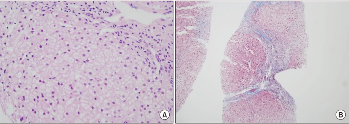
Fig. 1. Light microscopy examination of the liver biopsy tissue showed (A) diffuse microvesicular fatty changes in hepatocytes (H&E, ×200) and (B) septal fibrosis (Masson-trichrome, ×40).
previously misdiagnosed with GSD.
CASE REPORT
A 6-year-old boy was referred with abdominal distension. The patient was sent to an orphanage when he was 1 year old; therefore, his specific birth history is unknown. At the age of 2, he had a seizure but did not receive any evaluation. When he was 3 years old, he was brought to a hospital because of ab- dominal distension, poor weight gain and devel- opmental delay. A blood test showed elevated liver enzymes, and hepatomegaly was observed by ab- dominal ultrasonography. Liver biopsy revealed swollen hepatocytes with flocculent cytoplasm and septal fibrosis. He was diagnosed with GSD. At 6 years of age, he was transferred to Seoul National University Children’s Hospital for further diagnostic evaluation and proper management.
At this time, his height was 107.3 cm (<3rd per- centile), body weight was 16.9 kg (<3rd percentile) and head circumference was 49 cm (5-10th percen- tile). His blood pressure was 123/77 mmHg (>95th percentile), pulse rate was 102/min, respiratory rate was 24/min, and body temperature was 37.1oC. His abdomen was soft but distended. There was no ten- derness or rebound tenderness. The liver was pal- pated four finger breadth below the costal margin.
The spleen was palpated two finger breadth below the costal margin.
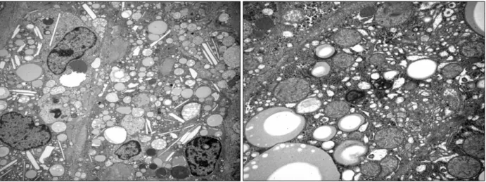
Liver magnetic resonance imaging (MRI) revealed a hepatic fat percentage of 6.35%. Aspartate amino- transferase (AST) and alanine aminotransferase (ALT) levels were elevated at 72 and 76 U/L, res- pectively. His lipid profile was also elevated, with a total cholesterol of 226 mg/dL and an LDL of 192 mg/dL. His high-density lipoprotein (HDL) level re- vealed a moderate reduction of 19 mg/dL. The pa- tient’s serum calcium level was slightly elevated at 11.9 mg/dL; however, the parathyroid hormone level was normal (8 pg/mL) . The findings of the liver biop- sy, through light microscopy, showed diffuse micro- vesicular fatty changes in hepatocytes and septal fib- rosis (Fig. 1). The portal spaces also showed some foamy macrophages. Ultrastructural examination revealed that the cytoplasm of the liver cells con- tained numerous lysosomes filled with a lipid mate- rial and intracytoplasmic cholesterol clefts, suggest- ing lipid metabolic disorder (Fig. 2). The patient’s LAL activity, determined by a dried blood spot test, was 10.2 pmol/punch/h, which was considerably lower than the normal value (controls, 265.4±78.3;
carriers, 107.1±30.7). Molecular genetic analysis of the LIPA gene was performed for confirmative diag- nosis. Direct sequencing analysis of LIPA identified a novel homozygous mutation, c530C>T (p.Thr177Ile),
Fig. 2. Electron microscopy examination of the liver tissue revealed macrophages with numerous lipid-filled lysosomes at the portal area and hepatocytes that contained many large glycogen particles and cholesterol clefts (×400).
Fig. 3. Direct sequencing of the LIPA gene. A novel homozygous mutation, c530C>T(p.Thr177Ile), was identified.
in exon 5 (Fig. 3). The Polyphen-2, Sorting intolerant from tolerant and Mutation Taster programs pre- dicted p.Thr177Ile to function as a pathogenic mutation. Two siblings of the patient were evaluated pre-symptomatically. Genetic analysis of the two sib- lings showed that one is a heterozygous carrier, while the other is unaffected.
Enzyme replacement therapy (sebelipase alfa 1 mg/kg every 2 weeks) was initiated, and the patient consequently showed improvements in transaminases and the lipid profile as well as a reduction in liver volume.
Marked decreases from baseline in AST (72 U/L at baseline, decreased to 44 U/L), ALT (76 U/L at base- line, decreased to 40 U/L), and LDL (192 mg/dL at baseline, decreased to 134 mg/dL) were seen after 10
months of treatment. In addition to the improve- ments in transaminases and the lipid profile, liver MRI revealed significant reduction in hepatic fat percentage (6.35% at baseline, decreased to 3.06%).
DISCUSSION
LALD is a disorder caused by deficiency in LAL ac- tivity, resulting in the accumulation of cholesteryl esters. Nearly all LALD patients present with hep- atosplenomegaly and growth retardation [2], and most patients show elevated hepatic transaminase (AST/ALT), serum total cholesterol and LDL levels, while HDL levels are decreased [3].
In the present case, the patient had been pre- viously diagnosed with GSD. GSD is an inherited dis- order of carbohydrate metabolism that is caused by deficiency in an enzyme involved in glycogen break- down, resulting in the accumulation of glycogen in hepatocytes. Similar to LALD patients, hepatos- plenomegaly, dyslipidemia, and growth retardation are the main findings in GSD [4,5]. However, unlike LALD patients, GSD patients may present with fast- ing hypoglycemia [6].
For the differential diagnosis of LALD and GSD, the findings of a liver biopsy are important.
Liver biopsy specimens collected from the current patient revealed diffuse microvesicular fatty changes
in hepatocytes and septal fibrosis; the portal spaces also showed some foamy macrophages. Ultrastruc- tural examination demonstrated that the liver cell cytoplasm contained numerous lysosomes filled with lipid material and intracytoplasmic cholesterol clefts. GSD patients also showed pale cytoplasmic changes in hepatocytes and occasional micro- vesicular steatosis in liver biopsies [3]. However, prominent nuclear hyperglycogenation and swollen hepatocyte are the key diagnostic features of GSD [7]. Additionally, the liver typically contains particles that are strongly stained with Periodic Acid-Schiff and can be decolorized using a bleaching agent; in addition, septal fibrosis is generally detected in GSD type 3. In contrast to GSD, periportal foamy macro- phage and intracytoplasmic cholesterol clefts are the main diagnostic features of LALD. Because of their overlapping clinical and histological findings, care- ful differential diagnosis is important.
The current patient had hypertension upon phys- ical examination when he was initially evaluated.
The cardiovascular manifestations of LALD patients include accelerated atherosclerosis, heart failure, aortic calcifications, myocardial infarction, and stroke. Hypertension was previously reported in 3 patients between 4 and 8 years of age [3].
The current patient’s serum calcium level was slightly elevated to 11.9 mg/dL, although an evalua- tion of hypercalcemia revealed no abnormality.
There have been no reports of hypercalcemia in LALD patients.
To date, over 40 LIPA mutations causing LALD have been identified. The most common LIPA gene mutation in Caucasians is an exon 8 splice-junction mutation (E8SJM-1G>A), which results in altered mRNA splicing and exon 8 skipping [3]. However, the LIPA gene mutations reported in Japanese pa- tients include Tyr22X, Val203 Leu and Leu264Pro [8,9]. Additionally, sequencing of the LIPA gene in this Korean patient revealed a novel homozygous mutation in exon 5 (p.Thr177Ile). These findings in Korean and Japanese patients suggest that LIPA gene mutations in Asians maybe different from those in Caucasians. Consequently, estimation of the
prevalence of LALD in Asian populations requires further study, as E8SJM-1G>A is not common in these racial groups [10].
Sebelipase alfa is a recently developed treatment that can modify the natural course of the illness. As shown in this case, sebelipase alfa produced a rapid decreases in serum transaminases, improvements in the serum lipid profile and a reduction in liver and spleen size [11,12] .
In summary, we report the first Korean case of LALD with a novel LIPA mutation. This patient had been previously misdiagnosed with GSD. Although rare, LALD should be considered in the differential diagnosis of a child with hepatosplenomegaly and dyslipidemia.
REFERENCES
1. Du H, Sheriff S, Bezerra J, Leonova T, Grabowski GA.
Molecular and enzymatic analyses of lysosomal acid li- pase in cholesteryl ester storage disease. Mol Genet Metab 1998;64:126-34.
2. Hoffman EP, Barr ML, Giovanni MA, Murray MF.
Lysosomal acid lipase deficiency. In: Pagon RA, Adam MP, Ardinger HH, Wallace SE, Amemiya A, Bean LJH, et al, eds. GeneReviews(R). Seattle (WA): University of Washington, 1993.
3. Bernstein DL, Hülkova H, Bialer MG, Desnick RJ.
Cholesteryl ester storage disease: review of the findings in 135 reported patients with an underdiagnosed disease. J Hepatol 2013;58:1230-43.
4. Heller S, Worona L, Consuelo A. Nutritional therapy for glycogen storage diseases. J Pediatr Gastroenterol Nutr 2008;47 Suppl 1:S15-21.
5. Burton BK, Deegan PB, Enns GM, Guardamagna O, Horslen S, Hovingh GK, et al. Clinical features of lyso- somal acid lipase deficiency. J Pediatr Gastroenterol Nutr 2015;61:619-25.
6. Barker CC, Butzner JD, Woodman RC, Parsons HG.
Crohn-like enteritis presenting as hypoglycemia in a patient with glycogen storage disease type 1b, treated with granulocyte colony-stimulating factor and splenectomy. J Pediatr Gastroenterol Nutr 2001;32:
197-200.
7. Ko JS, Moon JS, Seo JK, Yang HR, Chang JY, Park SS.
A mutation analysis of the AGL gene in Korean patients with glycogen storage disease type III. J Hum Genet 2007;59:42-5.
8. Fujiyama J, Sakuraba H, Kuriyama M, Fujita T, Nagata K, Nakagawa H, et al. A new mutation (LIPA Tyr22X) of lysosomal acid lipase gene in a Japanese pa- tient with Wolman disease. Hum Mutat 1996;8:377-80.
9. Kuranobu N, Murakami J, Okamoto K, Nishimura R, Murayama K, Takamura A, et al. Cholesterol ester stor- age disease with a novel LIPA mutation (L264P) that presented massive hepatomegaly: a case report. Hepa- tol Res 2016;46:477-82.
10. Scott SA, Liu B, Nazarenko I, Martis S, Kozlitina J, Yang Y, et al. Frequency of the cholesteryl ester storage disease common LIPA E8SJM mutation (c.894G>A) in
various racial and ethnic groups. Hepatology 2013;
58:958-65.
11. Burton BK, Balwani M, Feillet F, Barić I, Burrow TA, Camarena Grande C, et al. A phase 3 trial of Sebelipase alfa in lysosomal acid lipase deficiency. N Engl J Med 2015;373:1010-20.
12. Valayannopoulos V, Malinova V, Honzík T, Balwani M, Breen C, Deegan PB, et al. Sebelipase alfa over 52 weeks reduces serum transaminases, liver volume and im- proves serum lipids in patients with lysosomal acid li- pase deficiency. J Hepatol 2014;61:1135-42.