Korean J Perinatol Vol.26, No.1, Mar., 2015 http://dx.doi.org/10.14734/kjp.2015.26.1.35
� Original Article �
furosemide therapy in 1982. Among different studies, the prevalence of NC ranged from 7 to 41% in very low birth weight (VLBW) infants with a gestational age less than 32 weeks.3-9
The etiology of NC is multifactorial including low gestational age and birth weight. Male sex, family history, white race, mechanical ventilation, oxygen therapy, bronchopulmonary dysplasia (BPD), the use of furosemide, methylxanthine, dexamethasone, gentamicin, total parenteral nutrition (TPN), acidosis, hypercalcemia, hypophosphatemia, hypercalciuria, hyperoxaluria, hyperuricuria, and hypocitruria are associated with development of NC.2-14
Most cases of NC resolves within one year; how- Nephrocalcinosis (NC), defined as the deposition of
calcium crystals in the renal parenchyma, occurs due to an imbalance between promoters and inhibitors of crystallization in urine. After first description of NC in adult by Dimopoulos et al. in 1971,1 Hufnagle et al.2 reported 10 premature infants of NC after long-term
Risk Factors and Outcome of Nephrocalcinosis in Very Low Birth Weight Infants
Ho Sung Kim, M.D., Kumi Jeong, M.D., Young Youn Choi, M.D., Ph.D., Eun Song Song, M.D., Ph.D.
Department of Pediatrics, Chonnam National University Hospital, Gwangju, Korea
Purpose: The aim of this study was to determine the incidence, risk factors, and long-term outcome of nephrocalcinosis in very low birth weight (VLBW) infants.
Methods: A retrospective chart review was performed in VLBW infants between 2006 and 2012 in the neonatal intensive care unit.
Results: The incidence of nephrocalcinosis in VLBW infants was 10.2%. By univariate analysis, oligohydramnios and use of antenatal steroids were more frequent in the nephrocalcinosis group. In the nephrocalcinosis group, the gestational age and birth weight were lower and there were more number of female infants. Also, the initial blood pH, the lowest systolic blood pressure, and urine output on the first day of life were lower and bronchopulmonary dysplasia, sepsis, and urinary tract infection were more prevalent in the nephrocalcinosis group. The use of dexamethasone or ibuprofen and the lowest levels of phosphorus, protein and albumin were significantly lower in the nephrocalcinosis group. By binary logistic regression analysis, the use of antenatal steroids, female sex, 5-minute Apgar score, duration of oxygen therapy and total parenteral nutrition, and the lowest albumin level were found to be significant risk factors for nephrocalcinosis. Overall, the resolution rate was 64.1% and 88.6% within 12 months and 18 months, respectively.
Conclusions: The incidence of nephrocalcinosis in VLBW infants showed increasing trend. The risk factors of nephrocalcinosis were parameters for sick VLBW infants. Although the prognosis of nephrocalcinosis was relatively good, we should pay close attention to the development of complication.
Key Words: Nephrocalcinosis, Very low birth weight infants, Incidence, Risk factors, Outcome
Received: 9 August 2014 Revised: 5 October 2014 Accepted: 21 October 2014
Correspondence to: Eun Song Song, M.D., Ph.D., Department of Pediatrics, Chonnam National University Hospital, 42, jebong-ro, Dong-gu, Gwangju 501-757, Korea
Tel: +82-62-220-6649, Fax: +82-62-222-6103 E-mail: essong@jnu.ac.kr
Copyrightⓒ 2015 by The Korean Society of Perinatology
This is an Open Access article distributed under the terms of the Creative Commons Attribution Non-Commercial License (http://creativecommons.org/license/
by-nc/3.0/), which permits unrestricted non-commercial use, distribution, and reproduction in any medium, provided that the original work is properly cited.
The Korean Journal of Perinatology · pISSN 1229-2605 eISSN 2289-0432 · e-kjp.org
the medulla or cortex that was reproducible in both transverse and longitudinal directions on renal US.27
2. Date collection
Maternal data such as age, preterm premature rupture of membranes (PPROM), diabetic disease, hypertensive disease, histologic chorioamnionitis, and use of antenatal steroids were recorded. Gesta- tional age, birth weight, gender, Apgar scores at 1 minute and 5 minutes, pH in the first arterial or ven- ous blood gas analysis, lowest systolic blood pressure (measured by noninvasive blood pressure or invasive arterial blood pressure) and urine output on the first day after birth were investigated. Morbidity data include the presence of patent ductus arteriosus (PDA), BPD, bacterial sepsis, fungal sepsis, and UTI.
With respect to the treatment profile, duration of mechanical ventilation, oxygen therapy, TPN and phototherapy, and the postnatal day at initial trophic feeding were obtained.
The frequency of surfactant, dexamethasone, indomethacin, ibuprofen, and furosemide, and the duration of use of furosemide, gentamicin, cefota- xime, ampicillin, fluconazole, and theophylline were recorded. The highest and lowest levels of serum calcium and phosphate, blood urea nitrogen (BUN), creatinine, protein, and albumin levels were recorded from the data of every 2 week interval.
Follow-up renal US was performed every one to three months in patients with NC at the out-patient department. In addition, we obtained the data of the presence of UTI, renal function impairment, and hypertension in the medical records.
3. Statistical analysis
Data were analyzed by Statistical Program for the Social Science (SPSS) version 22.0, and P values less than 0.05 were considered statistically significant.
ever, in the minority of patients, NC can persist for several years.9, 15-18, 20 Short-term compli cations include urinary tract infection (UTI), renal stone, hydronephrosis, hematuria, and colicky pain. Delayed renal growth, renal function impairment, and hyper- tension have been reported as the long-term com- plications.16,17,21,22
A few studies that addressed the risk factors of NC have been reported in Korea,23-25 but there is no long-term follow-up data yet. The aim of this study was to determine the incidence, risk factors, and long-term outcome of NC in VLBW infants.
Subjects and Methods
1. Subjects
A retrospective chart review was performed in 688 infants of birth weight less than 1,500 g who were admitted between January 2006 and December 2012 in the neonatal intensive care unit of Chonnam National University Hospital, Gwangju, Korea. One hundred seventy-eight infants [43 infants who were discharged before 4 weeks of life, 11 infants who were transferred to other hospitals, 84 infants who died due to severe congenital anomaly or severe prematurity, and 40 infants who did not undergo renal ultrasonography (US)] were excluded. The remain- ing 510 infants were enrolled in this study; the NC group included 52 infants, and the control group included 458 infants.
Renal US was performed by a consultant radiolo- gist. The indication of US was hypercalcemia, or hypercalciuria (urinary calcium-creatinine ratio
>0.8),26 or presence of calcium crystals in urinalysis.
Additionally, renal US was routinely preformed in VLBW infants before discharge.
NC was defined as at least one bright reflection in
Continuous variables were compared using the Mann-Whitney U test. Non-continuous variables were compared using the X2 test or Fisher’s exact test. To identify statistically meaningful data in the univariate analysis, a binary logistic regression analysis was performed as the multivariate analysis.
Results
1. Incidence
The overall incidence of NC in VLBW infants was 10.2% with an increasing tendency in every year.
The incidence of NC differed depending on the birth weight, occurring in 15.4% below 750 g, 28.8%
between 750-999 g, 48.1% between 1,000-1,249 g, 7.7% between 1,250-1,499 g. The average time of diagnosis was 68.4 days of life (range 27-166 days of life). All cases in the NC group showed deposition of
calcium salts in the medulla of the kidney. Among the 52 VLBW infants in the NC group, 48 cases had bilateral NC (92.3%) and 4 cases had unilateral NC (7.7%, 2 cases in the left kidney, and 2 cases in the right kidney).
2. Univariate analysis of risk factors
1) Baseline characteristics of mothers and their infants
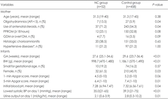
Compared with the control group, oligohydramnios and use of antenatal steroids were more frequent in the NC group. There was no significant difference in maternal age, PPROM, diabetic disease, histologic chorioamnionitis, and hypertensive disease in two groups (Table 1).
The NC group had lower gestational age and birth weight than the control group. NC developed predo- minantly in female infants in our study. Also, the
Table 1. Baseline characteristics of mothers and their infants
Variables NC group
(n=52) Control group
(n=458) P-value
Mother
Age (years), mean (range) 31.5 (19-40) 31.3 (17-45) 0.38
Oligohydramnios (AFI < 5), n (%) 7 (13.5) 27 (5.9) 0.04
Use of antenatal steroids, n (%) 37 (71.2) 243 (54.3) 0.04
PPROM (≥ 18 hours) 12 (23.1) 150 (32.8) 0.08
GDM or overt DM, n (%) 4 (7.7) 16 (3.5) 0.09
Histologic chorioamnionitis 20 (38.5) 151 (33.0) 0.65
Hypertensive diseases*, n (%) 11 (21.2) 97 (21.2) 1.00
Infants
GA (weeks), mean (range) 27.6 (25.1-34.6) 29.6 (23.7-36.4) <0.01
BW (g), mean (range) 998.7 (470-1,480) 1,186.1 (570-1,490) <0.01
Small for gestational age, n (%) 10 (19.2) 74 (16.2) 0.57
Female, n (%) 32 (61.5) 210 (45.9) 0.03
1- min Apgar score, mean (range) 4.5 (0-10) 5.2 (0-10) 0.06
5-min Apgar score, mean (range) 6.4 (1-10) 7.4 (1-10) 0.01
Initial blood pH, mean (range) 7.28 (6.94-7.47) 7.32 (6.56-7.61) 0.04
Lowest systolic BP on day 1 (mmHg), mean (range) 35.0(21-65) 39.3 (21-75) <0.01 Urine output on day 1 (ml/kg/hr), mean (range) 2.1 (0.6-3.9) 2.8 (0.3-10.2) 0.01 Abbreviations: NC, nephrocalcinosis; AFI, amniotic fluid index; PPROM, Preterm premature rupture of membranes; GDM, Gestational diabetes mellitus; DM, diabetes mellitus; GA, gestational age; BW, birth weight;BP, blood pressure.
*Hypertensive diseases include pregnancy induced hypertension, preeclampsia, eclampsia and chronic hypertension.
5-minute Apgar score, the initial blood, the lowest systolic BP and urine output on the first day of life were lower in the NC group (Table 1).
2) Morbidity
The incidence of BPD, bacterial sepsis, fungal sepsis, and UTI was significantly higher in the NC group, but there was no significant difference in the incidence of PDA between the two groups (Table 2).
3) Supportive care and drug treatment
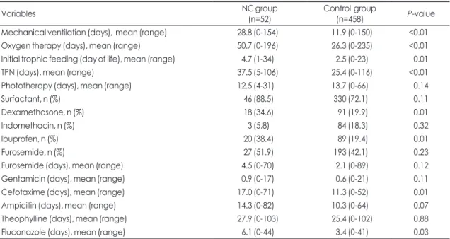
The duration of mechanical ventilation, oxygen therapy, and TPN support was significantly longer in the NC group. Compared to the control group, initial
trophic feeding was started later in the NC group.
The frequency of use of dexamethasone and oral ibuprofen was significantly higher in the NC group.
But, there was no difference in the frequency of use of surfactant, intravenous indomethacin, and furose- mide between two groups. The infants in the NC group were treated with cefotaxime and fluconazole for a significantly longer duration than the control group, but there was no significant difference in the duration of use of furosemide, gentamicin, and ampicillin between the two groups (Table 3).
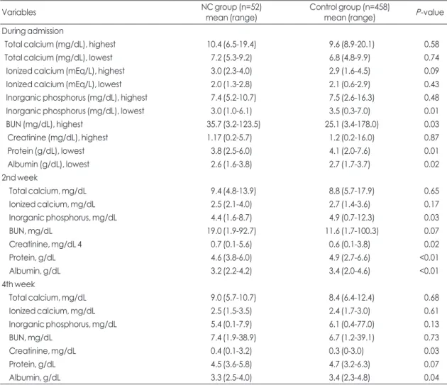
4) Laboratory findings on admission, 2nd weeks, and 4th weeks of life
During the hospitalization period, the lowest values of serum phosphate, protein, and albumin were significantly lower in the NC group, and the highest serum value of BUN was higher in the NC group than in the control group. At 2 weeks of life, the values of serum phosphate, protein, and albumin were significantly lower in the NC group, while the value of serum creatinine was higher in the NC group.
Table 2. Morbidities among very low birth weight infants
Variables NC group
(n=52) Control group (n=458) P-
value PDA, n (%) 40 (76.9) 269 (58.7) 0.11 BPD, n (%) 33 (63.5) 188 (41.0) 0.02 Bacterial sepsis, n (%) 25 (48.1) 156 (34.1) 0.03 Fungal sepsis, n (%) 13 (25.0) 39 (8.5) <0.01 Urinary tract infection,
n (%) 27 (51.9) 157 (34.3) 0.01
Abbreviations: NC, nephrocalcinosis; PDA, patent ductus arteriosus; BPD, bronchopulmonary dysplasia.
Table 3. Supportive care and drug treatment
Variables NC group
(n=52) Control group
(n=458) P-value
Mechanical ventilation (days), mean (range) 28.8 (0-154) 11.9 (0-150) <0.01
Oxygen therapy (days), mean (range) 50.7 (0-196) 26.3 (0-235) <0.01
Initial trophic feeding (day of life), mean (range) 4.7 (1-34) 2.5 (0-23) 0.01
TPN (days), mean (range) 37.5 (5-106) 25.4 (0-116) <0.01
Phototherapy (days), mean (range) 12.5 (4-31) 13.7 (0-66) 0.14
Surfactant, n (%) 46 (88.5) 330 (72.1) 0.11
Dexamethasone, n (%) 18 (34.6) 91 (19.9) 0.01
Indomethacin, n (%) 3 (5.8) 84 (18.3) 0.32
Ibuprofen, n (%) 20 (38.4) 89 (19.4) 0.01
Furosemide, n (%) 27 (51.9) 193 (42.1) 0.23
Furosemide (days), mean (range) 4.5 (0-70) 2.1 (0-89) 0.12
Gentamicin (days), mean (range) 0.9 (0-17) 0.6 (0-21) 0.11
Cefotaxime (days), mean (range) 17.0 (0-71) 11.3 (0-52) 0.01
Ampicillin (days), mean (range) 14.3 (0-82) 10.3 (0-64) 0.07
Theophylline (days), mean (range) 27.9 (0-103) 25.4 (0-102) 0.88
Fluconazole (days), mean (range) 6.1 (0-44) 3.4 (0-41) 0.03
Abbreviations: NC, nephrocalcinosis; TPN, total parenteral nutrition.
At 4 weeks of life, the value of serum albumin was significantly lower, while the value of serum crea- tinine was higher in the NC group than in the control group. But, neither the highest and lowest values of total and ionized calcium, nor the values of total and ionized calcium at 2 weeks and 4 weeks was different between the two groups (Table 4).
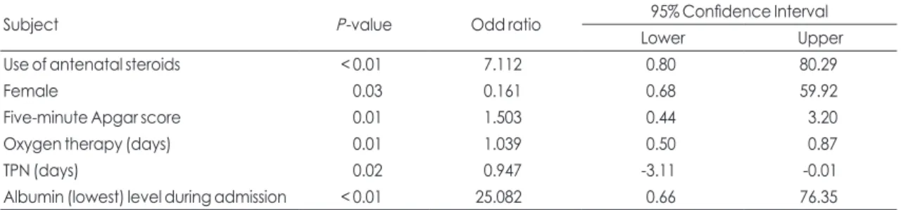
3. Significance of risk factors by multivariate analysis
Binary logistic regression was used as the multi- variate analysis. NC was selected as a dependent
variable, and the variables with statistical signifi- cance (P<0.05) were selected as independent vari- ables. As a result, use of antenatal steroids [P<0.01, odds ratio (OR) 7.112, 95% confidence interval (CI) 0.80-80.29], female sex (P=0.03, OR 0.161, 95% CI 0.69-59.92), 5-minute Apgar score (P=0.01, OR 1.503, 95% CI 0.44-3.20), duration of oxygen therapy (P=0.01, OR 1.039, 95% CI 0.50-0.87), duration of TPN support (P=0.02, OR 0.947, 95% CI -3.11-0.01), and lowest albumin level during the hospitalization period (P<0.01, OR 25.082, 95% CI 0.66-76.35) were the strongest risk factors for NC (Table 5).
Table 4. Laboratory findings on admission, 2nd weeks, and 4th weeks of life
Variables NC group (n=52)
mean (range) Control group (n=458)
mean (range) P-value
During admission
Total calcium (mg/dL), highest 10.4 (6.5-19.4) 9.6 (8.9-20.1) 0.58
Total calcium (mg/dL), lowest 7.2 (5.3-9.2) 6.8 (4.8-9.9) 0.74
Ionized calcium (mEq/L), highest 3.0 (2.3-4.0) 2.9 (1.6-4.5) 0.09
Ionized calcium (mEq/L), lowest 2.0 (1.3-2.8) 2.1 (0.6-2.9) 0.43
Inorganic phosphorus (mg/dL), highest 7.4 (5.2-10.7) 7.5 (2.6-16.3) 0.48
Inorganic phosphorus (mg/dL), lowest 3.0 (1.0-6.1) 3.5 (0.3-7.0) 0.01
BUN (mg/dL), highest 35.7 (3.2-123.5) 25.1 (3.4-178.0) 0.03
Creatinine (mg/dL), highest 1.17 (0.2-5.7) 1.2 (0.2-16.0) 0.87
Protein (g/dL), lowest 3.8 (2.5-6.0) 4.1 (2.0-7.6) 0.01
Albumin (g/dL), lowest 2.6 (1.6-3.8) 2.7 (1.7-3.7) 0.02
2nd week
Total calcium, mg/dL 9.4 (4.8-13.9) 8.8 (5.7-17.9) 0.65
Ionized calcium, mg/dL 2.5 (2.1-4.0) 2.7 (1.4-3.6) 0.17
Inorganic phosphorus, mg/dL 4.4 (1.6-8.7) 4.9 (0.7-12.3) 0.03
BUN, mg/dL 19.0 (1.9-92.7) 11.6 (1.7-100.3) 0.07
Creatinine, mg/dL 4 0.7 (0.1-5.6) 0.6 (0.1-3.8) 0.02
Protein, g/dL 4.6 (3.8-6.0) 4.9 (2.7-6.6) <0.01
Albumin, g/dL 3.2 (2.2-4.2) 3.4 (2.0-4.6) <0.01
4th week
Total calcium, mg/dL 9.0 (5.7-10.7) 8.4 (6.4-12.4) 0.68
Ionized calcium, mg/dL 2.5 (1.5-3.5) 2.4 (1.7-3.0) 0.61
Inorganic phosphorus, mg/dL 5.4 (0.1-7.9) 6.1 (0.4-77.0) 0.13
BUN, mg/dL 7.4 (1.9-38.9) 6.7 (1.2-39.1) 0.73
Creatinine, mg/dL 0.4 (0.1-3.2) 0.3 (0-3.0) 0.03
Protein, g/dL 4.5 (3.6-5.8) 4.7 (3.2-6.3) 0.07
Albumin, g/dL 3.3 (2.5-4.0) 3.4 (2.3-4.8) 0.04
Abbreviations: NC, nephrocalcinosis; BUN, blood urea nitrogen.
4. Prognosis
Follow-up renal US was performed in 45 VLBW infants. We couldn’t collect the data of 7 cases that were lost to follow-up. Among these 45 VLBW in- fants, NC was resolved in 13 infants (28.9%, 13/45) by 6 months after discharge. NC was resolved in other 12 and 7 infants by 12 months and 18 months, respectively, and the resolution rate at 12 months and 18 months was 55.6% (25/45) and 71.1% (32/45),
respectively. In 4 infants, NC resolved by 18 months of age, so the total resolution rate was 80.0% (36/45).
NC persisted but mildly decreased in size in the remaining 9 infants at the time of last follow-up. The average period of recovery was 10.3 months (44.1 weeks) in 36 VLBW infants.
During long-term follow-up, pyelonephritis and renal stones occurred in two VLBW infants each. One preterm female infant of 27 weeks gestational age and weighing 850 g had diagnosed the bilateral NC Table 5. Significance of risk factors by binary logistic regression analysis
Subject P-value Odd ratio 95% Confidence Interval
Lower Upper
Use of antenatal steroids < 0.01 7.112 0.80 80.29
Female 0.03 0.161 0.68 59.92
Five-minute Apgar score 0.01 1.503 0.44 3.20
Oxygen therapy (days) 0.01 1.039 0.50 0.87
TPN (days) 0.02 0.947 -3.11 -0.01
Albumin (lowest) level during admission < 0.01 25.082 0.66 76.35
Abbreviations: BPD, bronchopulmonary dysplasia; TPN, total parenteral nutrition.
Fig. 1. Renal ultrasonographic findings at two months of life showed nephrocalcinosis in both kidneys (a maximum diameter of 3.5 mm in the left kidney) with normal renal echogenicity and size (upper two). At four and a half months of life, diffuse nephrocalcinosis (large calculi in the left kidney) and decreased size of both kidneys, more marked in the left kidney (left 2.8x1.4 cm, right 8x8 cm) with loss of corticomedullary differentiation suggesting chronic renal parenchymal disease was noted (lower two).
with multiple stones (maximum diameter 3.5 mm) at 2 months of life (Fig. 1A). She experienced a cardiac tamponade but was successfully resuscitated at 6 days of life. Transiently, her renal function decreased.
Not only progression of renal atrophy was observed in the left kidney without regression of NC (Fig. 1B), but also renal function impairment was noted in the
99mTc-MAG3 renal scan. She was followed up at the out-patient department for 7 years with conservative medical treatment for hypertension (systolic blood pressure 96 mmHg / diastolic blood pressure 74 mmHg) and chronic renal failure (BUN 32.0 mg/dL, creatinine 2.3 mg/dL).
Discussion
The incidence of NC in this study is 10.2%, which was coincide with that previously reported.3-9 The varying prevalence of NC is considered to be due different study populations and era, development in the sensitivity of ultrasound equipment, and work- manship of the observer.27 In several reports, the incidence of NC showed a decreasing trend with development in care of preterm infants,6,20,24 but this study showed an increasing tendency for NC. This finding is suspected to be due to an increase birth rate in infants with low gestational age and birth weight and survival of them, and active evaluation of VLBW infants with close attention to NC. Renal US was performed by a consultant radiologist in charge of pediatric specialty, and the sonographic diagnostic criteria and the policy to check renal US remain constant above mentioned during study period.
The risk factors for NC are different among re- ports, and the most important risk factors are gesta- tional age and birth weight.5-12 At a low gestational age, the lower glomerular filtration ratio and the
shorter loop of Henle lead to slow urinary transition, while the sensitivity for heterogeneous crystallization is high, which result in decreased calcium excretion and increased calcification. Also, during the neonatal period, VLBW infants are exposed more frequently and for a longer time to multiple risk factors asso- ciated with treatment.28 In this study the NC group had lower gestational age and birth weight than the control group. The NC group also had lower 5 minute Apgar score, initial blood pH, the lowest systolic blood pressure and urine output on the first day of life than the control group. These findings are related clinical characteristics of low gestational age and low birth weight infants. Narendra et al.6 reported that male sex was associated with an increased risk of NC; on the other hand, NC occurred more frequently in female infants of this study.
Antenatal corticosteroids therapy is recommended for pregnant women at risk of preterm birth to pre- vent RDS and many preterm neonates receive gluco- corticoids for treatment or prevention of BPD. Steroid treatment generally resulted in an imbalance between bone resorption and osteogenesis, and so it can lead to osteopenia, hypercalciuria, and nephrolithiasis. In this study, the use of both antenatal steroids and postnatal steroids were associated with NC in the univariate analysis, but only the use of antenatal steroids was associated with NC in the multivariate analysis. An association between postnatal dexame- thasone treatment, high calcium excretion and NC is, indeed, found in preterm neonates.14,24,29 but there is no clear correlation between antenatal steroids and NC, so it is not possible to explain satisfactorily the connection in this study.
Short and Cooke5 found that the duration of oxygen administration was the strongest clinical indicator of occurrence of renal calcification. They confirmed
that severe respiratory disease, as indicated by duration of ventilation, duration of oxygen admini- stration and oxygen dependence at 36 weeks after conception, was significantly associated with NC. In this study, we found that duration of mechanical ventilation and oxygen therapy, and BPD were signi- ficantly associated with the development of NC, which was similar to that in the report by Shim et al.5,
24 Citrate is well known to be one such inhibitory molecule and, indeed, hypocitraturia is an established risk factor for NC.30 Murphy and Mendoza reported that the lowered urinary citrate of preterm infants with lung disease may predispose to NC by increased urinary lithogenesis.5
Narendra et al6 found that TPN was significantly associated with NC and that the association is through phosphate deficiency, which is typically associated with hypercalciuria, while phosphate supplementation decreased urinary calcium excretion. The recent approach to nutrition of the preterm infant is based on starting feeding as soon as possible after birth, sup- plying a parenteral of amino acids (AA) and energy to avoid cellular catabolism and to promote extra- uterine growth. Calcium-phosphorus homeostasis was especially influenced by the early AA intake. The plasma phosphate levels in infants receiving paren- teral nutrition were inversely associated to the AA intake. The abrupt interruption of the continuous placental flow of AA and energy promotes catabolism and ion release by the cell. But the parenteral supply of high AA and energy maintains the cell in an anabolic state and promotes its uptake of phosphorus and potassium in the absence of an adequate intake.
Simultaneously, calcium mobilization results in ex- cess calcium in the extracellular space, which can lead to hypercalcemia and hypercalciuria. So we should pay attention to add the optimal phosphorus in
TPN, according to the administered AA intake. In this study, the lowest phosphate value and TPN were significantly associated with NC. There was no difference in calcium gradient between the two groups, while the lowest phosphate level during hospitalization and phosphate gradient in the second week of life were higher and duration of TPN was longer in the NC group. In our center, AA intake was started in the first day of life (2.0 g/kg) with calcium and phosphate and incremented daily up to 3.5-4.0 g/
kg at few days of life. Early administration of AA is also associated with a positive nitrogen balance, increased BUN, hyperammonemia, and metabolic acidosis in preterm infants. High BUN could be relat- ed not only to AA oxidation and the infant’s imma- turity but also to additional combined factors other than AA intolerance, such as renal function and hydration status.
Calcium reabsorption is carried out passively due to the density difference between sodium chloride and calcium in the loop of Henle, and furosemide induces NC via inhibiting calcium reabsorption.31 Park et al.23 reported that furosemide is a risk factor for NC, while Short and Cooke5 reported no associa- tion between furosemide and NC. In this study, the duration and frequency of furosemide therapy were not significantly associated with NC in both univari- ate and multivariate analyses.
Caffeine and theophylline, methylxanthine agents, accelerate urinary calcium excretion from two to ten times and increase prostaglandin synthesis. As a result, the renal blood flow and glomerular filtration rate decrease, leading to the occurrence of NC.32,33 In this study, the duration of theophylline treatment was significantly longer in the NC group in the univariate analysis, but not in the multivariate analysis.
In this study, low albumin level was a significant
risk factor for NC. Albumin is protein compartment binding calcium and several reports suggested that hypoalbuminemia leads to NC via excretion of free calcium in the serum.31 On the other hand, albumin concentration in newborn babies is different as gestational age. Zlotkin and Casselman34 reported that values for total protein and albumin in the pre- term infant were lower than in the full term infant.
Serum albumin concentration rose from about 20 g/L in 28 weeks gestation babies to about 30 g/L in term babies. In babies of postnatal age up to 8 weeks the albumin concentration continues to rise as the same rate as the in utero rise in concentration with in- creas ing gestation.35 The NC group had lower gesta- tional age and the concentration of albumin and protein than the control group.
In this study, gestational age (P=0.35) and birth weight (P=0.07) were not significant risk factors by binary logistic regression analysis. It was difficult to identify to influential factors. The limitations of this study were the small number of enrolled VLBW infants and its retrospective design. Maybe further evaluation will be helpful to conduct a subgroup analysis according to gestational age and birth weight.
In the majority of preterm infants, NC was resolved spontaneously, but in some preterm infants, it per- sisted for a long time. Schell-Feith et al.20 reported that 34% and 15% of preterm infants with NC did not recover until 15 months and 30 months of life, res- pectively. Similar to the above study, the resolution rate was 28.9%, 55.6%, and 71.1% at 6 months, 12 months, and 18 months of life in this study, respec- tively. NC was resolved additionally by over 18 months in 4 infants, but NC persisted but mildly de- creased in size in the remaining 9 infants at the time of last follow-up, so we expect the final resolution
rate will be increase later.
Porter et al.18 followed up the 14 VLBW infants with NC up to at 5-7 years of age who showed the 75%
resolution rate and no renal function impairment in the long time, but in this study, one preterm infant with NC developed chronic renal failure due to renal stones.
The limitations of this study were, first, that family history of renal stone was not obtained in each pa- tient. Second, the serum levels of bicarbonate, uri- nary pH, urinary citrate, urinary calcium/creatinine ratio, and urinary oxalate/creatinine were not ob- tained in all patients. Third, we evaluated the long- term outcome as medical records, so there was some lacking regular data about renal function, renal growth, urine analysis, blood pressure.
In conclusion, in this study, the incidence rate of NC in VLBW infants was 10.2%, and the risk factors for NC were use of antenatal steroids, female sex, low 5 minute Apgar score, duration of oxygen therapy and TPN and lowest albumin level during hospitalization.
The resolution rate was 80.0% over 18 months of life and it was obvious that long-term observation is necessary and complications such as hypertension and chronic failure might arise in the long run.
Discussion
1) Dimopoulos C, Capetanakis D, Doïcas J, Grafas D. Nephro- calcinosis in adults: 10 cases. Acta Urol Belg 1971;39:293- 303.
2) Hufnagle KG, Khan SN, Penn D, Cacciarelli A, Williams P.
Renal calcifications: a complication of long-term furosemide therapy in preterm infants. Pediatrics 1982;70:360-3.
3) Schell-Feith EA, Kist-van Holthe JE, Conneman N, van Zwieten PH, Holscher HC, Zonderland HM, et al. Etiology of nephrocalcinosis in preterm neonates: association of nutritional intake and urinary parameters. Kidney Int 2000;58:2102-10.
4) Gimpel C, Krause A, Franck P, Krueger M, Von Schnaken- burg C. Exposure to furosemide as the strongest risk fac- tor for nephrocalcinosis in preterm infants. Pediatr Int 2010;52:51-6.
5) Short A, Cooke RW. The incidence of renal calcification in preterm infants. Arch Dis Child 1991;66:412-7.
6) Narendra A, White MP, Rolton HA, Alloub ZI, Wilkinson G, McColl JH, et al. Nephrocalcinosis in preterm babies. Arch Dis Child Fetal Neonatal Ed 2001;85:207-13.
7) Hein G, Richter D, Manz F, Weitzel D, Kalhoff H. Develop- ment of nephrocalcinosis in very low birth weight infants.
Pediatr Nephrol 2004;19:616-20.
8) Chang HY, Hsu CH, Tsai JD, Li ST, Hung HY, Kao HA, et al. Renal calcification in very low birth weight infants.
Pediatr Neonatol 2011;52:145-9.
9) Saarela T, Vaarala A, Lanning P, Koivisto M. Incidence, ultrasonic patterns and resolution of nephrocalcinosis in very low birthweight infants. Acta Paediatr 1999;88:655-60.
10) Jacinto JS, Modanlou HD, Crade M, Strauss AA, Bosu SK. Renal calcification incidence in very low birth weight infants. Pediatrics 1988;81:31-5.
11) Sheu JN, Chen CH, Lue KH, Chen JY, Tsau YK, Chen JH.
Renal calcification in very low birth weight infants. Am J Nephrol 1993;13:6-11.
12) Karlowicz GM, Katz ME, Adelman RD, Solhaug MJ.
Nephrocalcinosis in very low birth weight neonates: family history of kidney stones and ethnicity as independent risk factors. J Pediatr 1993;122:635-8.
13) Hoppe B, Hesse A, Neuhaus T, Fanconi S, Blau N, Roth B, et al. Influence of nutrition on urinary oxalate and calcium in preterm and term infants. Pediatr Nephrol 1997;11:687-90.
14) Kamitsuka MD, Williams MA, Nyberg DA, Fox KA, Lee DL, Hickok D. Renal calcification: a complication of dexamethasone therapy in preterm infants with bronch- opulmonary dysplasia. J Perinatol 1995;15:359-63.
15) Hoppe B, Duran I, Martin A, Kribs A, Benz-Bohm G, Michalk DV, et al. Nephrocalcinosis in preterm infants: a single center experience. Pediatr Nephrol 2002;17:264-8.
16) Downing GJ, Egelhoff JC, Daily DK, Thomas MK, Alon U. Kidney function in very low birth weight infants with furosemide-related renal calcifications at ages 1 to 2 years. J Pediatr 1992;120:599-604.
17) Jones CA, King S, Shaw NJ, Judd BA. Renal calcification in preterm infants: follow up at 4–5 years. Arch Dis Child Fetal Neonatal Ed 1997;76:185-9.
18) Porter E, McKie A, Beattie TJ, McColl JH, Aladangady N, Watt A, et al. Neonatal nephrocalcinosis: long term follow up. Arch Dis Child Fetal Neonatal Ed 2006;91:333-6.
19) Bonsante F, Lacobelli S, Latorre G, Rigo J, De Felice D, Robillard PY, et al. Initial amino acid intake influences phosphorus and calcium homeostasis in preterm infants-- it is time to change the composition of the early parenteral nutrition. PLoS One 2013;8:e72880.
20) Schell-Feith EA, Kist-van Holthe JE, van Zwieten PH, Zonderland HM, Holscher HC, Swinkels DW, et al. Preterm neonates with nephrocalcinosis: natural course and renal function. Pediatr Nephrol 2003;18:1102-8.
21) Ezzedeen F, Adelman RD, Ahlfors CE. Renal calcification in preterm infants: pathophysiology and long-term sequelae.
J Pediatr 1988;113:532-9.
22) Kist-van Holthe JE, van Zwieten PH, Schell-Feith EA, Zonderland HM, Holscher HC, Wolterbeek R, et al. Is nephrocalcinosis in preterm neonates harmful for long-term blood pressure and renal function? Pediatrics 2007;119:468- 75.
23) Park MJ, Park KI, Park MS, Namgung R, Lee C, Han DG, et al. Furosemide-induced nephrocalcinosis in very low birth weight infants. J Korean Pediatr Soc 1994;37:553-9.
24) Shim GH, Lee JA, Shin YJ, Kim EK, Park JD, Kim BI, et al. Risk factors of nephrocalcinosis in very low birth weight (VLBW) infants. J Korean Pediatr Soc 2004;47:275-81.
25) Lee HS, Sung IK, Kim SJ, Youn YA, Lee JY, Lim GY, et al.
Risk factors associated with nephrocalcinosis in preterm infants. Am J Perinatol 2014;31:279-86.
26) Porter CC, Avner ED. Idiopathic hypercalciuria. In:
Kliegman RM, Stanton BF, Schor NF, St. Geme JW III, Behrman RE. Nelson textbook of pediatrics. 19th ed.
Philadelphia: Elsevier Saunders Co:2011, p.1795.
27) Schell-Feith EA, Holscher HC, Zonderland HM, Kist- Van Holthe JE, Conneman N, van Zwieten PH, et al.
Ultrasonographic features of nephrocalcinosis in preterm neonates. Br J Radiol 2000;73:1185-91.
28) Schell-Feith EA, Kist-van Holthe JE, van der Heijden AJ.
Nephrocalcinosis in preterm neonates. Pediatr Nephrol 2010;25:221-30.
29) Cranefield DJ, Odd DE, Harding JE, Teele RL. High inci- dence of nephrocalcinosis in extremely preterm infants treated with dexamethasone. Pediatr Radiol 2004;34:138-42.
30) Murphy JL, Mendoza SA. Decreased urinary citrate in premature infants with lung disease. Child Nephrol Urol 1990;10:76-80.
31) Lee CT, Chen HC, Lai LW, Yong KC, Lien YH. Effects of furosemide on renal calcium handling. Am J Physiol Renal Physiol 2007;293:1231-7.
32) Mazkereth R, Laufer J, Jordan S, Pomerance JJ, Boichis H, Reichman B. Effects of theophylline on renal function in
premature infants. Am J Perinatol 1997;14:45-9.
33) Zanardo V, Dani C, Trevisanuto D, Meneghetti S, Guglie- lmi A, Zacchello G, et al. Methylxanthines increase re nal calcium excretion in preterm infants. Biol Neonate 1995;
68:169-74.
34) Zlotkin SH, Casselman CW. Percentile estimates of re-
ference values for total protein and albumin in sera of pre- mature infants (less than 37 weeks of gestation). Clin Chem 1987;33:411-3.
35) Reading RF, Ellis R, Fleetwood A. Plasma albumin and total protein in preterm babies from birth to eight weeks. Early Hum Dev 1990;22:81-7.
= 국 문 초 록 =
극소 저체중 출생아에서 신석회화증의 위험 요인과 예후
전남대학교병원 소아청소년과
김호성^정금희^최영륜^송은송
목적: 본 연구는 극소 저체중 출생아에서 발생한 신석회화증의 발생빈도, 위험인자 및 장기 예후를 알아보고자 하였다.
방법: 2006년부터 2012년까지 신생아 중환자실에 입원한 출생 체중 1,500 g 미만 극소 저체중 출생아의 의무기록지를 후향적으로 조사하였다.
결과: 극소 저체중 출생아에서 신석회화증의 발생빈도는 10.2%로 증가하는 추세를 보였다. 단변수 분석을 한 결과, 신
석회화증을 보인 환아에서 양수과소증과 산전 스테로이드 사용이 많았고, 임신나이와 출생 체중이 더 작고 여아가 많 았다. 또한 생후 첫날 동맥혈 가스 검사상 pH, 수축기 혈압의 최저치와 소변양이 의미있게 낮았다. 기관지폐 형성 이상과 패혈증, 그리고 요로감염이 더 많이 발생하였고, dexamethasone과 ibuprofen 사용이 더 많았으며, 입원기간 동안 인 최저치, 단백질과 알부민 최저치가 의미있게 낮았다. 이분형 로지스틱 회귀분석을 이용하여 다변수 분석결과, 산전 스테 로이드 사용, 여아, 5분 Apgar 점수, 산소 치료와 총정맥영양 기간 및 알부민 최저치가 유의성이 있었다. 신석회화증의 자 연호전은 생후 12개월과 18개월에 각각 55.6%, 71.1%이었다.
결론: 극소 저체중 출생아에서 신석회화증의 발생빈도는 증가하는 추세로, 여러 위험인자가 더 작고 아픈 환아들의 치
료 경과와 연관 있었다. 자연호전이 80.0%로 양호하더라도 합병증 발생 등을 포함한 장기적인 추적관찰이 필요할 것으 로 사료되었다.
중심 단어: 신석회화증, 극소 저체중 출생아, 발생률, 위험 요인, 예후