Background and Purpose Cognitive and cerebrovascular diseases are common in the el- derly, but differences in the plasma levels and associations of plasma biomarkers in these dis- eases remain elusive.
Methods The present study investigated differences in plasma fatty acids [eicosapentaenoic acid (EPA) and docosahexaenoic acid (DHA)], adiponectin, reptin, plasma markers of inflam- mation [high-sensitivity C-reactive protein (hsCRP) and serum amyloid A (serum AA)], and plasma lipids [high-density lipoprotein and low-density lipoprotein (LDL)] in patients with Al- zheimer’s disease (AD) (n=266), mild cognitive impairment (MCI) (n=44), vascular dementia (VaD) (n=33), and ischemic stroke (IS) (n=200) in comparison to normal controls (n=130).
Results The serological data showed that lower EPA and DHA levels and higher reptin and LDL levels were associated with AD and IS, the reptin/adiponectin ratio was strongly associated with IS, the hsCRP level was more strongly associated with VaD and IS, and the serum AA level was associated with all three cognitive diseases and IS.
Conclusions This is the first report of differences in the expression levels of plasma biomarkers and peripheral arterial tonometry among AD, MCI, VaD, and IS patients and normal controls.
These different associations indicate that diverse pathological mechanisms underlie these diseases.
Key Words Alzheimer’s disease, ischemic stroke, mild cognitive impairment, plasma biomarker, vascular dementia.
Different Associations of Plasma Biomarkers
in Alzheimer’s Disease, Mild Cognitive Impairment, Vascular Dementia, and Ischemic Stroke
INTRODUCTION
Neurological disorders have become serious long-term problems that affect more than 450 million individuals worldwide. The most common forms of neurological diseases include cognitive and cerebrovascular diseases, which lead to significant impairment in the activi- ties of daily living (ADL) and consequently decrease the quality of life, especially in the el- derly.1 Alzheimer’s disease (AD) constitutes 69% of dementia cases in those older than 75 years, and is characterized by gradual loss of cognitive, affective, and ADL capabilities.2 Vas- cular dementia (VaD) is the second most common dementia,3 and is characterized by de- cline in memory and executive functions. Mild cognitive impairment (MCI) precedes clini- cally evident dementia,4 which has minimal-to-mild cognitive impairment that does not significantly impact the ADL. Although the age-standardized mortality rate of ischemic stroke (IS) has clearly decreased in recent years, stroke is still one of the leading causes of death and disability in Japan.5 The complexity of blood as a source of biomarkers is reflected in the limitations of various proteomic techniques, but these may still allow the detection of the above four diseases earlier than when using classically recognized markers.
Jingwei Shang Toru Yamashita Yusuke Fukui Dongjing Song Xianghong Li Yun Zhai Yumiko Nakano Ryuta Morihara Nozomi Hishikawa Yasuyuki Ohta Koji Abe
Department of Neurology, Graduate School of Medicine, Dentistry and Pharmaceutical Sciences, Okayama University, Okayama, Japan
pISSN 1738-6586 / eISSN 2005-5013 / J Clin Neurol 2018;14(1):29-34 / https://doi.org/10.3988/jcn.2018.14.1.29
Received March 20, 2017 Revised August 5, 2017 Accepted August 10, 2017 Correspondence Koji Abe, MD, PhD Department of Neurology, Graduate School of Medicine, Dentistry and Pharmaceutical Sciences, Okayama University,
2-5-1 Shikata-cho, Okayama 700-8558, Japan Tel +81-86-235-7365 Fax +81-86-235-7368
E-mail [email protected]
cc This is an Open Access article distributed under the terms of the Creative Commons Attribution Non-Com- mercial License (http://creativecommons.org/licenses/by-nc/4.0) which permits unrestricted non-commercial use, distribution, and reproduction in any medium, provided the original work is properly cited.
JCN
Open Access ORIGINAL ARTICLEPlasma Biomarkers in AD, MCI, VaD, and IS
JCN
Omega-3 polyunsaturated fatty acids (ω-3 PUFAs) can in- hibit hepatic triglyceride synthesis, reduce platelet aggrega- tion, cause vascular relaxation, and reduce inflammation.6 Eicosapentaenoic acid (EPA, 20:5n-3) and docosahexaenoic acid (DHA, 22:6n-3) are ω-3 PUFAs that have shown benefi- cial effects on brain functions through improving episodic memory and learning functions in healthy adults.7 EPA and DHA are useful clinical biomarkers for identifying whether the fatty-acid balance in the body is optimal or suboptimal.
Reptin is essential for systemic viability and plays antagonistic roles in tissue growth and regulation of the tumor metastasis suppressor gene; several studies have found that it is overex- pressed in certain types of cancer, including hepatocellular carcinoma and colorectal cancer.8 Adiponectin, a hormone that is secreted solely by adipocytes, exerts significant effects on atherogenesis, endothelial function, and vascular remod- eling via the modulation of signaling cascades in cells of the vasculature. A plasma adiponectin concentration of less than 4.0 μg/mL was associated with a twofold increase in the in- cidence of coronary artery disease, and adiponectin is a useful marker for identifying individuals at risk of developing meta- bolic syndrome and early-stage atherosclerosis.9
Inflammation may contribute to cognitive decline and de- mentia. Higher levels of high-sensitivity C-reactive protein (hsCRP), a biomarker of inflammation, were associated with worse performance in cognitive tests after stroke.10 Serum am- yloid A (serum AA) is an acute-phase protein that acts as a biomarker of inflammation, and previous studies have pro- duced data on the local production of serum AA proteins in histologically normal, atherosclerotic, AD, inflammatory, and tumor tissues.11 Lipid biomarkers are frequently used to assess the risk of cardio-cerebrovascular disease, such as high-densi- ty lipoprotein (HDL) and low-density lipoprotein (LDL). HDL performs a wide range of functions, including antioxidation, anti-inflammation, and proendothelial functions and the mod- ulation of immune function.12 High levels of LDL lead to ath- erosclerosis, which increases the risks of heart attack and IS.13
The aim of the present study was to determine the differ- ent expression levels of and associations between plasma fat- ty acids (EPA and DHA), adiponectin and reptin, plasma markers of inflammation (hsCRP and serum AA), plasma lip- ids (HDL and LDL), and peripheral arterial tonometry among AD, MCI, VaD, and IS patients and normal controls. Data on
these different expression levels may represent useful informa- tion for analyzing the different pathological mechanisms un- derlying these diseases.
METhODS
Participants
This was a retrospective case–control study performed in the outpatient clinic of Okayama University Hospital and affili- ated hospitals from April 2011 to April 2016 that investigated AD (n=295), MCI (n=47), VaD (n=34), and IS (n=200). Two hundred and thirty age- and sex-matched individuals in whom no neurological or psychiatric diseases were found in medical examinations were included as normal controls (102 males and 98 females).
The clinical information on the normal controls and the patients is summarized in Table 1. A diagnosis of AD was es- tablished based on clinical criteria and diagnostic guidelines as described previously.14 MCI was diagnosed according to the well-known Petersen’s criteria. VaD was diagnosed based on recommendations of the Neuroepidemiology Branch of the National Institute of Neurological Disorders and Stroke and the Association Internationale pour la Recherche et l’Enseignement en Neurosciences. IS was diagnosed by neu- rologists based on the clinical history, general physical and neurological examinations, and ancillary tests such as radio- logical examinations (CT/MRI), according to the WHO Mon- itoring of Trends and Determinants in Cardiovascular Dis- ease projects.15
Ethical permission for this study was provided by the Eth- ics Committee on Epidemiological Studies of the Okayama University Graduate School of Medicine, Dentistry and Phar- maceutical Sciences (approval #777), and written informed consent was obtained from all participants prior to enrollment.
Serological laboratory tests
Blood samples were collected in the fasting state. Serologi- cal data for EPA, DHA, adiponectin, reptin, hsCRP, serum AA, HDL, and LDL were obtained from 696 participants (normal controls, n=130; AD, n=266; MCI, n=44; VaD, n=
33; IS, n=200). Serum samples were analyzed for routine biochemical parameters immediately after collection, while aliquots of the samples were also stored at -20°C for subse-
Table 1. Demographic and clinical characteristics of the normal controls and the patients
Patient characteristics Normal AD MCI VaD IS
n 200 295 47 34 200
Sex (males/females) 102/98 186/109 30/17 19/15 110/90
Age at examination (years) 75.4±9.1 79.8±7.5 76.6±7.5 76.9±9.6 72.5±10.2
AD: Alzheimer’s disease, IS: ischemic stroke, MCI: mild cognitive impairment, VaD: vascular dementia.
Shang J et al.
JCN
quent assays of EPA, DHA, adiponectin, reptin, and serum AA. EPA and DHA were assayed using gas chromatography/
mass spectrometry. Adiponectin, reptin, and serum AA were assayed by solid-phase sandwich ELISA using commercial kits.
Statistical analysis
Continuous demographic and clinical data are presented as mean±SD values in Table 1. Statistical analyses were performed using standard statistical software (SPSS 22.0, IBM Corp., Ar- monk, NY, USA). After checking for normality, we performed Kruskal-Wallis tests to compare eight serum data values (EPA, DHA, adiponectin, reptin, hsCRP, serum AA, HDL, and LDL) between the normal controls and the four disease groups. Dif- ferences with a probability value of p<0.05 were considered statistically significant.
RESUlTS
Participant characteristics
The demographic and clinical features of the normal controls and the four disease groups (AD, MCI, VaD, and IS patients) are presented in Table 1. The sex ratio was well matched across these five groups, and the age of the participants was mainly concentrated around the mid-70s.
Plasma fatty-acid changes
The serological tests indicated that the EPA levels were sig- nificantly lower in AD and IS patients than in the normal controls (Fig. 1A). On the other hand, DHA was remarkably lower in the four disease groups than in the normal controls (Fig. 1B). However, neither EPA nor DHA differed among the four disease groups.
Reptin and adiponectin changes
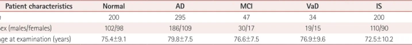
The reptin level was significantly higher in the AD and IS pa- tients than in the normal controls (Fig. 2A), while the adipo- nectin level was significantly lower in the IS patients than in the AD and MCI patients (Fig. 2B). The reptin/adiponectin ratio was significantly higher in the IS patients than in the nor- mal controls and the AD, MCI, and VaD patients (Fig. 2C), with no significant difference among the latter four groups.
Plasma markers of inflammation changes
The hsCRP level was significantly higher in the VaD and IS patients than in the normal controls (Fig. 3A), and higher in the VaD than the AD patients. The serum AA level was signifi- cantly higher in the four disease groups than in the normal controls (Fig. 3B), with no difference among the four disease groups.
Plasma lipid levels and peripheral arterial tonometry analyses
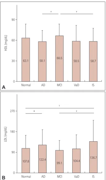
The HDL level did not differ significantly between the normal controls and the four disease groups, but it was significantly lower in the AD and IS patients than in the MCI patients (Fig.
4A). The LDL level was significantly higher in the AD and IS patients than in the normal controls (Fig. 4B), and higher in the IS than the MCI patients.
DISCUSSION
Blood-based biomarkers span a massive dynamic range of pro- teins in blood, but their use in clinical diagnostic practice is still far from optimal despite considerable research into their diagnostic utility in discriminating cognitive and cardiovas- cular diseases. In this study we examined differences in the
120
80
40
0
EPA (μg/μL)
Normal AD MCI VaD IS
†
†
70.2 56.7 68.8
59.1 65.8
A
180
120
60
0
DHA (μg/μL)
Normal AD MCI VaD IS
*
†
†
†
127.6
89.7 103.2
87.6 96.7
B
Fig. 1. Plasma fatty-acid levels in the normal controls and the four disease groups: AD, MCI, VaD, and IS. Intergroup comparisons of EPA (A) and DHA (B) levels. *p<0.05, †p<0.01 vs. normal. AD: Alzheimer’s disease, DHA: docosahexaenoic acid, EPA: eicosapentaenoic acid, IS: ischemic stroke, MCI: mild cognitive impairment, VaD: vascular dementia.
Plasma Biomarkers in AD, MCI, VaD, and IS
JCN
expression levels of several plasma biomarkers in elderly AD, MCI, VaD, and IS patients and elderly normal controls, and found differences in expression levels that are indicative of the diverse pathological mechanisms underlying these dis- eases.
Dietary habits have changed during the last decade, espe- cially in Western countries, with decreases in the intake of ω-3 PUFAs.16 EPA and DHA are considered the most important
ω-3 PUFAs, having anti-inflammatory, antioxidant, antiath- erogenic, antiamyloid, and neuroprotective properties in the brain.17 Higher intakes of EPA and DHA are associated with lower risks of AD and IS.18 The present study found that the plasma EPA level was lower in AD and IS patients, while the DHA level was markedly lower in all four disease groups (Fig.
1). These data indicate that both EPA and DHA were associ- ated with AD and IS, while DHA is more strongly associated than IS with memory tasks.
Reptin is involved in the regulation of gene transcription, remodeling of chromatin, DNA damage sensing and repair, and tumor biology.19 Higher adiponectin levels have been as- sociated with cognitive decline,20 and the present study found that a higher reptin level was associated with AD and IS (Fig.
18
12
6
0
Reptin (ng/mL)
*
†
Normal AD MCI VaD IS
6.1 8.3 7.2 5.9 8.6
A
30
20
10
0
Adiponectin (μg/mL)
‡
§
Normal AD MCI VaD IS
14.9 17.0 17.6 15.7 13.5
B
2.4
1.6
0.8
0 Reptin/adiponectin ratio (×10-3)
‡
‡
† §
Normal AD MCI VaD IS
0.55 0.68 0.61 0.54 0.89
C
Fig. 2. Comparison of reptin (A), adiponectin (B), and reptin/adipo- nectin ratio (C) levels in the normal controls and the four disease groups. *p<0.05, †p<0.01 vs. normal, ‡p<0.05, §p<0.01 within the disease groups. AD: Alzheimer’s disease, IS: ischemic stroke, MCI: mild cognitive impairment, VaD: vascular dementia.
18
12
6
0
hsCRP (μg/mL)
*
*
†
Normal AD MCI VaD IS
1.18 1.62 1.29 1.56
5.19
A
150
100
50
0
Serum AA (μg/mL)
* *
*
*
Normal AD MCI VaD IS
12.5 15.7 23.3 32.5
15.3
B
Fig. 3. Levels of plasma markers of inflammation in the normal con- trols and the four disease groups. Intergroup comparisons of hsCRP (A) and serum AA (B) levels. *p<0.01 vs. normal; †p<0.05 within the disease groups. AD: Alzheimer’s disease, hsCRP: high-sensitivity C-re- active protein, IS: ischemic stroke, MCI: mild cognitive impairment, se- rum AA: serum amyloid A, VaD: vascular dementia.
Shang J et al.
JCN
2A). Similar to a previous study,21 we found that the adipo- nectin level did not differ significantly between the normal controls and the four disease groups, while it was lower in IS patients than in AD and MCI patients (Fig. 2B). A recent study has shown that dramatic increases in serum adiponec- tin in AD are positively correlated with the severity of de- mentia, which is due to an elevated level of adiponectin be- ing associated with amyloid β-peptide toxicity, inflammatory reactions, and programmed cell death in the AD brain. In contrast, a cross-sectional study found that the adiponectin level was unaltered in AD patients.22 The actions of adiponec- tin in the central nervous system are still poorly understood, but it does play a protective role against atherosclerotic vascu- lar change and exerts a cerebroprotective effect via an endo-
thelial nitric oxide synthase-dependent mechanism, and the loss of these effects enhances endothelial dysfunction in IS patients.23 Hypoadiponectinemia is concomitantly involved in the pathogenesis of atherosclerosis in subjects with cere- bral infarction,24 which might reflect a close relationship be- tween adiponectin-metabolic-syndrome inflammation and the development of atherosclerosis.25 The present study is the first to find a strong association between the reptin/adiponec- tin ratio and IS (Fig. 2C), which suggests that IS is associated with high levels of DNA damage and repair in cells besides in- flammation.
While hsCRP may be a risk factor for cognitive impair- ment,26 the present results suggest that the hsCRP level is as- sociated with VaD and IS but not with AD (Fig. 3A). Some studies have emphasized the importance of serum AA in in- flammation, atherosclerosis, thrombosis, AA-induced amy- loidosis, rheumatoid arthritis, and neoplasia.11 Our results indicated that the serum AA level was significantly increased in AD, MCI, VaD, and IS (Fig. 3B). These data suggest that the hsCRP level is mainly associated with primary vascular diseases such as VaD and IS, and that the serum AA level is more generally associated with cognitive diseases and IS.
HDL modulates cognitive function in aging and age-relat- ed neurodegenerative disorders.12 High plasma levels of HDL can protect against IS.27 In the present study, the HDL level did not differ significantly between the normal controls and the four disease groups (Fig. 4A), which is probably due to the large amount of fish include in the Japanese diet.28 On the other hand, the LDL level was significantly higher both in AD and IS (Fig. 4B), which supports previous reports.29 Endo- thelial dysfunction is an early predictor of IS, and the reactive hyperemia index (RHI) is correlated with the risk of IS and cognitive dysfunction.30 Although the differences were not significant, the present study found a tendency for RHI to be lower in the four disease groups than in the normal controls.
This study was subject to several limitations. First, this study had a retrospective case–control design and was performed within a small area of Japan. Second, relatively few patients and controls were included, especially in the MCI and VaD groups, which had the effect of producing relatively wide SD values for the VaD group. Finally, subtypes of IS were not analyzed separately.
In summary, the present study is the first to compare differ- ences between plasma biomarkers in elderly AD, MCI, VaD, and IS patients and elderly normal controls. The findings sug- gest that lower EPA and DHA levels and higher reptin and LDL levels are both associated with AD and IS, the reptin/adi- ponectin ratio is strongly associated with IS, the hsCRP level is more strongly associated with VaD and IS, and the serum AA level is associated with all three cognitive diseases and IS.
90
60
30
0
HDL (mg/dL)
‡
‡
Normal AD MCI VaD IS 58.7 66.5 58.5
58.1 63.1
A
270
180
90
0
LDL (mg/dL)
*
†
‡
Normal AD MCI VaD IS
107.8 122.4
99.1 104.4 136.7
B
Fig. 4. Plasma lipid levels in the normal controls and the four disease groups. Intergroup comparisons of HDL (A) and LDL (B) levels. *p<0.05,
†p<0.01 vs. normal, ‡p<0.05 within disease groups. AD: Alzheimer’s disease, HDL: high-density lipoprotein, IS: ischemic stroke, LDL: low- density lipoprotein, MCI: mild cognitive impairment, VaD: vascular de- mentia.
Plasma Biomarkers in AD, MCI, VaD, and IS
JCN
Conflicts of Interest
The authors have no financial conflicts of interest.
Acknowledgements
This work was partly supported by Grant-in-Aid for Scientific Research (B) 25293202, (C) 15K09316 and Challenging Research 15K15527 and Young Research 15K21181, and by Grants-in-Aid from the Research Committees (Mizusawa H, Nakashima K, Nishizawa M, Sasaki H, and Aoki M) from the Ministry of Health, Labour and Welfare of Japan.
REFERENCES
1. Kraal JJ, Peek N, van den Akker-Van Marle ME, Kemps HM. Effects and costs of home-based training with telemonitoring guidance in low to moderate risk patients entering cardiac rehabilitation: The FIT@
Home study. BMC Cardiovasc Disord 2013;13:82.
2. Hishikawa N, Fukui Y, Sato K, Kono S, Yamashita T, Ohta Y, et al.
Characteristic features of cognitive, affective and daily living functions of late-elderly dementia. Geriatr Gerontol Int 2016;16:458-465.
3. Rizzi L, Rosset I, Roriz-Cruz M. Global epidemiology of dementia:
Alzheimer’s and vascular types. Biomed Res Int 2014;2014:908915.
4. Lopez OL. Mild cognitive impairment. Continuum (Minneap Minn) 2013;19(2 Dementia):411-424.
5. Takashima N, Arima H, Kita Y, Fujii T, Miyamatsu N, Komori M, et al.
Incidence, management and short-term outcome of stroke in a general population of 1.4 million Japanese–Shiga Stroke Registry. Circ J 2017;
81:1636-1646.
6. Kume A, Kurotani K, Sato M, Ejima Y, Pham NM, Nanri A, et al. Poly- unsaturated fatty acids in serum and homocysteine concentrations in Japanese men and women: a cross-sectional study. Nutr Metab (Lond) 2013;10:41.
7. Yurko-Mauro K. Cognitive and cardiovascular benefits of docosahexae- noic acid in aging and cognitive decline. Curr Alzheimer Res 2010;7:
190-196.
8. Diop SB, Bertaux K, Vasanthi D, Sarkeshik A, Goirand B, Aragnol D, et al. Reptin and Pontin function antagonistically with PcG and TrxG complexes to mediate Hox gene control. EMBO Rep 2008;9:260-266.
9. Stojanović S, Ilić MD, Ilić S, Petrović D, Djukić S. The significance of adiponectin as a biomarker in metabolic syndrome and/or coronary artery disease. Vojnosanit Pregl 2015;72:779-784.
10. Kliper E, Bashat DB, Bornstein NM, Shenhar-Tsarfaty S, Hallevi H, Auriel E, et al. Cognitive decline after stroke: relation to inflammatory biomarkers and hippocampal volume. Stroke 2013;44:1433-1435.
11. Urieli-Shoval S, Linke RP, Matzner Y. Expression and function of serum amyloid A, a major acute-phase protein, in normal and disease states.
Curr Opin Hematol 2000;7:64-69.
12. Hottman DA, Chernick D, Cheng S, Wang Z, Li L. HDL and cognition in neurodegenerative disorders. Neurobiol Dis 2014;72 Pt A:22-36.
13. Stampfer MJ. Cardiovascular disease and Alzheimer’s disease: com- mon links. J Intern Med 2006;260:211-223.
14. McKhann GM, Knopman DS, Chertkow H, Hyman BT, Jack CR Jr,
Kawas CH, et al. The diagnosis of dementia due to Alzheimer’s disease:
recommendations from the National Institute on Aging-Alzheimer’s Association workgroups on diagnostic guidelines for Alzheimer’s dis- ease. Alzheimers Dement 2011;7:263-269.
15. DeLaPaz RL, Wippold FJ 2nd, Cornelius RS, Amin-Hanjani S, Ang- tuaco EJ, Broderick DF, et al. ACR Appropriateness Criteria® on cere- brovascular disease. J Am Coll Radiol 2011;8:532-538.
16. Simopoulos AP. An increase in the omega-6/omega-3 fatty acid ratio increases the risk for obesity. Nutrients 2016;8:128.
17. Kim M, Nam JH, Oh DH, Park Y. Erythrocyte α-linolenic acid is as- sociated with the risk for mild dementia in Korean elderly. Nutr Res 2010;30:756-761.
18. Yurko-Mauro K, McCarthy D, Rom D, Nelson EB, Ryan AS, Blackwell A, et al. Beneficial effects of docosahexaenoic acid on cognition in age- related cognitive decline. Alzheimers Dement 2010;6:456-464.
19. Grigoletto A, Lestienne P, Rosenbaum J. The multifaceted proteins Reptin and Pontin as major players in cancer. Biochim Biophys Acta 2011;1815:147-157.
20. Song J, Lee WT, Park KA, Lee JE. Association between risk factors for vascular dementia and adiponectin. Biomed Res Int 2014;2014:261672.
21. Sener U, Uludag IF, Kose S, Ozcelik M, Zorlu Y. Is adiponectin a risk factor for transient ischaemic attacks? Endokrynol Pol 2015;66:214- 218.
22. Khemka VK, Bagchi D, Bandyopadhyay K, Bir A, Chattopadhyay M, Biswas A, et al. Altered serum levels of adipokines and insulin in prob- able Alzheimer’s disease. J Alzheimers Dis 2014;41:525-533.
23. Shimabukuro M, Higa N, Asahi T, Oshiro Y, Takasu N, Tagawa T, et al. Hypoadiponectinemia is closely linked to endothelial dysfunction in man. J Clin Endocrinol Metab 2003;88:3236-3240.
24. Sasaki M, Otani T, Kawakami M, Ishikawa SE. Elevation of plasma retinol-binding protein 4 and reduction of plasma adiponectin in sub- jects with cerebral infarction. Metabolism 2010;59:527-532.
25. Chen MP, Tsai JC, Chung FM, Yang SS, Hsing LL, Shin SJ, et al. Hypo- adiponectinemia is associated with ischemic cerebrovascular disease.
Arterioscler Thromb Vasc Biol 2005;25:821-826.
26. Hu SL, Xiong W, Dai ZQ, Zhao HL, Feng H. Cognitive changes during prolonged stay at high altitude and its correlation with C-reactive pro- tein. PLoS One 2016;11:e0146290.
27. Dullaart RP. Increased coronary heart disease risk determined by high high-density lipoprotein cholesterol and C-reactive protein: modula- tion by variation in the CETP gene. Arterioscler Thromb Vasc Biol 2010;
30:1502-1503.
28. Yokoyama S. Unique features of high-density lipoproteins in the Japa- nese: in population and in genetic factors. Nutrients 2015;7:2359-2381.
29. Zanchetti A, Liu L, Mancia G, Parati G, Grassi G, Stramba-Badiale M, et al. Blood pressure and low-density lipoprotein-cholesterol lowering for prevention of strokes and cognitive decline: a review of available trial evidence. J Hypertens 2014;32:1741-1750.
30. Lim SL, Gao Q, Nyunt MS, Gong L, Lunaria JB, Lim ML, et al. Vascu- lar health indices and cognitive domain function: Singapore longitu- dinal ageing studies. J Alzheimers Dis 2016;50:27-40.