*Correspondence to: Hongso Yang
Professor, Department of Prosthodontics, School of Dentistry, Chonnam National University, 33 Youngbong-ro, Buk-gu, Gwangju, 520757, Republic of Korea Tel: +82-62-530-5823, Fax: +82-62-530-0130, E-mail: [email protected] Received: October 22, 2018/Last Revision: December 2, 2018/Accepted: December 6, 2018
Copyright© 2018 The Korean Academy of Stomatognathic Function and Occlusion.
It is identical to Creative Commons Non-Commercial License.
cc
Introduction
To manage decayed lesions, conventional concept of cavity design for restoration not only focuses on dental caries prevention, but also focuses on reten- tion and resistance of the restoration. This means that the cavity must be deep enough with design fea- tures of parallel walls and flat floors. In addition, all unsupported enamel structure should be removed. A traditional approach to control caries inevitably leads to an excessive tooth reduction.
1The concept of minimal interventional dentistry has evolved due to improved understanding of car- ies processes and the development of adhesion restorative materials.
2With the paradigm shift from retentive restorations to conservative restorations, less invasive cavity preparation is increasingly empha- sized.
3-6From a clinical point of view, it is question- able whether fragile enamel walls without supporting dentin should be removed or preserved. It has been assumed that bonded composite will strengthen the tooth when the enamel has lost its dentin support.
Stress distribution of molars restored with minimal invasive and conventional technique: a 3-D finite element analysis
Sunmi Yang 1 , Seon-mi Kim 1 , Namki Choi 1 , Jae-hwan Kim 1 , Sung-Pyo Yang 2 , Hongso Yang 3 *
1
Department of Pedodontics, School of Dentistry, Chonnam National University, Gwangju, Republic of Korea
2
Department of Bio and Brain Engineering, KAIST, Daejon, Republic of Korea
3
Department of Prosthodontics, School of Dentistry, Chonnam National University, Gwangju, Republic of Korea
Purpose: This study aimed to analyze stress distribution and maximum von Mises stress generated in intracoronal restorations and in tooth structures of mandibular molars with various types of cavity designs and materials. Materials and Methods: Three- dimensional solid models of mandible molar such as O inlay cavity with composite and gold (OR-C, OG-C), MO inlay cavity with composite and gold (MR-C, MG-C), and minimal invasive cavity on occlusal and proximal surfaces (OR-M, MR-M) were designed. To simulate masticatory force, static axial load with total force of 200 N was applied on the tooth at 10 occlusal contact points. A finite element analysis was performed to predict stress distribution generated by occlusal loading. Results: Restorations with minimal cavity design generated significantly lower values of von Mises stress (OR-M model: 26.8 MPa; MR-M model: 72.7 MPa) compared to those with conventional cavity design (341.9 MPa to 397.2 MPa). In tooth structure, magnitudes of maximum von Mises stresses were similar among models with conventional design (372.8 - 412.9 MPa) and models with minimal cavity design (361.1 - 384.4 MPa). Conclusion: Minimal invasive models generated smaller maximum von Mises stresses within restorations. Within the enamel, similar maximum von Mises stresses were observed for models with minimal cavity design and those with conventional design. (J Dent Rehabil Appl Sci 2018;34(4):297-305)
Key words: finite element analysis; inlay; minimal cavity design; stress; composite
However, such assumption is based on clinical evalu- ations or clinical cases reports.
7-11Such biomechani- cal assumption that the composite can strengthen the teeth has not been fully verified.
Several studies have reported the effect of cavity design on fracture resistance of teeth and restora- tion by occlusal loading.
12-14Most works on fracture mechanism in restored teeth are related to in vivo or in vitro experimental analyses.
15-18Modern computer aided design and finite element analysis (CAD-FEA) methodologies play an essential role in biomedical investigations of clinical situations in various dental fields.
19-26When practiced in living subjects, some dental re- search studies are expensive and ethically doubtful.
Conversely, using virtual models and simulations can improve investigation performance, reduce the cost of in vitro and in vivo experiments, and improve profitability.
27The aim of this study was to compare stress dis- tribution and maximum von Mises stress generated in intracoronal restorations and tooth structures of mandibular molars using three-dimensional FEA method. The following independent variables were investigated: (1) type of cavity design (conventional versus minimal); and (2) type of restorative materials (composite resin versus gold alloy).
Materials and Methods
1. Three-dimensional solid model generation Mandibular molar tooth was scanned using a 3-D scanner (Freedom HD, DOF Inc, Seoul, Korea).
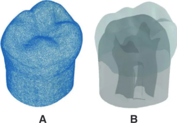
Obtained surface contours and meshes were then imported into SolidWorks 2015 software (Dassault Systems Solid-Works Corp, Waltham, USA). Three- dimensional solid model of intact mandibular molar was generated using a “SCANto3D” add-in module (Fig. 1A).
Interfacial surface between pulp chamber and den- tin and interfacial surface between dentin and enamel were made by lofting technique of the CAD program according to the anatomy of natural tooth (eHuman 3-D Tooth Atlas 7.6, eHuman Inc, Fremont, USA).
Once enamel, dentin, and pulp 3-D volumes were generated, Boolean operations were used to ensure congruence between related interfacial surfaces. For instance, dentin volume was created by subtracting pulp cavity volume. Enamel 3-D volume was then obtained by subtracting dentin volume (Fig. 1B). All solid models were derived from the three-dimension- al solid model of the intact mandibular molar.
272. Cavity preparation design
Based on the three-dimensional CAD model of un-restored mandibular molar tooth, inlays with conventional cavity model and composite filling with minimal invasive preparation model were made. Four 3-D experimental models were designed and created:
(1) O cavity with conventional design (OR-C, OG-C models); (2) MO cavity with conventional design (MR-C, MG-C models); (3) O cavity with minimal design (OR-M model); and (4) D cavity with minimal design (MR-M model). Shape and dimensions of intracoronal restorations were taken from the litera- ture.
28All inlays with conventional design cavities had pulpal and axial walls with at least 0.6 mm dentin thickness over the pulp while gingival walls of proxi- mal boxes were located 0.4 mm above cemento- enamel junction. The narrowest portion of the prep- aration was 1.0 mm faciolingually, which was located between buccal and lingual cusp tips. The cavity ex-
A B
Fig. 1. (A) Stereolithography (STL) scan image of
mandibular first molar. (B) 3D CAD model of pulp,
dentin, and enamel were generated and assembled.
tended the full length of the occlusal groove, includ- ing mesial and distal pits with their radiating grooves.
The pulpal wall was flat horizontally. The occlusocer- vical thickness of inlay was between 0.7 mm and 2.6 mm in O cavity. In MO inlay cavity, proximal boxes were extended proximally from the occlusal cavity.
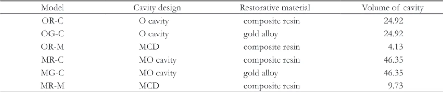
The shape of the proximal box was on straight lines or planes with thickness of at least 0.8 mm. Minimal- ly invasive models (OR-M, DR-M) preserved the un- supported enamel and removed the minimum tooth structure in spherical form, which was limited to the area of dental caries lesion (Fig. 2). Volumes of res- torations were 24.92 mm
3in conventional O cavity and 46.35 mm
3in conventional MO cavity. Those of OR-M model and MR-M model were 4.13 mm
3and 9.73 mm
3, respectively, in minimally invasive cavity (Table 1).
3. Finite element analysis
In minimal cavity design groups, restorative mate- rial was composite resin. In conventional cavity de- sign groups, two types of restorative materials were tested: (1) gold alloy (E = 95.6 GPa, υ = 0.35),
20and (2) composite resin (E = 9.5 GPa, υ = 0.24).
16Mate- rial properties of dentin (E = 18.6 GPa, υ = 0.32)
21and enamel (E = 84.1 GPa, υ = 0.3)
20were assigned.
All materials were assumed to have linear, elastic, and isotropic properties. The bonding interface between dentin and composite or enamel was considered to be perfect in this experiment. Three-dimensional solid models were meshed with tetrahedral elements.
The number of elements and nodes varied accord- ing to models (59,009 - 73,064 elements and 89,352 - 108,739 nodes). Fixed zero-displacement in three spatial dimensions (X, Y, and Z) was assigned to nodes at the bottom surface of the tooth, preventing rigid body displacement for all models. To simulate biting force, a total amount of 200 N load was ap- plied vertically on the tooth at 10 occlusal contact points (5 buccal cusp points, 3 central fossa points, and each point on both marginal ridges) (Fig. 3). A static finite element analysis (FEA) was performed to predict the stress distribution generated by occlusal loading.
Results
In order to analyze stress distribution and location, all structures created were isolated from the rest of the model. For each group, peak stresses on restor- ative materials and abutment teeth were evaluated separately.
Table 1. Experimental models and cavity volume for each restoration
Model Cavity design Restorative material Volume of cavity
OR-C O cavity composite resin 24.92
OG-C O cavity gold alloy 24.92
OR-M MCD composite resin 4.13
MR-C MO cavity composite resin 46.35
MG-C MO cavity gold alloy 46.35
MR-M MCD composite resin 9.73
MCD: minimal cavity design; Unit: mm
3.
Fig. 2. Two conventional inlay models of O cavity (CG-
C), MO cavity (MG-C), and two minimal invasive designs
for occlusal caries (OR-M) and proximal caries (MR-M)
models were made.
1. Stress distribution in restorations
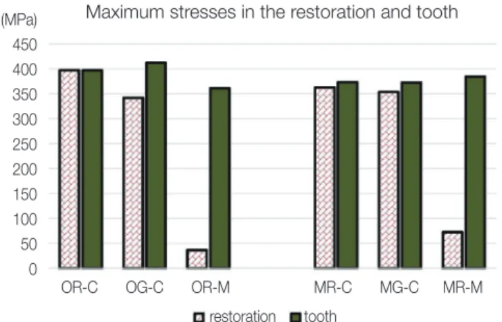
Maximum values of von Mises stress within res- torations with minimal cavity design generated were significantly lower (OR-M model: 26.8 MPa; MR-M model: 72.7 MPa) compared to those with conven- tional cavity design (OR-C model: 397.2 MPa; OG-C model: 341.9 MPa; MR-C model: 362.5 MPa; MG-C model: 352.6 MPa).
Fig. 3. A total amount of 200 N axial load was applied at 10 occlusal points: 5 points in buccal cusp area and 5 points in marginal ridges and central fossa area (model OG-C). Solid model with restoration, enamel, dentin, and pulp chamber of the mandibular molar was meshed with tetrahedral elements. Bottom of the model was fixed in all directions as a boundary condition.
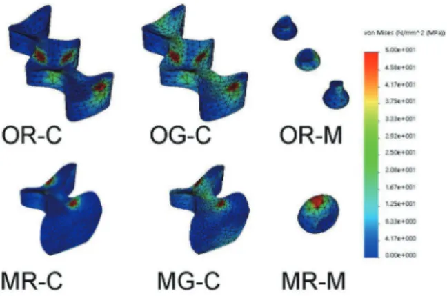
Regarding the effect of cavity design, minimal invasive designs (OR-M, MR-M) generated 5 to 10 times smaller maximum von Mises stresses than those with conventional inlay designs (Fig. 4). Re- garding the effect of dental material, composite resin (OR-C, MR-C) exhibited slightly higher maximum von Mises stresses than gold alloy (OG-C, MG-C) in restorations. Gold inlay (OG-C) showed more favorable and well distributed stresses in the restora- tion than composite resin inlay (OR-C) (Fig. 5, Fig.
6). Overall, the order of stress intensity in restora- tions with conventional inlay/minimal cavity filling designs was as follows: OR-C > MR-C > MG-C >
OG-C > MR-M > OR-M.
The differences in stress magnitudes over the adja- cent enamel along cavosurface margins of composite restorations (OR-C, OR-M, MR-M) were distin- guished. In terms of stress location, high concentra- tions of von Mises stress on surfaces of restorations were found near the occlusal contact areas where bit- ing forces were applied (Fig. 7).
2. Stress distribution in abutment teeth
In tooth structure, magnitudes of maximum von Mises stresses in models with conventional design were between 372.8 MPa and 412.9 MPa, while those in models with minimal cavity designs were between 361.1 MPa and 384.4 MPa. The gold O cavity inlay (OG-C) produced the highest von Mises stress (412.0
Fig. 4. Minimal invasive cavity designs (OR-M, MR-M) produced very small maximum von Mises stress magnitude compared to conventional inlay designs in the restoration. There were no significant differences in maximum stress magnitudes within the abutment tooth among models.
Maximum stresses in the restoration and tooth (MPa)
450 400 350 300 250 200 150 100 50
0 OR-C OG-C OR-M MR-C MG-C MR-M
restoration tooth
MPa) while the composite minimal O cavity design (OR-M) generated the lowest von Mises stress (361.1 MPa) (Fig. 4). When comparing all experimental models, m a x i mu m stress values generated in the abutment teeth were close to one another (i.e., be- tween conventional/minimal invasive cavity designs and tested restorative materials).
In terms of stress distribution patterns in enamel and dentin, similar results were observed for all ex- perimental models. High stress concentrations were found at the enamel surface near buccal cusp tips, central fossa, and marginal ridges where axial occlu- sal forces were applied (Fig. 6, Fig. 7, and Fig. 8).
Fig. 7. Cavosurface margin between resin restorations (OR-C, MR-M) and tooth structures showed significant difference in stress gradient and distribution. Peak stress was observed at the occlusal loading area and cemento-enamel junction in all models.
Fig. 8. Von Mises stress distribution by occlusal loading in the abutment tooth with M-D cross-sectional view.
Note stress concentration was observed around the occlusal contact area, pulp horns, and cemento-enamel junction.
Fig. 5. Von Mises stress distribution generated by occlusal loading in the restoration of each experimental model. Models with composite (OR-C, MR-C) showed stress concentration at the loading area. Models with gold alloy (OG-C, MG-C) showed widely distributed stresses within the restorations.
Fig. 6. Von Mises stress distribution by occlusal
loading in the restoration and the abutment tooth
with M-D cross-sectional view. Models of OG-C and
MG-M produced well distributed stress inside the gold
restoration.
Discussion
Preservation of sound tooth structures is the pri- mary goal of restorative dentistry. Even if removal of additional dental tissue is necessary, protecting the remaining tooth structure from undesirable mechani- cal responses should be considered. Tooth prepara- tion designs proposed for posterior inlay restorations have been based upon recommendations made by GV Black for cast metal and amalgam, resulting in considerable tooth structure removal with oppos- ing walls that are parallel.
28The preparation design for an indirect restoration must satisfy a balance between preserving the tooth structure and maximiz- ing the strength of the restoration. However, there is a problem within the concept of the original GV Black classification since it identifies the position of lesion and prescribes cavity design regardless of the size and extent of carious lesion. Recently, the Acad- emy of Operative Dentistry European Section has considered adhesively bonded resin composites for use in direct minimal intervention approaches to re- store posterior teeth, emphasizing the importance of the practice of evidence-based minimal intervention dentistry to extend the longevity of restorations.
3Masticatory loads in the posterior region are much higher than those in the anterior region. Stress con- centrations can manifest themselves in various forms of clinical failures such as tooth fracture and frac- ture of restorative body. The main purpose of this research was to evaluate the maximum stress values and stress distribution in intracoronal restorations and the tooth after occlusal loading to identify failure possibility under various types of cavity designs and materials.
St-Georges et al.
14have reported fracture resistance of prepared teeth restored with bonded inlay for MOD preparations can weaken the teeth by approxi- mately 59%. Under compressive load testing, com- posite and ceramic inlay restorations do not restore the original strength of the teeth. Removal of mar- ginal ridges, increase in the depth and width of inlay cavity, and increased preparation in the proximal box formation are main reasons for the decrease in resis- tance. The current minimally invasive dentistry advo-
cates conservative principles of cavity preparation.
Small isolated lesions should be treated individually, not interconnected, as common practice for conven- tional inlay preparations. Furthermore, the prepara- tion should not be extended beyond dimensions of the caries lesion so that the enamel unsupported by dentin is preserved.
The traditional approach to control caries inevita- bly leads to an excessive tooth reduction. FEA results from Wayne et al.
26have revealed that larger restora- tion volume proportion will result in higher dentin- enamel stresses under static loading. This result suggests that minimal invasive cavity can produce stresses that are more favorable biomechanically. In our study, tooth structure prepared with conventional inlay designs removed five times greater volume than that with minimal invasive cavity designs (Table 1).
Within restorations, minimal invasive models gener- ated smaller peak stress while similar magnitudes of stresses were produced within the abutment tooth.
Low magnitudes of von Mises stresses observed in our experiment models with minimal invasive cavity contradicted GV Black’s classical principles of cavity preparation from the biomechanical point of view.
Our findings could serve as a basis for preserving as many intact tooth structures as possible (Fig. 4, Fig.
5, Fig. 6, and Fig. 7). A strengthening effect of the enamel without dentin support in minimally invasive technique could be expected in clinical situation of bonded composite restoration.
Guven et al.
22have analyzed the influence of inlay cavity design by FEA and reported that cavities with rounded corners showed less stress than those with rectangular corners due to improved stress distribu- tion capabilities of rounded corners. The model of conventional inlay has a box-shaped cavity with sharp margins. This might have increased the maximum stress of the model in our study.
Currently, composite resin as well as metal alloy
and dental ceramic represent logical options for res-
torations in posterior teeth. The restorative material
is a factor that can affect the biomechanics during
occlusal loading. Gold restorative material tends to
concentrate more stress inside the inlay, resulting in
lower cusp deflection than the resin.
15In our study,
gold inlay (OG-C) showed well distributed smaller peak stresses in the restoration than the composite resin inlay (OR-C). Interestingly, in contrast to cav- ity design of inlays/filling, only a small difference was observed among maximum stresses by different restorative materials in each experimental group (Fig.
4). Thus, using proper cavity design may be more im- portant than using a particular restorative material.
The fracture resistance of teeth restored with in- lay/filling is very complex. It is impossible to include all variables encountered in the oral environment in a computer simulation.
19Although von Mises stress concentration cannot predict failure patterns in a computer simulation, higher stress concentrations are related to fracture of restorations and failure of teeth restored with inlays or filling. In oral cavity during function, teeth are loaded with complex and variable forces.
Low magnitudes stresses observed in our experi- ment models of minimal invasive cavity suggest pre- serving as many intact tooth structures as possible from a mechanical point of view. Several limitations and weaknesses of computer simulation need to be addressed in the future.
Conclusion
Finite element analysis was performed to investi- gate the effect of different cavity preparation designs with various restorative materials on mandibular mo- lar after exposure to a masticatory force. Within the limitations of this study, the following conclusions were drawn:
Models with minimal invasive designs (OR-M, MR-M) generated 5 to 10 times smaller maximum von Mises stress within restorations than those with conventional inlay designs when occlusal load was applied.
Peak stress was generated at the occlusal contact area around marginal ridges or central fossa in all models. Gold inlay (OG-C) showed well distributed and smaller stresses in the restoration than compos- ite resin inlay (OR-C).
Lower magnitudes of von Mises stresses observed in models with minimal invasive cavity design sug-
gest that bonded composite can strengthen the tooth when enamel has lost its dentin support.
Acknowledgments
This work was supported by a grant (CRI 18026-1) of the Chonnam National University Hospital Re- search Institute.
ORCID
Sunmi Yang https://orcid.org/ 0000-0002-9802-0282 Seon-mi Kim https://orcid.org/ 0000-0001-5103-767X Namki Choi https://orcid.org/ 0000-0003-4830-8568 Jae-hwan Kim https://orcid.org/ 0000-0001-8088-6216 Sung-Pyo Yang https://orcid.org/ 0000-0003-4928-1838 Hongso Yang https://orcid.org/ 0000-0002-9138-4817
References
1. Sabbagh J, McConnell RJ, McConnell MC. Poste- rior composites: Update on cavities and filling tech- niques. J Dent 2017;57:86-90.
2. Tyas MJ, Anusavice KJ, Frencken JE, Mount GJ.
Minimal intervention dentistry - a review. FDI Commission Project 1-97. Int Dent J 2000;50:1-12.
3. Lynch CD, Opdam NJ, Hickel R, Brunton PA, Gurgan S, Kakaboura A, Shearer AC, Vanherle G, Wilson NH. Guidance on posterior resin compos- ites: Academy of Operative Dentistry - European Section. J Dent 2014;42:377-83.
4. Banerjee A. Minimal intervention dentistry: part 7.
Minimally invasive operative caries management:
rationale and techniques. Br Dent J 2013;214:107- 11.
5. Ericson D, Kidd E, McComb D, Mjör I, Noack MJ. Minimally Invasive Dentistry - concepts and techniques in cariology. Oral Health Prev Dent 2003;1:59-72.
6. Tassery H, Levallois B, Terrer E, Manton DJ, Ot- suki M, Koubi S, Gugnani N, Panayotov I, Jacquot B, Cuisinier F, Rechmann P. Use of new minimum intervention dentistry technologies in caries man- agement. Aust Dent J 2013;58 Suppl 1:40-59.
7. Casagrande L, Laske M, Bronkhorst EM, Huys-
mans MCDNJM, Opdam NJM. Repair may in- crease survival of direct posterior restorations - A practice based study. J Dent 2017;64:30-6.
8. Staehle HJ, Wohlrab T, Saure D, Wolff D, Frese C. A 6.5-year clinical follow-up of direct resin composite buildups in the posterior dentition: In- troduction of a new minimally invasive restorative method. J Dent 2015;43:1211-7.
9. White JM, Eakle WS. Rationale and treatment ap- proach in minimally invasive dentistry. J Am Dent Assoc 2000;131:13S-9S.
10. Abu-Hanna AA, Mjör IA. Resin composite re- inforcement of undermined enamel. Oper Dent 2004;29:234-7.
11. Eidelman E. Composite resin support of under- mined enamel in amalgam restorations. Pediatr Dent 1999;21:118-20.
12. Fonseca RB, Fernandes-Neto AJ, Correr-Sobrinho L, Soares CJ. The influence of cavity preparation design on fracture strength and mode of fracture of laboratory processed composite resin restora- tions. J Prosthet Dent 2007;98:277-84.
13. Yang H, Park C, Shin JH, Yun KD, Lim HP, Park SW, Chung H. Stress distribution in premolars restored with inlays or onlays: 3D finite element analysis. J Adv Prosthodont 2018;10:184-90.
14. St-Georges AJ, Sturdevant JR, Swift EJ Jr, Thomp- son JY. Fracture resistance of prepared teeth re- stored with bonded inlay restorations. J Prosthet Dent 2003;89:551-7.
15. Costa A, Xavier T, Noritomi P, Saavedra G, Borges A. The influence of elastic modulus of inlay mate- rials on stress distribution and fracture of premo- lars. Oper Dent 2014;39:E160-70.
16. Yamanel K, Caglar A, Gülsahi K, Ozden UA.
Effects of different ceramic and composite ma- terials on stress distribution in inlay and onlay cavities: 3-D finite element analysis. Dent Mater J 2009;28:661-70.
17. Latino C, Troendle K, Summitt JB. Support of un- dermined occlusal enamel provided by restorative materials. Quintessence Int 2001;32:287-91.
18. Soares CJ, Martins LR, Pfeifer JM, Giannini M.
Fracture resistance of teeth restored with indirect- composite and ceramic inlay systems. Quintessence
Int 2004;35:281-6.
19. Dejak B, Mlotkowski A. Three-dimensional finite element analysis of strength and adhesion of com- posite resin versus ceramic inlays in molars. J Pros- thet Dent 2008;99:131-40.
20. Jiang W, Bo H, Yongchun G, LongXing N. Stress distribution in molars restored with inlays or on- lays with or without endodontic treatment: a three- dimensional finite element analysis. J Prosthet Dent 2010;103:6-12.
21. Zarone F, Sorrentino R, Apicella D, Valentino B, Ferrari M, Aversa R, Apicella A. Evaluation of the biomechanical behavior of maxillary central inci- sors restored by means of endocrowns compared to a natural tooth: a 3D static linear finite elements analysis. Dent Mater 2006;22:1035-44.
22. Guven S, Akdogan M, Oz C, Dogan MS, Unal M, Unal S, Sahbaz C. Three-dimensional finite-element analysis of two ceramic inlay restorations with dif- ferent cavity designs. Biotechnol Biotechnol Equip 2015;29:579-85.
23. Magne P. Efficient 3D finite element analysis of dental restorative procedures using micro-CT data.
Dent Mater 2007;23:539-48.
24. Heo KH, Lim YJ, Kim MJ, Kwon HB. Three- dimensional finite element analysis of the splinted implant prosthesis in a reconstructed mandible. J Adv Prosthodont 2018;10:138-46.
25. Zelic K, Vukicevic A, Jovicic G, Aleksandrovic S, Filipovic N, Djuric M. Mechanical weakening of devitalized teeth: three-dimensional Finite Element Analysis and prediction of tooth fracture. Int En- dod J 2015;48:850-63.
26. Wayne JS, Chande R, Porter HC, Janus C. Effect of restoration volume on stresses in a mandibu- lar molar: a finite element study. J Prosthet Dent 2014;112:925-31.
27. Ausiello P, Franciosa P, Martorelli M, Watts DC.
Numerical fatigue 3D-FE modeling of indirect composite-restored posterior teeth. Dent Mater 2011;27:423-30.
28. Thompson MC, Thompson KM, Swain M. The all-
ceramic, inlay supported fixed partial denture. Part
1. Ceramic inlay preparation design: a literature re-
view. Aust Dent J 2010;55:120-7.
*교신저자: 양홍서
(520757)광주광역시 북구 용봉로 33 전남대학교 치의학전문대학원
Tel: 062-530-5823|Fax: 062-530-0130|E-mail: [email protected] 접수일: 2018년 10월 22일|수정일: 2018년 12월 2일|채택일: 2018년 12월 6일
최소 침습적 충진 및 통상적 인레이 법으로 수복한 대구치의 응력 분포:
3-D 유한 요소 해석
양선미 1 , 김선미 1 , 최남기 1 , 김재환 1 , 양성표 2 , 양홍서 3 *
1
전남대학교 치의학전문대학교 소아치과학교실
2
KAIST 뇌공학 및 의공학과
3