236 책임저자:서영록, 100-715, 서울시 중구 필동 3가 26번지
동국대학교 바이오시스템대학 생명과학과 Tel: 02-2260-3321, Fax: 02-2760-0674 E-mail: [email protected]
김정민, 300-716, 대전시 동구 용운동 96-3 대전대학교 한의과대학 동서생명과학연구원 Tel: 042-286-7478, Fax: 042-286-7479 E-mail: [email protected]
접수일:2010년 4월 2일, 1차 수정일:2010년 7월 8일, 2차 수정일:2010년 7월 12일, 게재승인일:2010년 7월 15일
*공동 저자임.
Correspondence to:Young Rok Seo
Department of Life Science, College of Life Science and Biotechnology, Dong- guk University-Seoul, 26, Pil-dong 3-ga, Jung-gu, Seoul 100-715, Korea Tel: +82-2-2260-3321, Fax: +82-2-2760-0674
E-mail: [email protected]
Co-Corresponding author:Jung Min Kim
Institute of Traditional Medicine and Bioscience, College of Oriental Medicine, Daejeon University, 96-3, Yongun-dong, Dong-gu, Daejeon 300-716, Korea
Tel: +82-42-286-7478, Fax: +82-42-286-7479 E-mail: [email protected]
한국인 난소암 조직에서 발현된 miRNA의 타겟 mRNA를 중심으로 하는 p53 관련 신호전달 체계 분석
1동국대학교 바이오시스템대학 생명과학과, 2경희대학교 의과대학 약리학교실 의과학연구소,
3가톨릭대학교 의과대학 산부인과학교실, 4대전대학교 한의과대학 동서생명과학연구원
권지영1,2ㆍ배수미3ㆍ안웅식3ㆍ김정민4*ㆍ서영록1,2*
p53 Associated Signaling Pathway Analysis in Terms of microRNAs and Their Target mRNAs in Korean Ovarian Cancer Tissues
Jee Young Kwon1,2, Sumi Bae3, Woong Shick Ahn3, Jung Min Kim4* and Young Rok Seo1,2*
1Department of Life Science, College of Life Science and Biotechnology, Dongguk University, Seoul 100-715, 2Department of Pharmacology, Biomedical Science Institute, School of Medicine, Kyung Hee University, Seoul 130-701, 3Department of Obstetrics and Gynecology, College of Medicine, The Catholic University of Korea, Seoul 137-040, 4Institute of
Traditional Medicine and Bioscience, College of Oriental Medicine, Daejeon University, Daejeon 300-716, Korea Ovarian cancer has been considered as the mostly common cancer among women in worldwide.
Molecular mechanism of initiation and progression in this cancer has been well studied, however, diagnotic and therapeutic programs has not been successfully established yet. In order to improve screening program to detect the ovarian cancer at the early stage and subsequently develop therapeutic modalities for cancer treatment, research focusing on more insight of regulatory molecular mechanisms are urgently required.
Here, we analyze effect of microRNAs (miRNAs), recognized as non-coding RNAs that negatively regulate expression of target genes, on gene expression at genome wide level. Concerning with potential miRNA features, each miRNA is able to inhibit hundreds of transcripts directly or indirectly. Therefore, signaling network analysis of miRNA targeting on particular mRNA would provide novel molecular interactions for establishing alternative approach to treat the ovarian cancer patients. Our group has analyzed miRNA microarray in ovarian tissues from Korean patients. Based on our previous microarray study, we discovered miRNA-associated target mRNAs participating in p53-regulated signaling. As result, we have presented 26 target genes related with p53-controlling pathway. Interestingly, the identified three genes encoding ING1, NDRG1 and CDC14A might be further recognized as novel target molecules in ovarian cancer. This study might provide evidence for better understanding mechanism in relevance to governing ovarian cancer and consequently for improving diagnosis and therapy of this cancer. (Cancer Prev Res 15, 236-240, 2010)
Key Words: Ovarian cancer, microRNA, p53, Signaling pathway
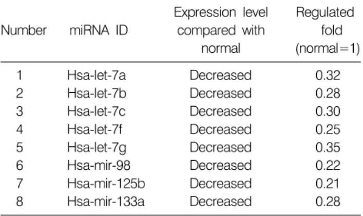
Table 1. Eight miRNAs identified in Korean ovarian cancer tissues
Expression level Regulated Number miRNA ID compared with fold
normal (normal=1)
1 Hsa-let-7a Decreased 0.32
2 Hsa-let-7b Decreased 0.28
3 Hsa-let-7c Decreased 0.30
4 Hsa-let-7f Decreased 0.25
5 Hsa-let-7g Decreased 0.35
6 Hsa-mir-98 Decreased 0.22
7 Hsa-mir-125b Decreased 0.21
8 Hsa-mir-133a Decreased 0.28
서 론
난소암은 부인과 암 중에서 치사율이 가장 높은 암종 으로 잘 알려져 있으며1) 여러 종류의 subtype 중 serous type이 가장 일반적인 것으로 보고되고 있다.2∼4) 미국 뿐 아니라, 서유럽 국가들의 여성암 사망률 중 5위를 차지 하고 있으며5) 한국인을 대상으로 한 통계학적인 연구결 과에 따르면 난소암에 걸린 환자의 수가 꾸준히 증가하 고 있다.6) 다른 종의 암에서와 마찬가지로 난소암 초기 에 진단을 통해서 암의 발병여부를 알게 될 경우, 생존율 은 매우 높아질 수 있다. 하지만, 여성의 70% 이상은 진 행된 단계에서 진단되어지며 이때부터의 5년 생존율은 30% 밖에 되지 않는 것으로 보고되고 있다.7) 이에 난소 암 초기에 암의 생성여부를 판단할 수 있는 보다 향상된 진단 프로그램 및 진행성 난소암의 효율적인 치료법이 절실히 요구된다.
Micro RNA (miRNA)라고 불리는 small noncoding RNAs 는 전사 후 단계에서 유전자의 발현을 조절하는 것으로 밝혀진 새로운 분자 그룹이며 타겟 mRNA의 3’ untrans- lated region에 binding 함으로서 translation 과정으로의 진 행을 막거나 mRNA의 degradation을 유도하다고 알려져 있다.8) 최근 miRNA가 세포의 분화, 성장, 사멸과 같은 세포사의 필수과정에서 매우 중요한 역할을 하고 있음 이 밝혀지고 있으며9,10) 뿐만 아니라, 비정상적인 발현 또 는 돌연변이 된 miRNA가 암에서 발견된다는 연구결과 가 보고되어지고 있다.11∼18) 이는 miRNA가 그들의 타겟 유전자의 특성에 따라 종양유전자(oncogenes) 또는 종양 억제유전자(tumor suppressor genes)로서의 가능성을 가지 고 있음을 의미하며 암 연구에 있어 새롭게 조명 받아야 하는 주제로 인식되고 있다. 지금까지 대표적으로 알려져 있는 miRNA의 타겟 유전자들로는 RAS,11) HMGA2,12,13) BCL214), c-Myc,15) p5317) 등이 있다.
이들 중 p53은 대표적인 항 종양억제인자로 난소암을 포함한 대부분의 암에서 돌연변이 되어 있는 것으로 알 려져 있다.19) p53은 전사인자로서 유전자 손상, 저산소 증, 종양인자 활성의 부적절 등과 같은 자극에 의해 활성 화되고 결과적으로 세포주기 정지(cell cycle arrest), 세포 사멸(apoptosis), 노화(senescence)와 같은 항증식성의 반응 (antiproliferative response)을 유도한다. 이 과정에서 p21, Gadd45a, Bax와 같은 타겟 유전자들을 활성화 시키는 것 으로 알려져 있으며20) p53을 중심으로 하는 세포반응의 이해는 난소암 발병의 명확한 기전 이해를 위해 필수적 이라고 할 수 있다. 이에 본 연구는 한국인 난소암 조직
에서 발굴된 miRNA의 타겟 mRNA를 중심으로 이와 관 련된 p53 신호전달체계를 분석하고자 하며 이들의 새로 운 난소암 타겟 분자로서의 가능성을 논하고자 한다.
재료 및 방법 1. miRNA의 타겟 mRNA 예측
본 연구에 앞서 수행된 한국인 난소암 조직에서의 miRNA microarray에서 발굴되고 qPCR를 거쳐 검증된 8개 의 miRNA의 타겟 mRNA는 Sanger Database (http://www.
mirbase.org)를 사용하여 추출하였다.
2. miRNA의 타겟 mRNA 중 p53 관련 신호전달체계 분석
miRNA의 타겟 유전자들 중 p53 관련 유전자는 Gene- cards database (http://www.genecards.org)로부터 추출하였으 며, DAVID (http://david.abcc.ncifcrf.gov/), Medline databases (http://www.ncbi.nlm.nih.gov/)를 이용하여 신호전달체계를 분석하였다.
결과 및 고찰 1. miRNA의 타겟 mRNA 유전자 예측
한국인 난소암 조직에서 발굴된 miRNA 중 qPCR를 통 해 검증된 8개의 miRNA (Table 1)의 타겟 mRNA를 Sang- er database를 사용하여 추출할 수 있었으며 그 결과 3,770 개의 타겟 mRNA를 확인할 수 있었다. 본 연구에서 발굴 된 8개 중 miR-98과 miR-133a는 본 연구를 통해 처음으로 밝혀진 난소암 관련 miRNA이며 let-7 family21)와 miR- 125b22)는 앞선 다른 연구그룹의 결과와 일치하는 것으로 확인되었다.
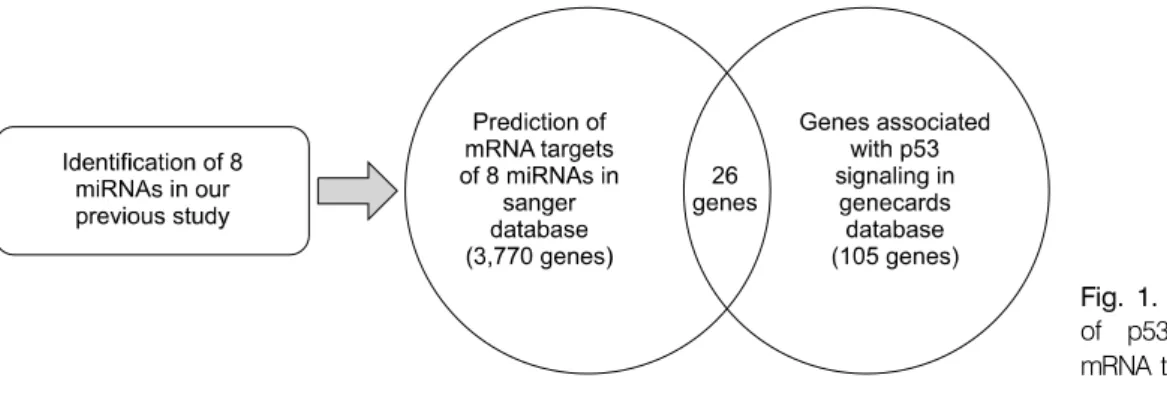
Fig. 1. A diagram for prediction of p53 associated genes in mRNA target genes of 8 miRNAs.
Table 2. p53 associated genes in mRNA target genes of 8 miRNAs
Number Genbank ID Gene symbol Description
1 NM_078467 CDKN1A Cyclin-dependent kinase inhibitor 1A (p21, Cip1)
2 NM_005427 TP73 Tumor protein p73
3 NM_032848 C12orf52 Chromosome 12 open reading frame 52 4 NM_198219 ING1 Inhibitor of growth family, member 1 5 NM_006096 NDRG1 N-myc downstream regulated gene 1 6 NM_138349 TP53I13 Tumor protein p53 inducible protein 13 7 NM_021145 DMTF1 Cyclin D binding myb-like transcription factor 1 8 NM_005225 E2F1 E2F transcription factor 1
9 NM_005427 TP73 Tumor protein p73
10 NM_003883 HDAC3 Histone deacetylase 3
11 NM_000389 CDKN1A Cyclin-dependent kinase inhibitor 1A (p21, Cip1)
12 NM_004060 CCNG1 Cyclin G1
13 NM_002392 MDM2 Mdm2, transformed 3T3 cell double minute 2, p53 binding protein (mouse) 14 NM_058197 CDKN2A Cyclin-dependent kinase inhibitor 2A (melanoma, p16, inhibits CDK4) 15 NM_001001683 MED11 Mediator of RNA polymerase II transcription, subunit 11 homolog (S. cerevisiae) 16 NM_003672 CDC14A CDC14 cell division cycle 14 homolog A (S. cerevisiae)
17 NM_152319 C12orf54 Chromosome 12 open reading frame 54
18 NM_022045 MTBP Mdm2, transformed 3T3 cell double minute 2, p53 binding protein (mouse) binding protein, 104 kDa
19 NM_006879 MDM2 Mdm2, transformed 3T3 cell double minute 2, p53 binding protein (mouse)
20 NM_138764 BAX BCL2-associated X protein
21 NM_015316 PPP1R13B Protein phosphatase 1, regulatory (inhibitor) subunit 13B 22 NM_033010 PCBP4 Poly (rC) binding protein 4
23 NM_001703 BAI2 Brain-specific angiogenesis inhibitor 2
24 NM_001188 BAK1 BCL2-antagonist/killer 1
25 NM_001702 BAI1 Brain-specific angiogenesis inhibitor 1
26 NM_032286 MED10 Mediator of RNA polymerase II transcription, subunit 10 homolog (NUT2, S.
cerevisiae) Let-7 family 중 let-7f는 난소암 외 유방암,23) 폐암,21) 갑 상선암,24) 전립선암25)에서도 저발현되는 것으로 알려져 있으며 oncogene으로 잘 알려진 RAS의 발현에 관여함으 로서 암의 생성과정에서 중요한 역할을 할 것으로 예상 되어지는 분자이다.11) 이에 난소암 억제를 위한 연구에 있어 let-7 유전자의 분자적 기전 이해를 통한 발현 조절 은 발암억제에 효과적인 타겟 분자로서 작용할 수 있을
것으로 예측된다. 또한, miR-125b는 세포의 분화, 성장에 관여하는 것으로 보고되어져 있으며26,27) ERBB2와 ERBB3 을 타겟 유전자로 이들을 조절하는 것으로 알려져 있다.27) 염색체 11q23-24에 위치하는 miR-125b는 난소암, 유방 암, 폐암에서 대부분 돌연변이 되어져 있는 것으로 나타 났으며28) 이는 miR-125b의 타겟 유전자인 ERBB2가 난소 암 환자에서 과발현된 것29,30)과 매우 밀접한 관계를 가
지고 있다고 할 수 있다. 이와 같이 miRNA의 발현 정도 에 따른 타겟 유전자의 발현은 난소암을 비롯한 다양한 암종에서 암의 형성 및 진행을 유도하는데 매우 연관이 깊은 것으로 사료되며 이에 한국인 난소암 환자에서의 발견된 miRNA의 타겟 유전자의 분석 및 이와 관련된 신 호전달체계 분석은 난소암의 분자적 기전을 이해하는데 필수적이라 할 수 있다.
2. miRNA의 타겟 mRNA에서의 p53 관련 신호전달체 계 분석
한국인 난소암 조직에서 발굴된 miRNA의 타겟 mRNA 들 중 p53 관련 신호전달체계 분석을 위하여 Genecard V3 database를 통해 얻은 p53 관련 유전자들과 본 연구에 서 얻은 3,770 개의 타겟 유전자를 비교분석(Fig. 1) 하였 으며 그 결과 26개의 p53관련 유전자(Table 2)를 추출할 수 있었다. 본 결과에서 얻어진 p53 관련 26개의 유전자 들 중 ING1, NDRG1, CDC14A 유전자는 아직까지 난소 암에 그 기능이 밝혀지지 않은 유전자이다.
ING1은 p53과 관련된 항 종양인자로 알려진 유전자31) 로서 최근에는 miRNA의 processor인 DGCR8을 조절한다 는 연구결과가 보고된 바 있다.32) 뿐만 아니라, 다양한 암 세포에서 발현된 CDC14A는 p53의 타겟 유전자로서 상호결합하여 Cdk1/cyclin B 복합체를 형성함으로서 발암 과정에 관여한다는 연구결과가 보고되었다.33) 이러한 타 겟 분자들은 향후 보다 심도 있는 검증 연구가 필요하지 만, 우선 일차적으로 한국인 난소암에서 특이적으로 나 타날 수 있는 miRNA의 타겟 분자들이며 p53 관련 sig- naling network를 새롭게 구축하는데 사용될 수 있을 것이 라 예상된다.
결 론
본 연구는 한국인 난소암 환자에서 특이적으로 나타 난 miRNA의 타겟 mRNA를 중심으로 하는 p53 관련 신 호전달체계를 분석함으로서 난소암 발병에 관여하는 새 로운 타겟 분자를 발굴하고 이를 바탕으로 하는 새로운 분자적 기전의 가능성을 제시하고자 하였으며 궁극적으 로 본 연구를 통해 난소암의 치료 뿐 아니라 조기 진단 의 마커를 개발하는데 도움을 줄 수 있을 것으로 기대하 는 바이다.
감사의 글
본 연구는 보건복지부 암정복추진연구개발사업 지원
으로 이루어진 것임(과제고유번호: 0820330).
참 고 문 헌
1) Willner J, Wurz K, Allison KH, Galic V, Carcia RL, Goff BA, Swisher EM. Alterate molecular genetic pathways in ovarian carcinoma of common histological types. Hum Pathol 38, 607-613, 2007.
2) Singer G, Kurman RJ, Chang HW, Cho SK, Shih IeM.
Diverse tumorigenic pathways in ovarian serous carcinoma.
Am J Pathol 160, 1223-1228, 2002.
3) Shih IeM, Kurman RJ. Ovarian tumorigenesis: a proposed model basedon morphological and molecular genetic analysis.
Am J Pathol 164, 1511-1518, 2004.
4) Singer G, Oldt R 3rd, Cohen Y, Wang BG, Sidransky D, Kurman RJ, Shih IeM. Mutations in BRAF and KRAS characterize the development of low-grade ovarian serous carcinoma. J Natl Cancer Inst 95, 484-486, 2003.
5) Society AC. Cancer facts and figures 2007. Atlanta: American Cancer Society, 2007.
6) Chung HH, Hwang SY, Jung KW, Won YJ, Shin HR, Kim JW, Lee HP. Ovarian cancer incidence and survival in Korea:
1993-2002. Int J Gynecol Cancer 17, 595-600, 2007.
7) David CC, Alexander YN. MicroRNA and ovarian cancer.
Histol Histopathol 23, 1161-1169, 2008.
8) He L, Hannon GJ. MicroRNAs: small RNAs with a big role in gene regulation. Nat Rev Genet 5, 522-531, 2004.
9) Miska EA. How microRNAs control cell division, differenti- ation, and death. Curr Opin Genet Dev 5, 563-568, 2005.
10) Zamore PD, Haley B. Ribo-gnome: the big world of small RNAs. Science 309, 1519-1524, 2005.
11) Johnson SM, Grosshans H, Shingara J, Byrom M, Jarvis R, Cheng A, Labourier E, Reinert KL, Brown D, Slack FJ. RAS is regulated by the let-7 microRNA family. Cell 120, 635-647, 2005.
12) Mayr C, Hemann MT, Bartel D. Disrupting the pairing be- tween let-7 and HMGA2 enhances oncogenic transformation.
Science 315, 1576-1579, 2007.
13) Lee YS, Dutta A. The tumor suppressor microRNA let-7 re- presses the HMGA2 oncogene. Genes Dev 21, 1025-1030, 2007.
14) Cimmino A, Calin GA, Fabbri M, Iorio MV, Ferracin M, Shimizu M, Wojcik SE, Aqeilan RI, Zupo S, Dono M, Rassenti L, Alder H, Volinia S, Liu CG, Kipps TJ, Negrini M, Croce CM. miR-15 and miR-16 induce apoptosis by targeting BCL2. Proc Natl Acad Sci USA 102, 13944-13949, 2005.
15) O’Donnell KA, Wentzel EA, Zeller KI, Dang CV, Mendell JT. c-Myc-regulated microRNAs modulate E2F1expression.
Nature 435, 839-833, 2005.
16) He L, Thomson JM, Hemann MT, Hernando-Monge E, Mu D, Goodson S, Powers S, Cordon-Cardo C, Lowe SW, Hannon
GJ, Hammond SM. A microRNA polycistron as a potential human oncogene. Nature 435, 828-833, 2005.
17) Voorhoeve PM, le Sage C, Schrier M, Gillis AJ, Stoop H, Nagel R, Liu YP, van Duijse J, Drost J, Griekspoor A, Zlotorynski E, Yabuta N, De Vita G, Nojima H, Looijenga LH, Agami R. A genetic screen implicates miRNA-372 and miRNA-373 as oncogenes in testicular germ cell tumors. Cell 124, 1169-1181, 2006.
18) Costinean S, Zanesi N, Pekarsky Y, Tili E, Volinia S, Heerema N, Croce CM. Pre-B cell proliferation and lymphoblastic leukemia/high-grade lymphoma in E(mu)-miR155 transgenic mice. Proc Natl Acad Sci USA 103, 7024-7029, 2006.
19) Greenblatt MS, Bennett WP, Hollstein M, Harris CC. Muta- tion in the p53 tumor suppressor gene: clues to cancer etiolo- gy and molecular pathogenesis. Cancer Res 54, 4855-4878, 1994.
20) Helton ES, Chen X. p53 modulation of the DNA damage response. J Cell Biochem 100, 883-896, 2007.
21) Calin GA, Ferracin M, Cimmino A, Di Leva G, Shimizu M, Wojcik SE, Iorio MV, Visone R, Sever NI, Fabbri M, Iuliano R, Palumbo T, Pichiorri F, Roldo C, Garzon R, Sevignani C, Rassenti L, Alder H, Volinia S, Liu CG, Kipps TJ, Negrini M, Croce CM. A MicroRNA signature associated with prog- nosis and progression in chronic lymphocytic leukemia. N Engl J Med 353, 1793-1801, 2005.
22) Roldo C, Missiaglia E, Hagan JP, Falconi M, Capelli P, Bersani S, Calin GA, Volinia S, Liu CG, Scarpa A, Croce CM.
MicroRNA expression abnormalities in pancreatic endocrine and acinar tumors are associated with distinctive pathologic features and clinical behavior. J Clin Oncol 24, 4677-4684, 2006.
23) Iorio MV, Ferracin M, Liu CG, Veronese A, Spizzo R, Sabbioni S, Magri E, Pedriali M, Fabbri M, Campiglio M, Ménard S, Palazzo JP, Rosenberg A, Musiani P, Volinia S, Nenci I, Calin GA, Querzoli P, Negrini M, Croce CM.
MicroRNA gene expression deregulation in human breast cancer. Cancer Res 65, 7065-7070, 2005.
24) Visone R, Pallante P, Vecchione A, Cirombella R, Ferracin M, Ferraro A, Volinia S, Coluzzi S, Leone V, Borbone E, Liu CG, Petrocca F, Troncone G, Calin GA, Scarpa A, Colato C, Tallini
G, Santoro M, Croce CM, Fusco A. Specific microRNAs are downregulated in human thyroid anaplastic carcinomas. Oncogene 26, 7590-7595, 2007.
25) Porkka KP, Pfeiffer MJ, Waltering KK, Vessella RL, Tam- mela TL, Visakorpi T. MicroRNA expression profiling in pro- state cancer. Cancer Res 67, 6130-6135, 2007.
26) Lee YS, Kim HK, Chung S, Kim KS, Dutta A. Depletion of human micro-RNA miR-125b reveals that it is critical for the proliferation of differentiated cells but not for the down- regulation of putative targets during differentiation. J Biol Chem 280, 16635-16641, 2005.
27) Scott GK, Goga A, Bhaumik D, Berger CE, Sullivan CS, Benz CC. Coordinate suppression of ERBB2 and ERBB3 by en- forced expression of micro-RNA miR-125a or miR-125b. J Biol Chem 282, 1479-1486, 2007.
28) Martin ES, Cesari R, Pentimalli F, Yoder K, Fishel R, Himelstein AL, Martin SE, Godwin AK, Negrini M, Croce CM. The BCSC-1 locus at chromosome 11q23-q24 is a candi- date tumor suppressor gene. Proc Natl Acad Sci USA 100, 11517-11522, 2003.
29) Shih IeM, Kurman RJ. Ovarian tumorigenesis: a proposed model based on morphological and molecular genetic analysis.
Am J Pathol 164, 1511-1518, 2004.
30) Lassus H, Leminen A, Vayrynen A, Cheng G, Gustafsson JA, Isola J, Butzow R. ERBB2 amplification is superior to protein expression status in predicting patient outcome in serous ovarian carcinoma. Gynecol Oncol 92, 31-39, 2004.
31) Abad M, Menéndez C, Füchtbauer A, Serrano M, Füchtbauer EM, Palmero I. Ing1 mediates p53 accumulation and chroma- tin modification in response to oncogenic stress. J Biol Chem 282, 31060-31067, 2007.
32) Gomez-Cabello D, Callejas S, Benguría A, Moreno A, Alonso J, Palmero I. Regulation of the microRNA processor DGCR8 by the tumor suppressor ING1. Cancer Res 70, 1866-1874, 2010.
33) Paulsen MT, Starks AM, Derheimer FA, Hanasoge S, Li L, Dixon JE, Ljungman M. The p53-targeting human phospha- tase hCdc14A interacts with the Cdk1/cyclin B complex and is differentially expressed in human cancers. Mol Cancer 5, 1-11, 2006.