• 검색 결과가 없습니다.

A Novel Treatment of Recurrent Temporomandibular Joint Dislocation with Intermaxillary Fixation Using Microimplant: A Case Report JOMP

N/A
N/A
Protected

Academic year: 2021

Share "A Novel Treatment of Recurrent Temporomandibular Joint Dislocation with Intermaxillary Fixation Using Microimplant: A Case Report JOMP"

Copied!
7
0
0

로드 중.... (전체 텍스트 보기)

전체 글

(1)

pISSN 2288-9272 eISSN 2383-8493 J Oral Med Pain 2014;39(4):156-162 http://dx.doi.org/10.14476/jomp.2014.39.4.156

A Novel Treatment of Recurrent Temporomandibular Joint Dislocation with Intermaxillary Fixation Using Microimplant: A Case Report

In-Kyung Kee*, Jin-Seok Byun*, Jae-Kap Choi

Department of Oral Medicine, School of Dentistry, Kyungpook National University, Daegu, Korea

Received October 8, 2014 Revised October 16, 2014 Accepted October 31, 2014

Dislocation of temporomandibular joint (TMJ) is troublesome condition which was most com- monly shown far anteriorly displaced mandibular condyle to the articular eminence, thereby make it impossible to close one’s mouth. It is often referred to as ‘open lock’ in clinically.

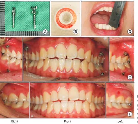
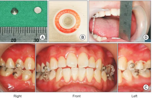
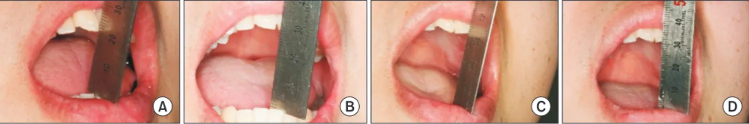
Although anatomical modification of the articular eminence through eminectomy has been considered most satisfactory and efffective treatment for managing the recurrent dislocation, it seldom performs clinically due to its invasiveness and patient’s reluctance. We thought a short- term intermaxillary fixation could be of benefit to the patient suffering from recurrent dislo- cation. A 21-year-old male patient with recurrent TMJ dislocation which had developed after excessive mouth opening, was successfully treated with intermaxillay fixation using microim- plant for 2 months. It is more conservative and less complicated method than eminectomy in treating recurrent TMJ dislocation. Transient intermaxillary fixation using microimplant and elastics could be one of treatment options for recurrent TMJ dislocation.

Key Words: Dental implants; Dislocations; Temporomandibular disorders; Temporomandibular joint

*These authors equally contributed to this study.

Correspondence to:

Jae-Kap Choi

Department of Oral Medicine, School of Dentistry, Kyungpook National University, 2177, Dalgubeol-daero, Jung-gu, Daegu 700-412, Korea Tel: +82-53-600-7321 Fax: +82-53-426-2195 E-mail: [email protected]

injured joint lose its function as passive restrictors and the masticatory muscles lose coordination during mouth open- ing and closing. In addition, specific anatomic characteris- tics of TMJ such as steepness of the articular eminence and joint laxity may predispose a patient to TMJ dislocation. A TMJ articular eminence which showed “short steep posterior slope” and “long flat anterior slope” was generally regarded vulnerable to dislocation.

2)

In treating recurrent TMJ dislocation, behavioral modifi- cation is the primary treatment option. However, if the pa- tient’s hazardous habit could not be managed, additional methods should be considered to restrict mouth opening precisely. Although many treatment modalities have been suggested in the literature,

3,4)

only surgical alteration of TMJ called eminectomy or eminoplasty has been regarded as definitive in treating TMJ dislocation.

5)

No satisfactory

INTRODUCTION

Temporomandibular joint (TMJ) dislocation is character- ized when the mandibular condyle moves far anteriorly during opening and passes the articular eminence eas- ily. Subsequently the ligaments around the joint are of- ten stretched by an intra-articular effusion, which induces various physical and social problems such as pain, closing limitation, difficulty in speech and chewing. According to Diagnostic Criteria for Temporomandibular Disorders 2014 guideline,

1)

if dislocated condyle was voluntarily reduced, it is called “subluxation,” and if not, the term “luxation” was used. In general, dislocation of TMJ clinically designated

“luxation” of TMJ.

Macrotraumatic event by extensive mouth opening is re- garded main cause of TMJ dislocation. The ligaments of

JOMP

Journal of Oral Medicine and Pain

Copyright Ⓒ 2014 Korean Academy of Orofacial Pain and Oral Medicine. All rights reserved.

CC This is an open-access article distributed under the terms of the Creative Commons Attribution Non-Commercial License (http://creativecommons.org/licenses/by-nc/3.0/), which permits unrestricted non-commercial use, distribution, and reproduction in any medium, provided the original work is properly cited.

(2)

could not fully open the mouth on daytime due to fear of TMJ dislocation.

On clinical examination, he showed 23 mm of maxi- mum pain free mouth opening with “soft-end feel.” He re- ported palpational tenderness on the left preauricular area and also on the temporalis and the masseter. Panoramic radiograph showed steep slope of articular eminence with- out entire translational movement (Figs. 1, 2). Cone beam computed tomography of TMJ showed steep posterior slope of articular eminence (Fig. 3). TMJ magnetic resonance im- aging showed normal engagement of articular disc and condyle on both TMJ during opening and closing (Fig. 4).

Collectively, it seems that the recurrent TMJ dislocation was caused by excessive displacement of the mandible after forced mouth opening as well as anatomic predisposition.

For the initial treatment, we first tried to modify the pa- tient’s behaviors. The patient was instructed not to open wide and restrict the condylar movement within a rotation- al movement. The appropriate range of mouth opening was guided by placing two fingers in vertical into the patient’s mouth. In addition, nonsteroidal anti-inflammatory drugs and physical therapies on both masticatory muscles and left TMJ were prescribed to reduce the pain. Two weeks later, even though he did his best not to open widely, TMJ dis- location still happened by unconscious yawning and dur- ing sleeping. Therefore, we had to consider another method nonsurgical or conservative methods for recurrent TMJ dis-

location has been available so far. In this study, we present a novel conservative trials for the management of recurrent dislocation of the TMJ.

CASE REPORT

A 21-year-old military man was referred from Korean Armed Forces Capital Hospital to the Department of Oral Medicine, Kyungpook National University Hospital. He is complaining about recurrent TMJ dislocation and pain on left preauricular area. The symptoms are began 2 weeks ago immediately after a military assistant instructor forcefully push his fist into the patient’s mouth. He said TMJ disloca- tion occurred mainly during sleeping and yawning, and he

Right close Right open Left open Left close

Fig. 2. Panoramic radiograph of temporomandibular joint showed steep posterior slope of articular eminence and limited condylar movement Fig. 1. Panoramic radiograph of the patient showed steep posterior

slope of articular eminence.

(3)

achieve limitation of mouth opening, two elastic power chains (Energy Chain J-120; Rocky Mountain Morita Corp., Tokyo, Japan) were installed between were tied between ip- silateral upper 1st premolar and lower 2nd premolar (Fig.

5). Although, the elastic power chains were frequently de- tached, the total numbers of TMJ dislocation were decreased during first 2 weeks. However, the patient was complaining which would be effective in unconscious condition like

sleeping and yawning.

We decided to perform the intermaxillary fixation which passively limits the condylar movement only to the ro- tation. Four metal buttons (lingual attachment, 601-05;

Tomy Inc., Tokyo, Japan) were attached on buccal surface of both upper 1st premolars and lower 2nd premolars. To

Fig. 3. Cone-beam computed tomo graphy of temporomandibular joint showed steep posterior slope and long flat ante rior slope of articular eminence.

Right Left

Fig. 4. Magnetic resonance image of tem- poromandibular joint (TMJ) showed normal engagement of articular disk in both TMJ;

however, there were no fully translational movement in both TMJ.

Right Left

Close

Open

(4)

was installed for resilient intermaxillary fixation which is ranged 27 mm of maximum opening (Fig. 6). During the first week, the pain on left preauricular area was decreased and the patient did not experience dislocation of the jaw.

Elastic power chain was changed daily and the patient was reassured behavioral modification and opening exercise to reinforce the TMJ ligaments and masticatory muscles. After 4 weeks of intermaxillary fixation, the patient reported TMJ dislocation twice a week and satisfied with the treatment outcome especially for the absence of previous tooth pain during the application of button-assisted intermaxillary fix- ation. The pain on left preauricular area was gradually de- creased and maximum mouth opening without dislocation was increased at 33 mm. After 8 weeks of intermaxillary fixation, the patient reported the decreased occurrence of TMJ dislocation and comfortable maximum mouth open- ing was maintained at 33 mm. Although one of microim- plant was detached during the treatment, there was no re- bound of dislocation. Microimplant was finally removed after 8 weeks, and the patient was reassured to maintain behavioral modification and opening exercise. On 12 weeks about dull pain on button-attached teeth and sensation of

malocclusion. Accordingly, considering his anatomic char- acteristic of articular eminence, we recommended the emi- nectomy at this point. However, he refused eminectomy in fear of surgical treatment and possible complications of surgery.

Although the button-assisted intermaxillary fixation was effective in reducing TMJ dislocation, the patients was in- tolerated to the treatment due to tooth pain and discomfort of occlusion. Therefore we tried to change the method of anchorage from tooth itself to alveolar bone. We implanted two microimplants (AbsoAnchor BH1514-06; Dentos Inc., Daegu, Korea) on interdental gingiva between both upper 2nd premolars and 1st molars, and additional two microim- plants (AbsoAnchor BH1413-06; Dentos) between both low- er 2nd premolars and 1st molars. The patient was instructed about general precaution after implantation, and prescribed antibiotics, anti-inflammatory drugs. After 1 week, im- plantation site healed well and showed no signs of mobil- ity or infection except mild gingival inflammation around microimplants. Elastic power chain (Energy Chain J-120)

Fig. 5. Microimplant-assisted intemaxillary fixation. (A) Microimplants. (B) Elastic power chain. (C) Intraoral photo of micro- implant-assisted intermaxillary fixation.

(D) Maximum mouth opening after inter- maxillary fixation. (E) Intraoral photo after removal of microimplant.

A B

C D

E

Right Front Left

(5)

joint laxity, ligaments act as passive restrainer which lim- it the excessive joint movement. Of the ligaments in TMJ, temporomandibular (TM) and stylomandibular ligaments are known to function as main restrictors against exces- sive opening of the condyle. TM ligament is consist of outer oblique and inner horizontal portion. Until the translation of the condyle is initiated, the outer oblique TM ligament keep the condyle not to open wide. Stylomandibular liga- ment is important in protrusive movement of mandible.

Once the outer oblique TM ligament and stylomandibular ligament were excessively elongated by the accidental ex- cessive mouth opening, mandibular condyle are more vul- nerable to dislocate because ligamentous elongation was considered irreversible.

In general, cognitive-behavioral modification by patient education is considered to be the first and most important step in treating recurrent anterior dislocation of TMJ. The patient have to be educated the possible causes and patho- physiology of condylar dislocation with careful consider- ation of all habitual and anatomic factors based on anam- nesis, clinical and radiographic examination. We explained that the previous accident and anatomic properties of the patient might be related with the onset and progress of TMJ dislocation. He was instructed to restrict mouth opening not to reach the critical point of dislocation, combined with physical therapies. However, the dislocation was mainly follow-up, the patient was satisfied with the current condi-

tion and discharged.

DISCUSSION

Dislocation of TMJ is troublesome condition which was most commonly shown far anteriorly displaced mandibular condyle to the articular eminence, thereby make it impos- sible to close one’s mouth. It occurs in forms of “acute or chronic,” or “temporary or recurrent,” and is often referred to as “open lock” in clinically. Although the severity was different, most dentist would occasionally experience this condition with their patient during the practice, especially after prolonged mouth opening. Generally, any macrotrau- mas which induces extensive mouth opening such as yawn- ing singing and singing are regarded as main cause of on- set of TMJ dislocation. In addition, some researchers have proposed that certain anatomic features in the joint such as

“steepness of the articular eminence” or “joint laxity” were closely linked with vulnerability of TMJ dislocation.

2,6)

In detail, articular eminence with “short and steep posterior slope” and “long and flat anterior slope” is tend to dislo- cate, and integrity of the ligaments, musculatures and bony structures are essential for the dynamic stability of TMJ.

According to diagnostic images, the patient in this case also showed typical trait of TMJ anatomy. In an aspect of

Fig. 6. Button-assisted intemaxillary fixa- tion. (A) Button. (B) Elastic power chain.

(C) Intraoral photo of button-assisted inter - maxillary fixation. (D) Maximum mouth opening after intermaxillary fixation.

A B

C D

Right Front Left

(6)

orthodontic treatment for tooth movement.

8)

Microimplant is implanted on interdental periodontium and removed af- ter the proper tooth movement was accomplished. Micro- implant has many advantages in oral cavity; 1) easy to im- plant and remove, 2) no effects on tooth itself, and 3) less irritation to movable soft tissue. In the present case, it was already confirmed that intermaxillary fixation itself was ef- fective in reducing the patient symptoms. Therefore we just modified the technic to get anchorage of the elastics. We change the structure of getting anchorage of intermaxil- lary fixation from teeth itself to alveolar bone. Compare with previous button-assisted method, novel microimplant- assisted method have several advantages as follows. First, microimplant inserted in periodontium was free of tooth-re- lated symptoms. Second, because microimplant was placed near the root apex in vertical dimension, it induces less ir- ritation to the movable soft tissue. Third, because microim- plant could be placed more posteriorly than the button, it has advantage of aesthetic aspect. Last, microimplant could be used in edentulous patient. Only one problem using mi- croimplant in the present case was spontaneous detachment of microimplant before the end of treatment.

The mechanism of intermaxillary fixation on recurrent TMJ dislocation was generally understood by myostatic contracture of elevator muscle at the point of dislocation.

2)

We maintained microimplant-assisted intermaxillary fixa- tion for 8 weeks with the range of mouth opening at 33 mm because it was enough to get myostatic contracture to prevent dislocation. In fact, after the removal of microim- plant, the patient showed similar frequency of TMJ disloca- tion in our case (Fig. 7). The unloading time after implan- tation is also considered. Actually, because microimplant might not be integrated to the bone, there is no need to be waiting in orthodontics. However, in this case, we allowed occurred during sleeping and yawning which was not un-

der his voluntary control. Moreover, he was getting nervous and afraid about aggravation of the symptoms, thereby we tried to consider more active treatment modalities.

If it is clear that recurrent or chronic TMJ dislocation is mainly due to an anatomical feature of articular eminence, the treatment strategies should be focused on therapeutic alteration of anatomic structure using eminectomy or emi- noplasty.

3)

Many studies reported that eminectomy of TMJ might be a satisfactory and effective treatment for man- aging the recurrent dislocation in patient with the appar- ent anatomic causes.

7)

In our patient, the morphology of articular eminence which showed “short steep posterior slope” and “long flat anterior slope,” is probably indicat- ing disadvantage to dislocation. Therefore, we considered TMJ eminectomy as one of applicable treatment modalities.

However, our patient refused the surgery in fear of surgery and associated complications.

We next tried to restrict the opening within an appropri- ate range which could prevent the jaw from dislocation.

Intermaxillary fixation may be used alone or in combina- tion of other treatments in order to immobilize the TMJ.

2)

Resilient intermaxillary fixation with the maximum rota- tional movement of 27 mm was performed using metal but- tons and elastic power chains. The method was immediately effective in reducing the frequency of dislocation during sleeping and yawning. However as the patient uses the de- vice longer, he began to complain with the pain on button- attached teeth and malocclusion. That is because extrusive forces during the opening were directly transferred to the supporting teeth through elastic chain. Consequentially, the patient was intolerant of button-assisted intermaxillary fix- ation method.

Microimplant (mini implant) has been widely used in

Fig. 7. Change of comfortable mouth opening range after microimplant-assisted intermaxillary fixation. (A) One week after microimplant. (B)

A B C D

(7)

REFERENCES

1. Schiffman E, Ohrbach R, Truelove E, et al; International RDC/

TMD Consortium Network, International Association for Dental Research; Orofacial Pain Special Interest Group, International Association for the Study of Pain. Diagnostic Criteria for Tem- poromandibular Disorders (DC/TMD) for Clinical and Research Applications: recommendations of the International RDC/TMD Consortium Network and Orofacial Pain Special Interest Group. J Oral Facial Pain Headache 2014;28:6-27.

2. Okeson JP. Management of temporomandibular disorders and oc- clusion. 6th ed. St. Louis: Mosby, Inc.; 2008.

3. Akinbami BO. Evaluation of the mechanism and principles of management of temporomandibular joint dislocation. Systematic review of literature and a proposed new classification of tem- poromandibular joint dislocation. Head Face Med 2011;7:10.

4. Caminiti MF, Weinberg S. Chronic mandibular dislocation: the role of non-surgical and surgical treatment. J Can Dent Assoc 1998;64:484-491.

5. Holmlund AB, Gynther GW, Kardel R, Axelsson SE. Surgical treatment of temporomandibular joint luxation. Swed Dent J 1999;23:127-132.

6. Shorey CW, Campbell JH. Dislocation of the temporomandibular joint. Oral Surg Oral Med Oral Pathol Oral Radiol Endod 2000;89:

662-668.

7. Martins WD, Ribas Mde O, Bisinelli J, França BH, Martins G. Re- current dislocation of the temporomandibular joint: a literature review and two case reports treated with eminectomy. Cranio 2014;32:110-117.

8. Chen CH, Chang CS, Hsieh CH, et al. The use of microimplants in orthodontic anchorage. J Oral Maxillofac Surg 2006;64:1209- 1213.

1 week of loading free due to difference of forces.

Though recurrent TMJ dislocation is remaining trouble- some problem to the dentist, there has been no definitive treatment except extensive surgical interventions includ- ing eminectomy or eminoplasty when conservative treat- ments failed to manage the recurrence of TMJ dislocation.

However, considering that most patient would not eas- ily accepted the surgery, less invasive treatment modalities was required in many cases. Our case report showed that a 21-year-old male patient with recurrent TMJ anterior dis- location which developed after accidental excessive mouth opening was successfully treated with intermaxillay fixa- tion using microimplant. Through this case report, we sug- gest that microimplant-assisted intermaxillary fixation could be a novel treatment option for recurrent TMJ ante- rior dislocation.

CONFLICT OF INTEREST

No potential conflict of interest relevant to this article

was reported.

수치

Fig. 2. Panoramic radiograph of temporomandibular joint showed steep posterior slope of articular eminence and limited condylar movement Fig
Fig. 4. Magnetic resonance image of tem- tem-poromandibular joint (TMJ) showed normal  engagement of articular disk in both TMJ;
Fig. 5. Microimplant-assisted intemaxillary  fixation. (A) Microimplants. (B) Elastic  power chain
Fig. 6. Button-assisted intemaxillary fixa- fixa-tion. (A) Button. (B) Elastic power chain
+2

참조

관련 문서

Purpose: The purpose of this study is to report the safety and effectiveness of arthroscopic-assisted implantation of Cartistem®, an articular cartilage

The slip system ( =Slip plane + Slip direction) depends on the crystal structure of the metal and is such that the atomic distortion that accompanies the motion of a dislocation

3,4 Therefore, to date, studies have been conducted to examine the absorbable fixation plate manufactured using polymer and a fixation screw that can

Conclusion: In Neer type II distal clavicle fracture treatment, both groups showed satisfactory result in clinical and radiological outcomes, but the hook plate

Just as we achieved good control with a joint-based controller that was based on a linearizing and decoupling model of the arm, we can do the same for the Cartesian case.

Biological treatment of wastewater is an economical green technology, however, the effluent water quality of a biological wastewater treatment facility is

So, we evaluated the ideal screw fixation by setting a study model to compare a position of screw at the same time the screw diameter, number of

This study aims to suggest a basic data for the activation of aromatherapy pimple treatment program with the pimple skin care and treatment by testing the actual