http://dx.doi.org/10.5090/kjtcs.2012.45.3.171 ISSN: 2233-601X (Print) ISSN: 2093-6516 (Online)
1
Department of Thoracic and Cardiovascular Surgery, Chungnam National University Hospital, Chungnam National University School of Medicine,
2Department of Otorhinolaryngology, Chungnam National University Hospital, Chungnam National University School of Medicine
†This study was financially supported by research fund of Chungnam National University in 2010.
Received: October 14, 2011, Revised: October 20, 2011, Accepted: October 22, 2011
Corresponding author: Seung Pyung Lim, Department of Thoracic and Cardiovascular Surgery, Chungnam National University Hospital, Chungnam National University School of Medicine, 282 Munhwa-ro, Jung-gu, Daejeon 301-721, Korea
(Tel) 82-42-280-7376 (Fax) 82-42-280-7373 (E-mail) [email protected]
C
The Korean Society for Thoracic and Cardiovascular Surgery. 2012. All right reserved.
CC
This is an open access article distributed under the terms of the Creative Commons Attribution Non-Commercial License (http://creative- commons.org/licenses/by-nc/3.0) which permits unrestricted non-commercial use, distribution, and reproduction in any medium, provided the original work is properly cited.
Clinical Features of Deep Neck Infections and Predisposing Factors for Mediastinal Extension
Shin Kwang Kang, M.D.
1, Seokkee Lee, M.D.
1, Hyun Kong Oh, M.D.
1,
Min-Woong Kang, M.D., Ph.D.
1, Myung Hoon Na, M.D., Ph.D.
1, Jae Hyeon Yu, M.D., Ph.D.
1, Bon Seok Koo, M.D., Ph.D.
2, Seung Pyung Lim, M.D., Ph.D.
1Background: Deep neck infections (DNI) can originate from infection in the potential spaces and fascial planes of the neck. DNI can be managed without surgery, but there are cases that need surgical treatment, especially in the case of mediastinal involvement. The aim of this study is to identify clinical features of DNI and analyze the pre- disposing factors for mediastinal extension. Materials and Methods: We reviewed medical records of 56 patients suffering from DNI who underwent cervical drainage only (CD group) and those who underwent cervical drainage combined with mediastinal drainage for descending necrotizing mediastinitis (MD group) from August 2003 to May 2009 and compared the clinical features of each group and the predisposing factors for mediastinal extension.
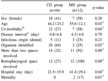
Results: Forty-four out of the 56 patients underwent cervical drainage only (79%) and 12 patients needed both cervical and mediastinal drainage (21%). There were no differences between the two groups in gender (p=0.28), but the MD group was older than the CD group (CD group, 44.2±23.2 years; MD group, 55.6±12.1 years;
p=0.03). The MD group had a higher rate of co-morbidity than the CD group (p=0.04). The CD group involved more than two spaces in 14 cases (32%) and retropharyngeal involvement in 12 cases (27%). The MD group in- volved more than two spaces in 11 cases (92%) and retropharyngeal involvement in 12 cases (100%). Organism identification took place in 28 cases (64%) of the CD group and 3 cases of (25%) the MD group (p=0.02). The mean hospital stay of the CD group was 21.5±15.9 days and that of the MD group was 41.4±29.4 days (p=0.04).
Conclusion: The predisposing factors of mediastinal extension in DNI were older age, involvement of two or more spaces, especially including the retropharyngeal space, and more comorbidities. The MD group had a longer hospi- tal stay, higher mortality, and more failure to identify causative organisms of causative organisms than the CD group.
Key words: 1. Infection 2. Neck 3. Mediastinitis
INTRODUCTION
Deep neck infections (DNI) can originate from infection in
the potential spaces and fascial planes of the neck. The pri-
mary origin of DNI is unknown in most cases, but it can be
caused by dental problems, cervical lymphadenitis, sialoadeni-
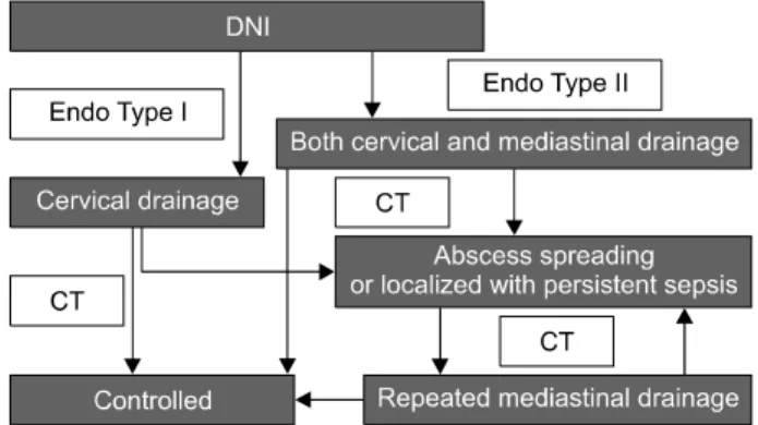
Fig. 1. Algorithm of surgical management for deep neck infections (DNI) and descending necrotizing mediastinitis. CT, computed to- mography.
tis, acute tonsillitis, and peritonsillar abscess [1]. Most pa- tients of DNI can be treated with a broad spectrum of intra- venous antibiotics without complications. However, complica- tions such as airway obstruction in patients with lateral phar- yngeal cavity or peritonsilar abscess, deep vein thrombosis, mediastinitis, pneumonitis, laryngeal edema, and encephalitis can occur [1]. Some DNI require surgical treatment, espe- cially in cases of mediastinal involvement, known as descend- ing necrotizing mediastinitis (DNM). Many acute mediastinal infections result from esophageal perforation or infection fol- lowing a trans-sternal cardiac procedure. However, they may result from oropharyngeal infection spreading along the fas- cial planes into the mediastinum [2-5]. Criteria of diagnosing DNM were clearly established by Estrera in 1983 [2]. These included clinical manifestations such as severe infection, char- acteristic radiology features, necrotizing mediastinal infection revealed at operation or autopsy, and the relationship of or- opharyngeal infection to the development of DNM. Endo et al. [3] classified DNM into three types according to the ex- tension of DNM using computed tomography (CT). Type I is a localized disease above the carina, Type II A is charac- terized by diffuse anterior mediastinal involvement, and Type II B involves both the anterior and posterior mediastinum [3].
We report the surgical results of the cervical drainage group in DNI compared with the group to which mediastinal drainage was added. The clinical features and predisposing factors of DNI progressing to DNM were analyzed.
MATERIALS AND METHODS
The medical records of patients who underwent surgical treatment for DNI and DNM from August 2003 to May 2009 were reviewed retrospectively. Their demographics, etiologies associated with systemic diseases (hypertension, diabetes mel- litus, liver cirrhosis, and chronic renal failure), preoperative condition (sepsis), disease interval, infectious origin, bacteriol- ogy, radiology, duration of hospitalization, and outcomes were reviewed. Sepsis was defined as high fever (>38.2
oC), unstable vital signs (blood pressure <90 mmHg, respiratory rate >25/min), leucopenia (white blood cell <4,000/mm
3), thrombocytopenia (platelet <100,000/mm
3), oliguria (0.5 mL/kg/hr), or prolonged coagulation time (activated partial
thromboplastin time >60 sec or international normalized ra- tio >1.5). In the present article, 56 patients were suffering from DNI; 44 patients received cervical drainage only (CD group) and 12 patients needed both cervical and mediastinal drainage for mediastinal involvement (MD group).
Neck and chest CT scans were performed on all patients clinically suspected of DNI. Shortly after the criteria of Estrera were fulfilled, nil per os and fluid therapy was initiated. Empiric antibiotics, including third-generation cepha- losporin and clindamycin, were administered immediately.
Appropriate antibiotics for causative organisms were given based on the results of culture identification.
Type I DNM was managed with cervical drainage only and Type II DNM was treated with both cervical and mediastinal drainage by Endo’s classification. For the cervical drainage, we used an anterior approach to the DNI and maintained open drainage until the wound was clear. When follow-up CT scans showed that the disease had improved, treatment was terminated. However, when the CT scan showed an abscess spreading or localized persistent septic manifestation, repeated mediastinal drainage was performed. The CT scans were per- formed at the third, seventh, and fourteenth postoperative day routinely, or whenever routine chest X-rays showed abnormal findings (Fig. 1).
All data are expressed as mean±standard deviation. SPSS
ver. 11.5 (SPSS Inc., Chicago, IL, USA) was used to perform
the analysis. Statistical significance was defined as p<0.05.
Table 1. Bacteriology of the CD group
a)and MD group
b)CD group (n=44) MD group (n=12)
Streptococcus 13 (29.5) 3 (25)
Staphylococcus 7 (15.9) 0
Klebsiella 5 (11.4) 0
Pseudomonas 3 (6.8) 0
Not identified 16 (36.4) 9 (75)
Values are presented as number (%).
a)
CD group: cervical drainage only.
b)
MD group: both cervical and mediastinal drainage.
Table 2. Surgical results of the CD group
a)and MD group
b)CD group
(n=44)
MD group
(n=12) p-value Sex (female)
Age
Co-morbidity
d)Disease interval
e)(day) Infectious origin (dental) Organism identified More than two spaces
involved
Retropharyngeal space involved
Hospital stay (day) Mortality
18 (41) 44.2±23.2
12 (27) 6.8±6.8
5 (11) 28 (64) 14 (32)
12 (27)
21.5±15.9 0
7 (58) 55.6±12.1
7 (58) 6.3±4.0
3 (25) 3 (25) 11 (92)
12 (100)
41.4±29.4 2 (17)
0.28 0.03
c)0.04
c)0.79 0.23 0.02
c)0.00
c)0.00
c)0.04
c)0.01
c)Values are presented as mean±standard deviation or number (%).
a)
CD group: cervical drainage only.
b)
MD group: both cervical and mediastinal drainage.
c)
Statistically significant.
d)
Hypertension, diabetes mellitus, liver cirrhosis, or chronic re- nal failure.
e)