ABSTRACT
Background: Rheumatoid arthritis (RA) treatment may differ according to hepatitis B state and consequently may bring about different arthritis outcomes. However, whether hepatitis B affects treatment outcome remains unclear. We investigated differences in change in arthritis activity between RA patients according to concomitant hepatitis B virus infection.
Methods: A retrospective medical chart review was performed by two rheumatologic fellows using single center data, from January 2000 to March 2015. Among RA patients older than 18 years, patients with comorbidities that could affect RA treatment aside from hepatitis B were excluded. Using 1:3 propensity score matching, 40 hepatitis B virus surface antigen (HBsAg)-positive patients and 112 HBsAg-negative patients were included in the study.
Data were collected longitudinally using standardized electronic forms. The longitudinal relationship between HBsAg-positivity and RA activity was analyzed using generalized estimating equations.
Results: RA activity showed time-dependent improvement. Reductions of swollen joint count over time were significantly larger in the HBsAg-negative group. However, changes in disease activity score in 28 joints with three variables (DAS28-3), tender joint count, erythrocyte sedimentation rate and C-reactive protein level did not differ between the groups. There were no differences in alanine aminotransferase level. HBsAg-positive patients were less likely to receive methotrexate (odds ratio [OR], 0.09; 95% confidence interval [CI], 0.04–0.19;
P < 0.001) and more likely to receive sulfasalazine (OR, 3.67; 95% CI, 1.94–6.95; P < 0.001).
Conclusion: RA medication use varied according to HBsAg-positivity. However, improvement in RA activity was not significantly affected by concomitant hepatitis B infection.
Keywords: Rheumatoid Arthritis; Hepatitis B; Disease Activity Score
Original Article
Received: Dec 13, 2017 Accepted: Apr 20, 2018 Address for Correspondence:
Hoon-Suk Cha, MD, PhD
Department of Medicine, Samsung Medical Center, Sungkyunkwan University School of Medicine, 81, Irwon-ro, Gangnam-gu, Seoul 06351, Korea.
E-mail: [email protected]
© 2018 The Korean Academy of Medical Sciences.
This is an Open Access article distributed under the terms of the Creative Commons Attribution Non-Commercial License (https://
creativecommons.org/licenses/by-nc/4.0/) which permits unrestricted non-commercial use, distribution, and reproduction in any medium, provided the original work is properly cited.
ORCID iDs Yeonghee Eun
https://orcid.org/0000-0002-0758-2627 In Young Kim
https://orcid.org/0000-0003-1565-9038 Hyemin Jeong
https://orcid.org/0000-0001-6227-681X Hyungjin Kim
https://orcid.org/0000-0003-3036-7332 Jaejoon Lee
https://orcid.org/0000-0003-4793-5949 Moon Seok Choi
https://orcid.org/0000-0002-9690-9301 Eunmi Koh
https://orcid.org/0000-0002-6457-6792 Hoon-Suk Cha
https://orcid.org/0000-0001-5391-5376
Yeonghee Eun ,1 In Young Kim ,1 Hyemin Jeong ,2 Hyungjin Kim ,1 Jaejoon Lee ,1 Moon Seok Choi ,1 Eunmi Koh ,1 and Hoon-Suk Cha 1
1Deparment of Medicine, Samsung Medical Center, Sungkyunkwan University School of Medicine, Seoul, Korea
2Department of Medicine, Soonchunhyang University College of Medicine, Bucheon, Korea
Disease Characteristics and Change in Arthritis Activity according to Treatment in Hepatitis B Surface
Antigen-positive Rheumatoid Arthritis Patients: a Retrospective Chart
Review Study
Immunology, Allergic
Disorders & Rheumatology
Disclosure
The authors have no potential conflicts of interest to disclose.
Author Contributions Conceptualization: Eun Y, Cha HS.
Methodology: Eun Y, Kim IY. Data curation:
Eun Y, Jeong H. Formal analysis: Kim H, Lee J.
Writing - original draft: Eun Y, Kim IY, Jeong H.
Writing - review & editing: Kim H, Lee J, Choi MS, Koh E, Cha HS.
INTRODUCTION
Hepatitis B virus (HBV), a DNA virus with a human-only reservoir, is a serious global health concern. The worldwide prevalence of HBV infection is estimated to be 3.61%, and it is similar in rheumatoid arthritis (RA) patients.1,2 Coexistence of RA with hepatitis B makes the treatment of RA risky because some disease-modifying antirheumatic drugs (DMARDs) such as methotrexate and leflunomide may cause hepatotoxicity and some drugs that affect immune response can lead to reactivation of HBV.3-6 Consequently, RA treatment may differ according to hepatitis B status and this could lead to different arthritis outcomes.
Previous studies of RA patients with concomitant hepatitis B tended to focus on the safety of arthritis medications and reactivation of hepatitis.7-11 Only a small number of studies addressed differences in disease characteristics and treatment patterns of RA in relation to hepatitis B status.12 However, whether hepatitis B affects treatment outcomes such as RA disease activity remains unclear.
In this study, we investigated potential variation in changes in disease activity markers upon treatment between RA patients based on their concomitant hepatitis B status. We employed hepatitis B virus surface antigen (HBsAg) as a serological marker of HBV infection.
METHODS
Study population
A retrospective medical chart review was performed by two rheumatologic fellows using single center data, from 1 January 2000 to 31 March 2015. Patients with overlapping autoimmune diseases, liver diseases other than hepatitis B, or other comorbidities that could affect RA treatment (e.g., cardiovascular diseases, chronic kidney diseases, interstitial lung disease, diabetes, or previous malignancy within five years) were excluded. Forty patients with HBsAg- positive RA were finally enrolled in the study. We included a control group that comprised 112 age-matched, sex-matched, and entry-time-matched RA patients with negative HBsAg and negative hepatitis B core antibody (HBcAb) using propensity score matching.
Collection of study data
Baseline and longitudinal data of each participant were retrospectively collected at all visits from the date of entry to 31 March 2015. An electronic standardized abstraction form was used for data collection. Baseline data included demographic data, date of RA symptom onset, presence of erosions, and previous medication use. The modified Sharp/van der Heijde (mSvH) scoring system was used to quantify radiologic damage.13 Because X-ray data were insufficient, only hand X-rays at baseline were used in the assessment. Rheumatoid factor (RF) was measured by immunoturbidimetric assay (Roche/Hitachi Modular P. Roche Diagnostics, Mannheim, Germany; positivity ≥ 15 IU/mL). Anti-cyclic citrullinated peptide antibodies (ACPA) were detected using a chemiluminescent microparticle immunoassay (Architect, Abbott Laboratories, Abbott Park, IL, USA; positivity ≥ 5 U/mL). The disease activity score for 28 joints with three variables (DAS28-3), tender joint counts, swollen joint counts, erythrocyte sedimentation rate (ESR), C-reactive protein (CRP), liver function test as measured by alanine aminotransferase (ALT) in units/liter, and RA medication prescription were collected at baseline and updated at every visit. Because patient global assessment data were not collected, DAS28-3 was used as a disease activity marker.14,15
In HBsAg-positive RA patients, prescription of antiviral agents for hepatitis B, purpose of antiviral use (prophylactic or therapeutic), HBsAg positivity, and HBV DNA data were collected from follow-up visits. Characterization of hepatitis B was done at baseline and at every visit to the hepatology clinic. The “inactive HBsAg carrier state” is characterized by HBsAg positivity for > 6 months, serum HBV DNA titer of < 20,000 IU/mL in HBeAg-positive patients or of
< 2,000 IU/mL in HBeAg-negative patients and persistently normal ALT levels. “Chronic hepatitis B (CHB)” is characterized by HBsAg positivity for > 6 months, serum HBV DNA titer of > 20,000 IU/mL in HBeAg-positive patients or of > 2,000 IU/mL in HBeAg-negative patients, and persistent or intermittent elevation in ALT levels.16 Development of hepatocellular carcinoma and reactivation of hepatitis B were also assessed. HBV reactivation was defined by a > 10-fold (1 log10 IU/mL) rise in HBV DNA level when compared with the baseline value.17 Statistical analysis
For selection of control group, propensity score matching was performed using age, sex, and entry time, with a 1:3 case-control ratio. The Kolmogrov-Smirnov and Shapiro-Wilk tests were used to check normality of metric data. For data with a normal distribution, the mean ± standard deviation (SD) is shown, and we applied Student's t-tests for comparisons. For non-normally distributed data, we indicate the median (interquartile range) and used the Wilcoxon rank sum test. Categorical variables were compared using χ2 or Fisher's exact tests. Paired t-test for normally distributed data and Wilcoxon signed rank test for non-normally distributed data were used to compare RA disease activity markers and ALT level at the end of follow-up with those at study entry. Generalized estimating equations (GEEs) were used to assess the association between HBsAg positivity and RA disease activity markers/medication use over time. In GEE models, data were adjusted for visit date and further adjusted for age, sex, and duration of RA. All statistical analyses were performed using SAS, version 9.4 (SAS Institute Inc., Cary, NC, USA).
Ethics statement
The present study protocol was reviewed and approved by the Institutional Review Board of Samsung Medical Center (IRB No. 2015-09-069). The requirement for informed consent was waived since the study involved a minimum risk to the enrolled patients in this retrospective study and no identifiable information was used.
RESULTS
The study sample consisted of 40 patients with HBsAg-positive RA and 112 patients with HBsAg-negative RA. A description of patient characteristics at baseline is provided in Table 1.
Median age was 43 (interquartile range [IQR], 37–53) years, and women constituted 76.3% of the total study population. There were no significant differences between groups with regard to demographics RA disease characteristics including RF positivity, ACPA positivity, and the presence of erosions; instruments reflecting RA disease activity; or previous medication use at baseline. Methotrexate tended to be less frequently prescribed for HBsAg-positive patients, but the difference was not statistically significant. Leflunomide, cyclosporine, azathioprine, and biologic agents including tumor necrosis factor α (TNF-α) inhibitors were less commonly used in the study population, and there were no significant differences in the use of these medications between groups at baseline.
At baseline, the HBsAb-positive rate was significantly higher in the HBsAg-negative group than the HBsAg-positive group (49.1% vs. 7.5%; P < 0.001). No differences were noted in
the level of ALT. In HBsAg-positive patients, the positive rate for HBcAb was 96.9% (n = 31) and that for HBeAg was 19.4% (n = 7). HBV DNA data were available in 35 patients (87.5%
of HBsAg-positive patients); HBV DNA positivity was 37.1% (n = 13). Ten percent of HBsAg- positive patients received antiviral treatment (n = 4). Among 40 patients who had HBsAg, 28 patients were classified as inactive carriers and 9 patients were classified as having CHB.
Three patients had been diagnosed with liver cirrhosis.
The median number of visits per subject was 28 (IQR, 19–61) in the HBsAg-positive group and 27 (IQR, 16–45) in the HBsAg-negative group (P = 0.175). The median follow-up duration per subject was 2,361 (IQR, 1,462–4,326) days in the HBsAg-positive group and 1,863 (IQR, 1,133–3,659) days in the HBsAg-negative group, was similar between these groups (P = 0.119).
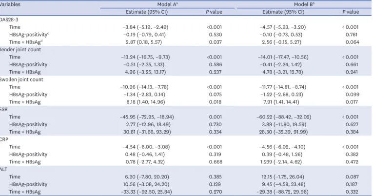
We used GEE regression analysis to determine the effects of time and HBsAg-positivity on RA disease activity markers and ALT (Table 2). All RA disease activity markers were negatively associated with time (P < 0.05 for all models). Swollen joint count was the only instrument that showed a significant interaction between time and HBsAg-positivity in both unadjusted and adjusted models (β-estimate, 8.18; 95% confidence interval [CI], 1.40–14.96; P = 0.018 in the unadjusted model; β-estimate, 7.91; 95% CI, 1.41–14.41; P = 0.017 in the adjusted model).
We observed a significant effect of HBsAg-positivity on the relationship between time and DAS28-3 (β-estimate, 2.79; 95% CI, 0.18–5.57; P = 0.026), but this interaction did not remain significant after adjustment for age, sex, and duration of RA. For tender joint count, ESR, and CRP levels, there were no interaction between time and HBsAg-positivity. ALT levels did not change significantly with time or with HBsAg-positivity.
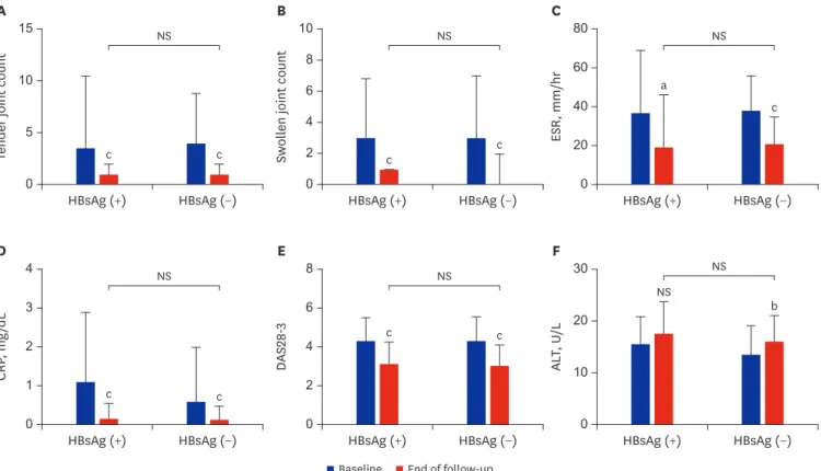
At the end of follow-up, disease activity markers were improved compared with baseline (Fig. 1).
There were no significant differences in disease activity markers between HBsAg-positive and HBsAg-negative patients.
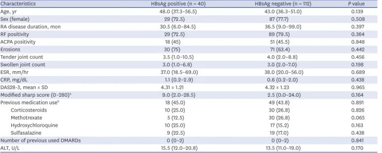
Table 1. Baseline characteristics of HBsAg-positive and HBsAg-negative RA patients
Characteristics HBsAg positive (n = 40) HBsAg negative (n = 112) P value
Age, yr 48.0 (37.3–56.5) 43.0 (36.3–51.0) 0.139
Sex (female) 29 (72.5) 87 (77.7) 0.508
RA disease duration, mon 30.5 (6.0–84.5) 36.5 (9.0–99.0) 0.397
RF positivity 29 (72.5) 89 (79.5) 0.364
ACPA positivity 18 (45) 51 (45.5) 0.848
Erosions 30 (75) 71 (63.4) 0.442
Tender joint count 3.5 (1.0–10.5) 4.0 (2.0–8.8) 0.456
Swollen joint count 3.0 (1.0–6.8) 3.0 (2.0–7.0) 0.198
ESR, mm/hr 37.0 (18.5–69.0) 38.0 (20.0–56.0) 0.689
CRP, mg/dL 1.1 (0.2–2.9) 0.6 (0.2–2.0) 0.438
DAS28-3, mean ± SD 4.31 ± 1.21 4.32 ± 1.23 0.965
Modified sharp score (0–280)a 9.0 (2.0–28.5) 2.5 (0.0–24.0) 0.164
Previous medication useb 18 (45.0) 49 (43.8) 0.891
Corticosteroids 10 (25.0) 30 (26.8) 0.826
Methotrexate 5 (12.5) 30 (26.8) 0.065
Hydroxychloroquine 10 (25.0) 17 (15.2) 0.163
Sulfasalazine 9 (22.5) 19 (17.0) 0.438
Number of previous used DMARDs 0 (0–2) 0 (0–2) 0.841
ALT, U/L 15.5 (12.0–20.8) 13.5 (11.0–19.0) 0.170
Data are presented as median with IQR or number (%).
HBsAg = hepatitis B virus surface antigen, RA = rheumatoid arthritis, RF = rheumatoid factor, ACPA = anti-cyclic citrullinated peptide antibodies, ESR = erythrocyte sedimentation rate, CRP = C-reactive protein, DAS28-3 = disease activity score for 28 joints with three variables, DMARD = disease-modifying antirheumatic drug, ALT = alanine aminotransferase, IQR = interquartile range.
aData were available for 33 and 100 patients in the HBsAg-positive group and the HBsAg-negative group, respectively; bPatients who had already been diagnosed and treated with RA at another hospital.
When all study visits were analyzed, methotrexate was less frequently used in HBsAg- positive patients (odds ratio [OR], 0.09; 95% CI, 0.04–0.19; P < 0.001; Fig. 2). HBsAg- positive patients were more likely to be prescribed sulfasalazine (OR, 3.67; 95% CI, 1.94–6.95; P < 0.001) and cyclosporine (OR, 3.57; 95% CI, 1.33–9.54; P = 0.011). On unadjusted analysis, tacrolimus was more frequently used in HBsAg-positive patients, but after adjusting for age, sex, and duration of RA, this difference was not significant.
Hydroxychloroquine was prescribed somewhat more often in HBsAg-positive patients, but this difference was not statistically significant. With regard to corticosteroids, leflunomide, and biologic agents including TNF-α inhibitors, there were no significant differences.
Combination therapy with conventional DMARDs was significantly more frequently applied in HBsAg-positive patients than HBsAg-negative patients (OR, 2.22; 95% CI, 1.19–4.14;
P = 0.012); however, there was no significant difference in the use of combination therapy with biologic agents between the two groups (OR, 0.76; 95% CI, 0.14–4.14; P = 0.755).
The frequency of intraarticular injection of corticosteroids was higher in HBsAg-positive patients than HBsAg-negative patients (median 2, IQR, 1–8 vs. 1, IQR, 0–3; P = 0.001) during the study period.
Among 40 patients with HBsAg-positivity, 15 patients (37.5%) received antiviral treatment.
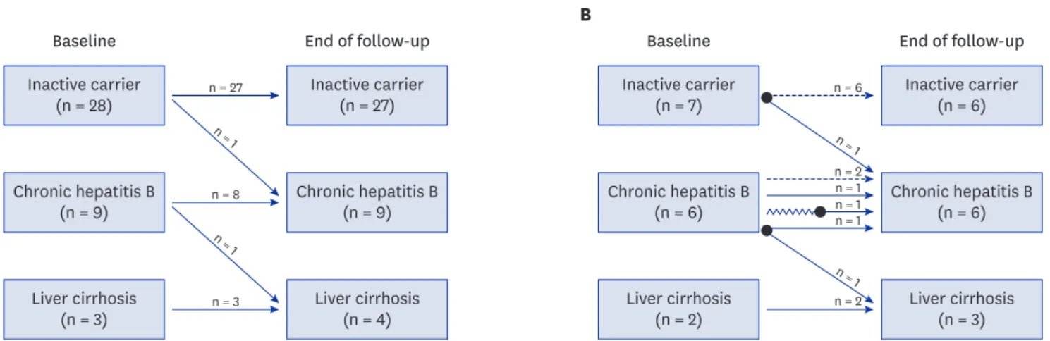
The purpose of antiviral treatment was prevention of hepatitis reactivation in 8 patients and therapy for hepatitis or cirrhosis in 7 patients. During the study period, one patient with CHB at baseline was newly diagnosed with liver cirrhosis and another patient who was an inactive carrier at baseline was newly diagnosed with CHB (Fig. 3). Two incident cases of hepatocellular carcinoma were observed.
Table 2. Regression analysis (GEE) of the effects of time and HBsAg-positivity on disease activity markers and ALT, adjusting for within-subject correlations
Variables Model Aa Model Bb
Estimate (95% CI) P value Estimate (95% CI) P value
DAS28-3
Time −3.84 (−5.19, −2.49) <0.001 −4.57 (−5.93, −3.20) < 0.001
HBsAg-positivityc −0.19 (−0.79, 0.41) 0.530 −0.10 (−0.73, 0.53) 0.761
Time × HBsAgd 2.87 (0.18, 5.57) 0.037 2.56 (−0.15, 5.27) 0.064
Tender joint count
Time −13.24 (−16.75, −9.73) <0.001 −14.01 (−17.47, −10.56) < 0.001
HBsAg-positivity −0.51 (−2.35, 1.33) 0.586 −0.41 (−2.24, 1.42) 0.661
Time × HBsAg 4.96 (−3.25, 13.17) 0.237 4.78 (−3.21, 12.78) 0.241
Swollen joint count
Time −10.96 (−14.13, −7.78) <0.001 −11.77 (−14.81, −8.74) < 0.001
HBsAg-positivity −1.34 (−2.83, 0.14) 0.075 −1.22 (−2.68, 0.23) 0.099
Time × HBsAg 8.18 (1.40, 14.96) 0.018 7.91 (1.41, 14.41) 0.017
ESR
Time −45.95 (−72.95, −18.94) 0.001 −60.22 (−88.42, −32.02) < 0.001
HBsAg-positivity 2.77 (−12.96, 18.49) 0.730 3.89 (−11.80, 19.59) 0.627
Time × HBsAg 30.81 (−31.66, 93.29) 0.334 28.30 (−35.39, 91.99) 0.384
CRP
Time −4.54 (−6.00, −3.08) <0.001 −4.56 (−6.02, −4.10) < 0.001
HBsAg-positivity 0.48 (−0.46, 1.41) 0.319 0.39 (−0.48, 1.26) 0.382
Time × HBsAg 0.78 (−2.77, 4.32) 0.668 1.239 (−2.14, 4.62) 0.472
ALT
Time 6.20 (−7.80, 20.20) 0.385 12.15 (−1.75, 26.04) 0.087
HBsAg-positivity 10.56 (−3.08, 24.20) 0.129 9.45 (−4.58, 23.48) 0.187
Time × HBsAg −33.33 (−92.50, 25.84) 0.270 −29.38 (−88.72, 29.96) 0.332
GEE = generalized estimating equation, HBsAg, hepatitis B virus surface antigen, ALT = alanine aminotransferase, CI = confidence interval, DAS28-3 = disease activity score for 28 joints with three variables, ESR = erythrocyte sedimentation rate, CRP = C-reactive protein, RA = rheumatoid arthritis.
aModel A adjusted for time only; bModel B adjusted for time, age, sex, and duration of RA; cThe HBsAg-negative group was coded as 0 and the HBsAg-positive group was coded as 1; dTime × HBsAg refers to the interaction term between time and HBsAg positivity.
Hepatitis reactivation occurred in 4 HBsAg-positive RA patients (10%, Table 3). One patient was an inactive carrier, and the other patients had CHB at baseline. Among the patients who experienced HBV reactivation, only one patient had received previous antiviral treatment at baseline, but the patient exhibited lower adherence to drug. The HBV DNA level decreased
c
NS
c
a
NS
c
0 10
5
Tender joint count
HBsAg (+) c
NS
c
HBsAg (−) HBsAg (+) HBsAg (−) HBsAg (+) HBsAg (−)
A 15
0 8 6
en joint countSwoll 24
B 10
0 60
ESR, mm/hr 20 40
C 80
c
NS
c
NS
NS
b
0 2
, mgCRP/dL 1
HBsAg (+) c
NS
c
HBsAg (−) HBsAg (+) HBsAg (−) HBsAg (+) HBsAg (−)
4
3
D
0 6
2
DAS28-3
4
E 8
0 20
ALT, U/L
10
F 30
Baseline End of follow-up
Fig. 1. RA disease activity markers and liver function test of HBsAg-positive and HBsAg-negative RA patients at baseline and at the end of follow-up; (A) Tender joint count, (B) Swollen joint count, (C) ESR, (D) CRP, (E) DAS28-3, and (F) ALT.
The P values > 0.05 marked NS.
RA = rheumatoid arthritis, HBsAg = hepatitis B surface antigen, ESR = erythrocyte sedimentation rate, CRP = C-reactive protein, DAS28-3 = Disease Activity Score for 28 joints with three variables, ALT = alanine aminotransferase, NS = not significant.
aP = 0.01–0.05, bP = 0.001–0.01, cP < 0.001.
Less frequent use in
HBsAg-positive RA patients More frequent use in HBsAg-positive RA patients
OR Leflunomide
Cyclosporine Biologics
0.01 0.1 1 10 100
Methotrexate Hydroxychloroquine Sulfasalazine Corticosteroids
Fig. 2. Adjusted OR and 95% CIs (lines) for use of RA medications over all study visits. An OR > 1 represents more frequent use in HBsAg-positive patients.
OR = odds ratio, CI = confidence interval, RA, = rheumatoid arthritis, HBsAg = hepatitis B surface antigen.
below the detection limit after antiviral treatment in three of these patients except one patient lost to follow-up. There were no cases of hepatic failure or death.
DISCUSSION
In this study, we investigated the differences in disease characteristics, treatment patterns and changes in disease activity according to concomitant HBV infection. Although previous studies have described disease activity in RA with HBV infection, these studies were carried out at a single time point, examined use of specific drugs, or did not include a control group.18-20 In translational research on patients with RA and concomitant hepatitis B infection, the functional activity of complement was further reduced in patients with hepatitis, and levels of complement were associated with the course of RA.21 Therefore, we conducted this study to determine whether HBV infection affects RA disease activity. To the best of our knowledge, Baseline
A B
End of follow-up Inactive carrier
(n = 28)
Chronic hepatitis B (n = 9)
Liver cirrhosis (n = 3)
Inactive carrier (n = 27)
Chronic hepatitis B (n = 9)
Liver cirrhosis (n = 4)
n = 27 n = 1
n = 8
n = 3 n = 1
Baseline End of follow-up
Inactive carrier (n = 7)
Chronic hepatitis B (n = 6)
Liver cirrhosis (n = 2)
Inactive carrier (n = 6)
Chronic hepatitis B (n = 6)
Liver cirrhosis (n = 3)
n = 6
n = 2 n = 1 n = 1 n = 1
n = 2 n = 1 n = 1
Fig. 3. Changes in hepatitis B status during the study period. (A) Status changes in all HBsAg-positive RA patients (n = 40). (B) Status changes in HBsAg-positive RA patients receiving antiviral agents (n = 15). Antiviral drugs were used for prophylaxis (broken line) and for therapy (solid line). Black circles (●) indicate reactivation of hepatitis B. One patient who had received previous antiviral agent therapy and showed poor adherence to the drug regimen (as indicated by a zigzag line) experienced reactivation.
HBsAg = hepatitis B surface antigen, RA = rheumatoid arthritis.
Table 3. Characteristics of patients who experienced HBV reactivation
Characteristics Patient 1 Patient 2 Patient 3 Patient 4
Age, yr 63 22 40 38
Sex Female Female Male Female
Hepatitis B state at baseline CHB Inactive carrier CHB CHB
HBsAb Negative Negative Negative Negative
HBeAg Negative Positive Negative Negative
HBeAb Positive Negative Positive Positive
HBV DNA level at baseline, IU/mL 6.7 × 10⁵ 4.5 × 10⁷ 8.2 × 10⁵ 6.1 × 10⁵
HBV DNA level at reactivation, IU/mL 2.0 × 10⁶ > 1.7 × 10⁸ 8.5 × 10⁶ > 1.7 × 10⁸
ALT at baseline, U/L 42 25 12 8
ALT at reactivation, U/L 191 234 156 538
RA medications HCQ, SSZ HCQ, SSZ, MTX HCQ, SSZ HCQ, SSZ, TAC
PD 2.5 mg/day PD 7.5 mg/day PD 5 mg/day
Antiviral usage before reactivation No No No Yes, but poor adherence
Treatment Entecavir Tenofovir Entecavir Entecavir
Changes in hepatitis state Undetectable HBV DNA Undetectable HBV DNA Follow-up loss Undetectable HBV DNA
LC, HCC CHB
HBV = hepatitis B virus, RA = rheumatoid arthritis, CHB = chronic hepatitis B, HBsAb = hepatitis B surface antibody, HBeAg = hepatitis B envelope antigen, HBeAb
= hepatitis B envelope antibody, ALT = alanine aminotransferase, HCQ = hydroxychloroquine, SSZ = sulfasalazine, PD = predinosolone, MTX = methotrexate, TAC = tacrolimus, LC = liver cirrhosis, HCC = hepatocellular carcinoma.
this is the first study comparing differences in disease activity markers and treatment pattern according to HBsAg-positivity in RA patients using long-term follow-up data.
One study reported that HBV infection did not have any significant association with disease activity or joint destruction in RA.16 Similarly, our analysis of baseline disease activity data revealed no differences between HBsAg-positive and HBsAg-negative patients. On regression analysis of longitudinal data, RA disease activity had a significant negative correlation with time, which likely resulted from effective RA treatment during the study period. Of the markers representing disease activity, tender joint counts, ESR, and CRP levels showed no significant differences between groups, while swollen joint counts were significantly different. Although joint counts are used as the primary measure of disease activity of RA, a number of limitations of joint counts have been described in several publications.22 Joint counts may have more noise and more instability than would be expected due to poor reducibility.23,24 Furthermore, joint counts showed relatively lower efficiency than global estimates and physical function in comparing active and control treatments in clinical trials.25 Thus, variation in swollen joint counts, without observed differences in any other measures, was not considered to indicate a meaningful difference in RA disease activity between HBsAg-positive and HBsAg-negative patients.
When all study visits were analyzed, methotrexate was prescribed significantly less frequently for HBsAg-positive patients. Such results were interpreted as reflecting real- world practice based on the ACR guidelines, in which methotrexate is contraindicated for CHB.26,27 Sulfasalazine was used more frequently in HBsAg-positive patients because it was considered relatively safer than other DMARDs. Hydroxychloroquine was prescribed for almost all HBsAg-positive patients, but was also frequently prescribed for HBsAg-negative patients, and thus there was no significant difference between groups. The observed difference in combination therapy with conventional DMARDs according to HBsAg- positivity is likely related to the difference in drugs frequently used, as demonstrated in this study. Sulfasalazine and hydroxychloroquine are frequently used as a combination therapy because these drugs are thought to have less disease-modifying effects than methotrexate monotherapy. The use of systemic corticosteroids was similar in both groups, whereas the frequency of intraarticular corticosteroid injection was higher in the HBsAg-positive group. Intraarticular corticosteroid injection may have systemic effects, but the risk of HBV reactivation is substantially less than 1% of cases.28,29 For this reason, local control using intraarticular corticosteroids seems to have been preferred in cases where systemic immunosuppressants are difficult to use, such as hepatitis B carriers.
The results of this study did not show any differences in the use of biologic agents based on the presence of HBsAg. However, since the number of patients included in the study was small, further studies are warranted on the effects of HBsAg positivity on biologic treatment.
Recent retrospective studies have reported that biologic DMARDs are not significantly different from conventional DMARDs in terms of HBV reactivation with no significant differences in clinical outcomes even after HBV DNA reactivation.30,31 However, one review did classify biologics such as TNF inhibitors as a high-risk group for HBV reactivation, so further large-scale studies are needed to confirm the effects of biologic agents on HBV reactivation.32
There were no statistical differences in serum transaminase level, according to HBsAg- positivity. These results suggest that safe treatment of RA is possible in patients with
concomitant chronic HBV infection, if close monitoring of liver function and frequent assessment of hepatitis state are combined with treatment modification.
HBV reactivation was reported in 4 patients who were positive for HBV DNA at baseline. A high level of baseline HBV DNA is a risk factor for HBV reactivation after immunosuppressive therapy.17,33,34 Previous studies demonstrated that preemptive antiviral therapy was effective in HBsAg-positive patients.6,35,36 Current guidelines recommend that HBsAg-positive patients should receive preemptive antiviral administration during immunosuppressive therapy regardless of HBV DNA levels.37-39 In this study, 37.5% of patients received antiviral therapy, lower than the expected proportion. There are two possible explanations for this situation.
First, there is the problem of the expense of antiviral treatment, because Korean national health insurance does not cover preemptive use of antiviral agents. Second, in some patients, drugs with low hepatotoxicity, like hydroxychloroquine and sulfasalazine, were sufficient for control of disease activity and prophylactic antiviral medication was not necessary.
Among the 15 patients receiving antiviral agents, one patient experienced HBV reactivation.
The patient who experienced reactivation while taking an antiviral agent showed poor compliance with the prescribed regimen, whereas no patients experienced HBV reactivation during adequate antiviral therapy. These results support previous publications, which described the increased HBV reactivation risk after discontinuation of antiviral agents.40 To maximize the effect of antiviral agent, medication adherence must be monitored and interventions to improve adherence should be considered.
Our study has several limitations that should be considered. First, since we conducted the study in a retrospective manner, the duration of follow-up and the interval of visits were quite variable, and some data were missing. For the detection of relationships, regression analysis using GEE models was conducted. GEE has several advantages in the analysis of longitudinal data, including that it allows the use of all available data. Second, patient-reported outcomes and global
assessments were not included in analysis due to insufficient information. Differences in patient- reported outcomes according to concomitant HBV infection need to be addressed. Third, because this study was conducted at a single center and exclusion criteria were strictly applied, there are limitations with respect to generalizability. Nonetheless, the effect of chronic HBV infection itself was forcefully evaluated by excluding other comorbidities that may affect the treatment of RA.
In conclusion, our findings suggest that HBsAg-positivity affected the selection of medication to treat RA, but did not show a clear association with changes in disease activity. In RA patients with concomitant hepatitis B infection, safe and effective treatment of RA is possible through medication adjustment and close monitoring of liver function and disease activity.
REFERENCES
1. Schweitzer A, Horn J, Mikolajczyk RT, Krause G, Ott JJ. Estimations of worldwide prevalence of chronic hepatitis B virus infection: a systematic review of data published between 1965 and 2013. Lancet 2015;386(10003):1546-55.
PUBMED | CROSSREF
2. Dougados M, Soubrier M, Antunez A, Balint P, Balsa A, Buch MH, et al. Prevalence of comorbidities in rheumatoid arthritis and evaluation of their monitoring: results of an international, cross-sectional study (COMORA). Ann Rheum Dis 2014;73(1):62-8.
PUBMED | CROSSREF
3. Aithal GP. Hepatotoxicity related to antirheumatic drugs. Nat Rev Rheumatol 2011;7(3):139-50.
PUBMED | CROSSREF
4. Ruderman EM. Overview of safety of non-biologic and biologic DMARDs. Rheumatology (Oxford) 2012;51 Suppl 6:vi37-43.
PUBMED | CROSSREF
5. Curtis JR, Beukelman T, Onofrei A, Cassell S, Greenberg JD, Kavanaugh A, et al. Elevated liver enzyme tests among patients with rheumatoid arthritis or psoriatic arthritis treated with methotrexate and/or leflunomide. Ann Rheum Dis 2010;69(1):43-7.
PUBMED | CROSSREF
6. Tamori A, Koike T, Goto H, Wakitani S, Tada M, Morikawa H, et al. Prospective study of reactivation of hepatitis B virus in patients with rheumatoid arthritis who received immunosuppressive therapy:
evaluation of both HBsAg-positive and HBsAg-negative cohorts. J Gastroenterol 2011;46(4):556-64.
PUBMED | CROSSREF
7. Tan J, Zhou J, Zhao P, Wei J. Prospective study of HBV reactivation risk in rheumatoid arthritis patients who received conventional disease-modifying antirheumatic drugs. Clin Rheumatol 2012;31(8):1169-75.
PUBMED | CROSSREF
8. Ryu HH, Lee EY, Shin K, Choi IA, Lee YJ, Yoo B, et al. Hepatitis B virus reactivation in rheumatoid arthritis and ankylosing spondylitis patients treated with anti-TNFα agents: a retrospective analysis of 49 cases. Clin Rheumatol 2012;31(6):931-6.
PUBMED | CROSSREF
9. Urata Y, Uesato R, Tanaka D, Kowatari K, Nitobe T, Nakamura Y, et al. Prevalence of reactivation of hepatitis B virus replication in rheumatoid arthritis patients. Mod Rheumatol 2011;21(1):16-23.
PUBMED | CROSSREF
10. Lan JL, Chen YM, Hsieh TY, Chen YH, Hsieh CW, Chen DY, et al. Kinetics of viral loads and risk of hepatitis B virus reactivation in hepatitis B core antibody-positive rheumatoid arthritis patients undergoing anti-tumour necrosis factor alpha therapy. Ann Rheum Dis 2011;70(10):1719-25.
PUBMED | CROSSREF
11. Burton MJ, Curtis JR, Yang S, Chen L, Singh JA, Mikuls TR, et al. Safety of biologic and nonbiologic disease-modifying antirheumatic drug therapy in veterans with rheumatoid arthritis and hepatitis B virus infection: a retrospective cohort study. Arthritis Res Ther 2015;17(1):136.
PUBMED | CROSSREF
12. Shin K, Jang H, Jang WM, Lee JS, Song YW. Treatment of rheumatoid arthritis patients with chronic hepatitis B: analysis of Korean National Health Insurance claims data. J Rheum Dis 2013;20(1):24-9.
CROSSREF
13. van der Heijde D. How to read radiographs according to the Sharp/van der Heijde method. J Rheumatol 2000;27(1):261-3.
PUBMED
14. Balsa A, Carmona L, González-Alvaro I, Belmonte MA, Tena X, Sanmartí R, et al. Value of disease activity score 28 (DAS28) and DAS28-3 compared to American College of Rheumatology-defined remission in rheumatoid arthritis. J Rheumatol 2004;31(1):40-6.
PUBMED
15. Prevoo ML, van 't Hof MA, Kuper HH, van Leeuwen MA, van de Putte LB, van Riel PL. Modified disease activity scores that include twenty-eight-joint counts. Development and validation in a prospective longitudinal study of patients with rheumatoid arthritis. Arthritis Rheum 1995;38(1):44-8.
PUBMED | CROSSREF
16. Lok AS, McMahon BJ. Chronic hepatitis B. Hepatology 2007;45(2):507-39.
PUBMED | CROSSREF
17. Hoofnagle JH. Reactivation of hepatitis B. Hepatology 2009;49(5 Suppl):S156-65.
PUBMED | CROSSREF
18. Zou CJ, Zhu LJ, Li YH, Mo YQ, Zheng DH, Ma JD, et al. The association between hepatitis B virus infection and disease activity, synovitis, or joint destruction in rheumatoid arthritis. Clin Rheumatol 2013;32(6):787-95.
PUBMED | CROSSREF
19. Kim PS, Ho GY, Prete PE, Furst DE. Safety and efficacy of abatacept in eight rheumatoid arthritis patients with chronic hepatitis B. Arthritis Care Res (Hoboken) 2012;64(8):1265-8.
PUBMED
20. Ming-Xu H, Chen M, Cai Y, Yan-Jia H. Clinical outcomes of low-dose leflunomide for rheumatoid arthritis complicated with hepatitis B virus carriage and safety observation. Pak J Med Sci 2015;31(2):320-4.
PUBMED | CROSSREF
21. Balabanova RM, Shekshina EV, Kozlov LV. Immunological features of rheumatoid arthritis in patients infected with viruses of hepatitis B, C and in patients with cryoglobulinemia. Ter Arkh 2004;76(11):74-7.
PUBMED
22. Pincus T. Limitations of a quantitative swollen and tender joint count to assess and monitor patients with rheumatoid arthritis. Bull NYU Hosp Jt Dis 2008;66(3):216-23.
PUBMED
23. Eberl DR, Fasching V, Rahlfs V, Schleyer I, Wolf R. Repeatability and objectivity of various measurements in rheumatoid arthritis. A comparative study. Arthritis Rheum 1976;19(6):1278-86.
PUBMED | CROSSREF
24. Lassere MN, van der Heijde D, Johnson KR, Boers M, Edmonds J. Reliability of measures of disease activity and disease damage in rheumatoid arthritis: implications for smallest detectable difference, minimal clinically important difference, and analysis of treatment effects in randomized controlled trials.
J Rheumatol 2001;28(4):892-903.
PUBMED
25. Pincus T, Amara I, Segurado OG, Bergman M, Koch GG. Relative efficiencies of physician/assessor global estimates and patient questionnaire measures are similar to or greater than joint counts to distinguish adalimumab from control treatments in rheumatoid arthritis clinical trials. J Rheumatol 2008;35(2):201-5.
PUBMED
26. Saag KG, Teng GG, Patkar NM, Anuntiyo J, Finney C, Curtis JR, et al. American College of Rheumatology 2008 recommendations for the use of nonbiologic and biologic disease-modifying antirheumatic drugs in rheumatoid arthritis. Arthritis Rheum 2008;59(6):762-84.
PUBMED | CROSSREF
27. Singh JA, Furst DE, Bharat A, Curtis JR, Kavanaugh AF, Kremer JM, et al. 2012 update of the 2008 American College of Rheumatology recommendations for the use of disease-modifying antirheumatic drugs and biologic agents in the treatment of rheumatoid arthritis. Arthritis Care Res (Hoboken) 2012;64(5):625-39.
PUBMED | CROSSREF
28. Habib GS. Systemic effects of intra-articular corticosteroids. Clin Rheumatol 2009;28(7):749-56.
PUBMED | CROSSREF
29. Perrillo RP, Gish R, Falck-Ytter YT. American Gastroenterological Association Institute technical review on prevention and treatment of hepatitis B virus reactivation during immunosuppressive drug therapy.
Gastroenterology 2015;148(1):221-244.e3.
PUBMED | CROSSREF
30. Chen MH, Chen MH, Liu CY, Tsai CY, Huang DF, Lin HY, et al. Hepatitis B virus reactivation in rheumatoid arthritis patients undergoing biologics treatment. J Infect Dis 2017;215(4):566-73.
PUBMED | CROSSREF
31. Nakamura J, Nagashima T, Nagatani K, Yoshio T, Iwamoto M, Minota S. Reactivation of hepatitis B virus in rheumatoid arthritis patients treated with biological disease-modifying antirheumatic drugs. Int J Rheum Dis 2016;19(5):470-5.
PUBMED | CROSSREF
32. Loomba R, Liang TJ. Hepatitis B reactivation associated with immune suppressive and biological modifier therapies: current concepts, management strategies, and future directions. Gastroenterology 2017;152(6):1297-309.
PUBMED | CROSSREF
33. Yeo W, Zee B, Zhong S, Chan PK, Wong WL, Ho WM, et al. Comprehensive analysis of risk factors associating with hepatitis B virus (HBV) reactivation in cancer patients undergoing cytotoxic chemotherapy. Br J Cancer 2004;90(7):1306-11.
PUBMED | CROSSREF
34. Shouval D, Shibolet O. Immunosuppression and HBV reactivation. Semin Liver Dis 2013;33(2):167-77.
PUBMED | CROSSREF
35. Lau GK, Yiu HH, Fong DY, Cheng HC, Au WY, Lai LS, et al. Early is superior to deferred preemptive lamivudine therapy for hepatitis B patients undergoing chemotherapy. Gastroenterology 2003;125(6):1742-9.
PUBMED | CROSSREF
36. Long M, Jia W, Li S, Jin L, Wu J, Rao N, et al. A single-center, prospective and randomized controlled study: can the prophylactic use of lamivudine prevent hepatitis B virus reactivation in hepatitis B s-antigen seropositive breast cancer patients during chemotherapy? Breast Cancer Res Treat 2011;127(3):705-12.
PUBMED | CROSSREF
37. Lok AS, McMahon BJ. Chronic hepatitis B: update 2009. Hepatology 2009;50(3):661-2.
PUBMED | CROSSREF
38. European Association for the Study of the Liver. EASL clinical practice guidelines: management of chronic hepatitis B virus infection. J Hepatol 2012;57(1):167-85.
PUBMED | CROSSREF
39. Korean Association for the Study of the Liver. KASL clinical practice guidelines: Management of chronic hepatitis B. Clin Mol Hepatol 2012;18(2):109-62.
PUBMED | CROSSREF
40. Mo YQ, Liang AQ, Ma JD, Chen LF, Zheng DH, Schumacher HR, et al. Discontinuation of antiviral prophylaxis correlates with high prevalence of hepatitis B virus (HBV) reactivation in rheumatoid arthritis patients with HBV carrier state: a real-world clinical practice. BMC Musculoskelet Disord 2014;15(1):449.
PUBMED | CROSSREF