Vitamin D Status and Bone Mineral Density in Children with Inflammatory Bowel Disease Compared to Those with
Functional Abdominal Pain
Low vitamin D has been implicated in reduced bone mineral density (BMD) in children with inflammatory bowel disease (IBD). Our study aimed to evaluate differences in serum 25-hydroxyvitamin D (25[OH]D) and total body less head (TBLH) BMD z-scores in children with Crohn’s disease (CD), ulcerative colitis (UC), and those with abdominal pain-related functional gastrointestinal disorder (AP-FGID) as the control group. We also examined the correlation between serum 25(OH)D and TBLH BMD z-score, and factors that affect each of these parameters. A total of 105 children were included and divided into 3 groups: AP- FGID (n = 45), CD (n = 43), and UC (n = 17). Among the 3 study groups, TBLH BMD z-scores were found to be significantly different (0.5 ± 0.8 in CD vs. 0.1 ± 0.8 in UC vs.
−0.1 ± 1.1 in FGID; P = 0.037), despite similar levels of serum 25(OH)D. Within each study group, correlation between serum 25(OH)D and TBLH BMD z-score was not observed.
Factors found to affect the TBLH BMD z-score were sex (P = 0.018), age (P = 0.005) and serum hemoglobin (P = 0.041), while factors influencing serum 25(OH)D were sex (P = 0.018), CD with reference to AP-FGID (P = 0.020), and serum phosphorus
(P = 0.018). Based on our results, vitamin D is a relatively small contributor to bone loss in pediatric IBD and clinicians should consider female sex, older age, and low hemoglobin as risk factors for low BMD in children with IBD.
Keywords: Bone Mineral Density; Vitamin D; Inflammatory Bowel Disease; Children; Asian Jenny Sohn,1,2 Eun Jae Chang,2
and Hye Ran Yang2,3
1Melbourne Medical School, Univeristy of Melbourne, Melbourne, Australia; 2Department of Pediatrics, Seoul National University Bundang Hospital, Seongnam, Korea; 3Department of Pediatrics, Seoul National University College of Medicine, Seoul, Korea
Received: 9 December 2016 Accepted: 11 February 2017 Address for Correspondence:
Hye Ran Yang, MD, PhD
Department of Pediatrics, Seoul National University Bundang Hospital, 82 Gumi-ro 173-beon-gil, Bundang-gu, Seongnam 13620, Republic of Korea
E-mail: [email protected]
https://doi.org/10.3346/jkms.2017.32.6.961 • J Korean Med Sci 2017; 32: 961-967
INTRODUCTION
Inflammatory bowel disease (IBD) is a chronic relapsing-remit- ting condition whose 2 major subtypes are Crohn’s disease (CD) and ulcerative colitis (UC). CD has the potential to affect any part of the gastrointestinal tract, while UC is typically confined to the colonic mucosa. IBD is a systemic disease with multiple extraintestinal symptoms and complications, including osteo- penia and osteoporosis. In adults, factors contributing to this bone loss have been reviewed well and include persistent pro- duction of proinflammatory cytokines, low body weight, low lean mass, minimal physical activity, extended treatment with systemic steroids, micronutrient deficiencies, and variations in interleukin (IL)-6 or IL-ra gene (1,2). By contrast, data on low bone mineral density (BMD) in pediatric IBD are still limited.
While the exact pathophysiology of impaired bone health in this population remains to be determined, vitamin D has gar- nered some interest in recent years (3).
Vitamin D is a well-known regulator of calcium homeostasis.
After dietary intake or cutaneous synthesis, it is hydroxylated in the liver to form 25-hydroxyvitamin D (25[OH]D), the function- al marker of vitamin D status in clinical practice. Further hydro-
xylation produces 1-25-dihydroxyvitamin D, which assists in regulating osteoblast differentiation, production of bone matrix components, gastrointestinal absorption of calcium, and reab- sorption of calcium and phosphate in the kidneys (4). Young patients with IBD are predisposed to vitamin D deficiency due to limited sunlight exposure from bed rest, reduced absorption of vitamin D consequent to mucosal inflammation or bowel re- section, increased uptake by inflammatory cells in the affected sites, and inadequate dietary intake from restricted diet or in- tentional avoidance (5-7). However, previous studies have re- ported vastly different prevalence rates of vitamin D deficiency in pediatric IBD patients, ranging from 19% to 62% (8,9).
To our knowledge, 6 studies have investigated the potential relationship between vitamin D deficiency and low BMD in chil- dren with IBD (10-15). However, these studies have minimal to no inclusion of non-Caucasian subjects and no study has in- cluded a local control group for the comparison of BMD. More- over, there is currently no published study that has examined the relationship between vitamin D and BMD among children with IBD in Asia, despite the dramatic rise in the incidence of pediatric IBD in this region in recent years (16-18).
Therefore, our study aimed to evaluate differences in serum ORIGINAL ARTICLE
Pediatrics
2017-03-16 https://crossmark-cdn.crossref.org/widget/v2.0/logos/CROSSMARK_Color_square.svg
25(OH)D and BMD z-scores in Korean children with CD, UC, and those with abdominal pain-related functional gastrointes- tinal disorder (AP-FGID) as the control group. We also aimed to examine the correlation between 25(OH)D and BMD z-score in these children, and to identify factors that impact on BMD z- score or 25(OH)D.
MATERIALS AND METHODS Study subjects
Electronic medical record database at Seoul National Universi- ty Bundang Hospital (SNUBH) was reviewed for this retrospec- tive study. A total of 105 patients, aged 5 to 20 years, who under- went upper gastrointestinal endoscopy and colonoscopy for ab- dominal pain at SNUBH between July 2011 and July 2015 were included. Subjects were divided according to their diagnosis:
AP-FGID (n = 45), CD (n = 43), and UC (n = 17). All children underwent relevant investigations including laboratory tests and BMD measurement at initial presentation and prior to com- mencing any steroid treatment.
Exclusion criteria were diagnosis of IBD-unclassified, incom- plete data, history of vitamin D supplementation, steroid thera- py or orthopedic surgery, and concurrent diagnosis of any oth- er condition known to cause malabsorption syndrome.
Anthropometric data
Anthropometric measurements of all patients were collected from the hospital’s medical records. Height was measured in centimetres to 1 decimal place using a standard stadiometer and weight was measured kilograms to 1 decimal place using a digital scale. Body mass index (BMI) was expressed as kg/m2.
Laboratory tests
Laboratory tests included in this study were serum hemoglo- bin, hematocrit, platelet count, white blood cell (WBC), abso- lute neutrophil count (ANC), calcium, phosphorus, alkaline phosphatase (ALP), total serum protein, albumin, erythrocyte sedimentation rate (ESR), high-sensitivity C-reactive protein (hs-CRP), and 25(OH)D. Serum 25(OH)D concentration was measured in nanograms per millilitre using liquid chromatog- raphy-mass spectrometry (LC-MS) (LC-MS/MS; Waters, Mil- ford, MA, USA).
BMD measurement
BMD of the whole body was measured using dual-energy X-ray absorptiometry (DXA; software enCORE 2011 version 13.60;
Lunar Prodigy, GE Medical systems, Madision, WI, USA). The scan was performed on all children at initial presentation and within 3 days of laboratory tests. Total BMD and total body less head (TBLH) BMD were expressed as z-scores using sex-specif- ic and age-matched reference data provided by the manufacturer.
Statistical analysis
SPSS 20.0 software program (IBM Corp., Armonk, NY, USA) was used to perform statistical analyses. Data were expressed as mean ± standard deviation. Parametric analysis was performed using one-way analysis of variance (ANOVA). Chi-square test was applied to assess the difference in sex among all subjects.
Pearson’s correlation was used to determine the correlation be- tween serum 25(OH)D and TBLH BMD z-score in all patients, while Spearman’s correlation was used to determine the corre- lation within each study group. Multiple regression analysis (analysis of covariance; ANCOVA) was used to evaluate various factors affecting TBLH BMD z-score or serum 25(OH)D con- centration. A P value less than 0.05 was regarded as statistically significant.
Ethics statement
This study was approved by the Institutional Review Board of Seoul National University Bundang Hospital (IRB No. B-1601- 330-109). In light of the observational nature of the study, informed consent was waived.
RESULTS
Patient characteristics
Clinical and biochemical profiles of pediatric patients with AP- FGID, CD, and UC are listed in Table 1. Among the 3 study groups, there were significant differences in hemoglobin, hematocrit, platelet count, WBC, ANC, ESR, hs-CRP, albumin, and calcium.
No significance was observed for serum 25(OH)D concentra- tion among the 3 groups (16.5 ± 5.3 ng/mL in AP-FGID; 16.3 ± 9.3 ng/mL in CD; 19.9 ± 7.2 ng/mL in UC; P = 0.224), despite a significant difference between the CD group and the UC group (P = 0.028). Among the 3 groups, TBLH BMD z-score was found to be significantly different (−0.1 ± 1.1 in AP-FGID; −0.5 ± 0.8 in CD; 0.1 ± 0.8 in UC; P = 0.037).
Correlation between serum 25(OH)D and BMD
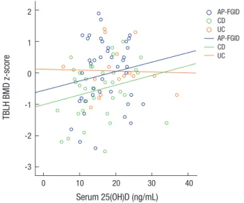
Correlation between serum 25(OH)D concentration and TBLH BMD z-score is shown in Fig. 1. Serum 25(OH)D was positively correlated with TBLH BMD z-score when all subjects were in- cluded in the analysis (rs= 0.231; P = 0.021). However, no sig- nificant correlation was observed within each study group (r = 0.151; P = 0.323 in AP-FGID; r = 0.249; P = 0.132 in CD; r = −0.032;
P = 0.906 in UC).
Factors affecting TBLH BMD z-score in all subjects
When multiple regression analysis of TBLH BMD z-score was performed, sex (P = 0.018), age (P = 0.005), and hemoglobin (P = 0.041) were found to be significant factors (Table 2). TBLH BMD z-score was lower in girls and older children, with a BMD z-score decrement of 0.118 for one-year increase in age. In ad-
dition, hemoglobin was positively correlated with TBLH BMD z-score in children with CD (r = 0.377; P = 0.018), but not in the other 2 study groups.
Table 1. Clinical and biochemical profiles of children with AP-FGID, CD and UC
Variables AP-FGID (n = 45) CD (n = 43) UC (n = 17) P value
Age, yr 13.6 ± 2.3 14.4 ± 2.8 14.5 ± 2.8 0.239
Gender (boys:girls) 26:19 32:11 10:7 0.225
Height, cm 159.1 ± 13.5 159.0 ± 15.6 161.3 ± 10.6 0.833
Weight, kg 50.2 ± 14.2 48.6 ± 13.6 48.6 ± 9.3 0.831
BMI, kg/m2 19.5 ± 3.6 18.9 ± 3.4 18.5 ± 2.1 0.532
Hemoglobin, g/dL 13.6 ± 1.6 12.8 ± 1.7 11.8 ± 2.3 0.001*
Hematocrit, % 40.1 ± 4.2 38.5 ± 4.4 35.6 ± 5.8 0.003*
Platelet (103/μL) 265.3 ± 110.4 359.3 ± 120.2 374.4 ± 180.1 0.001*
WBC (103/μL) 6.0 ± 1.4 7.7 ± 2.9 7.5 ± 3.1 0.012*
ANC (103/μL) 3.0 ± 1.2 5.0 ± 2.8 5.1 ± 3.8 0.000*
ESR, mm/hr 6.8 ± 5.5 27.7 ± 25.7 22.4 ± 15.9 0.000*
hs-CRP, mg/dL 0.2 ± 0.2 2.1 ± 3.7 1.1 ± 2.0 0.003*
Total protein, g/dL 7.2 ± 0.4 7.0 ± 0.7 7.1 ± 0.4 0.319
Albumin, g/dL 4.6 ± 0.3 4.0 ± 0.7 4.2 ± 0.4 0.000*
ALP, IU/L 189.2 ± 116.4 142.9 ± 74.3 177.4 ± 117.0 0.098
Calcium, mg/dL 9.2 ± 0.4 8.9 ± 0.6 8.9 ± 0.5 0.004*
Phosphorus, mg/dL 4.5 ± 0.7 4.4 ± 0.7 4.4 ± 0.8 0.741
25(OH)D, ng/mL 16.5 ± 5.3 16.3 ± 9.3 19.9 ± 7.2 0.224
Total BMD z-score −0.1 ± 1.1 −0.5 ± 0.8 −0.1 ± 0.8 0.071
TBLH BMD z-score −0.1 ± 1.1 −0.5 ± 0.8 0.1 ± 0.8 0.037*
Data presented as mean ± standard deviation.
AP-FGID = abdominal pain-related functional gastrointestinal disorder, BMI = body mass index, CD = Crohn's disease, UC = ulcerative colitis, WBC = white blood cell, ANC = absolute neutrophil count, ESR = erythrocyte sedimentation rate, hs-CRP = high-sensitivity C-reactive protein, ALP = alkaline phosphatase, 25(OH)D = 25-hydroxyvitamin D, BMD = bone mineral density, TBLH = total body less head.
*P value less than 0.05 is regarded as statistically significant.
Table 2. Multiple regression analysis of TBLH BMD z-score according to disease group, patient demographics, and laboratory results
Variables Coefficient SE P value 95% CI (lower, upper) Disease group (FGID-UC) −0.433 0.684 0.529 (−1.795, 0.929) Disease group (FGID-CD) 0.617 1.024 0.548 (−1.422, 2.657) Sex (girls-boys) 0.533 0.219 0.018* (0.096, 0.970)
Age −0.118 0.040 0.005* (−0.199, −0.037)
Hemoglobin 0.148 0.071 0.041* (0.006, 0.291)
WBC 0.007 0.043 0.878 (−0.079, 0.092)
Albumin −0.193 0.313 0.539 (−0.817, 0.431)
ESR −0.004 0.027 0.690 (−0.057, 0.049)
ESR × (FGID-UC) 0.011 0.027 0.690 (−0.043, 0.065) ESR × (FGID-CD) 0.016 0.031 0.596 (−0.045, 0.078)
Calcium 0.122 0.318 0.702 (−0.511, 0.755)
Phosphorus −0.051 0.146 0.729 (−0.342, 0.240)
25(OH)D × (FGID-UC) −0.010 0.035 0.784 (−0.080, 0.061) 25(OH)D × (FGID-CD) −0.028 0.044 0.529 (−0.115, 0.060) ANCOVA was used for statistical analysis; × symbol denotes interaction between the variables.
TBLH = total body less head, BMD = bone mineral density, SE = standard error, 95%
CI = 95% confidence interval, FGID = abdominal pain-related functional gastrointes- tinal disease, UC = ulcerative colitis, CD = Crohn’s disease, FGID-UC = UC in com- parison to FGID, FGID-CD = CD in comparison to FGID, girls-boys = boys in compari- son to girls, WBC = white blood cell, ESR = erythrocyte sedimentation rate, 25(OH) D = 25-hydroxyvitamin D, ANCOVA = analysis of covariance.
*P value less than 0.05 is regarded as statistically significant.
Factors affecting serum 25(OH)D in all subjects
Multiple regression analysis of serum 25(OH)D revealed that sex (P = 0.018), CD group with reference to AP-FGID (P = 0.020), and serum phosphorus (P = 0.018) were significant factors in Fig. 1. Correlation between serum 25(OH)D and TBLH BMD z-score in the 3 study
groups. There was no significant correlation in the AP-FGID group (r = 0.151; P = 0.323), CD group (r = 0.249; P = 0.132), or the UC group (r = −0.032, P = 0.906).
25(OH)D = 25-hydroxyvitamin D, TBLH = total body less head, BMD = bone mineral density, AP-FGID = abdominal pain-related functional gastrointestinal disorder, CD = Crohn’s disease, UC = ulcerative colitis.
TBLH BMD z-score
Serum 25(OH)D (ng/mL)
0 10 20 30 40
2 1 0 -1 -2 -3
AP-FGID CD UC AP-FGID CD UC
these children (Table 3). Lower serum 25(OH)D levels were as- sociated with female sex, CD group status, and lower serum phos- phorus levels. Moreover, serum phosphorus was positively cor- related with 25(OH)D in children with CD (r = 0.386; P = 0.012).
DISCUSSION
On the background of the rising incidence of pediatric IBD in Asia, this study is the first to formally investigate vitamin D sta- tus and BMD in Korean children with IBD. Our results demon- strated that in this cohort of children with CD, UC, AP-FGID, and TBLH BMD z-scores were significantly different and the lowest in children with CD. According to our study, the impor- tant determinants of TBLH BMD z-score in these children are sex, age, and hemoglobin concentration, rather than vitamin D status. Therefore, it seems that vitamin D may not be a useful predictor of bone loss in children with IBD as it is for their adult counterparts.
Our study revealed similar levels of serum 25(OH)D among children with CD, UC, and AP-FGID. This is possibly due to the AP-FGID group’s low 25(OH)D levels, which were similar to those of the CD group. AP-FGID is a functional gastrointestinal con- dition without any evidence of organic disease, but our results indicate that patients with severe symptoms may also develop nutrient deficiencies. Moreover, our results may reflect the fact that Korean children and adolescents in general are prone to vitamin D insufficiency from their largely sedentary indoor life- style and the consequent lack of exposure to sunlight (19).
TBLH BMD, along with lumbar spine BMD, was recently rec- ommended as the preferred skeletal site for DXA evaluation of pediatric subjects (20). Excluding the cranium minimizes con-
tribution of the child’s proportionally large head that may mask potential BMD deficits at other sites (21). In our study, TBLH BMD z-scores were significantly different among the 3 groups, with BMD considerably lower in children with CD compared to those with UC or AP-FGID. Therefore, our study supports previ- ous studies on children that have reported higher prevalence of low BMD in CD compared to UC (10,13,22,23), and higher prev- alence of growth failure and vertebral fractures in CD (24-26).
Higher risk of poor bone health in CD patients can be explained by small intestine involvement causing malabsorption or pro- tein-losing enteropathy, and the higher systemic inflammatory load that confers both direct effects on osteoblast activity and indirect effects such as more frequent use of steroids (25).
A major finding of our study was that sex, age, and hemoglo- bin concentration significantly influenced TBLH BMD z-scores in this cohort of children. Girls were found to have lower TBLH BMD z-scores, in line with the recent study by Schmidt et al. (14) who found TBLH BMD z-scores to be lower in girls at both base- line and 2 years later. Other studies that have contradicted this observation were cross-sectional studies with CD patients only (11,25), or included femoral neck BMD in their analysis (10) al- though this region has been deemed inappropriate for DXA scan- ning in children (27). It is well known that boys have greater bone mass after the onset of puberty, but the comparatively lower BMD in girls across all ages may also be explained by lower body wei- ght, lower lean mass, and lower levels of physical activity.
Older age was associated with lower BMD z-score in these children, echoing the results reported by Herzog et al. (28) who found that among children with CD in remission, those older were more likely to have low BMD. However, disease duration would have been responsible to an extent in this study, whereas all of our subjects were evaluated at initial presentation. Our re- sults could therefore be related to higher disease severity in chil- dren with earlier onset of symptoms, or characteristics of older children and adolescents that predispose them to low BMD, such as sedentary lifestyle or reduced compliance to treatment.
To our knowledge, this is the first study to report hemoglobin as a determinant of BMD in children with IBD. We observed a positive correlation between hemoglobin and BMD in children with CD, which may be reflective of poor overall nutritional sta- tus and lack of hematopoietic nutrients such as iron, vitamin B12, and folate. Alternatively, lower hemoglobin may be related to higher disease activity, since low hematocrit is one of the com- ponents of the Pediatric Crohn’s Disease Activity Index. It would be interesting to see if future studies replicate this novel finding and aid in characterising more predictive markers of diminished bone mass in pediatric IBD.
In accordance with other published studies (10-15), we found no significant correlation between serum 25(OH)D and BMD z-score. Although the lack of correlation is somewhat counter- intuitive, this was observed in both UC and CD groups with the Table 3. Multiple regression analysis of serum 25(OH)D according to disease group,
patient demographics, and laboratory results
Variables Coefficient SE P value 95% CI (lower, upper) Disease group (FGID-UC) −0.593 2.081 0.776 (−4.729, 3.543) Disease group (FGID-CD) 6.946 2.928 0.020* (1.126, 12.767) Sex (girls-boys) 3.139 1.302 0.018* (0.552, 5.727)
Age −0.075 0.233 0.747 (−0.539, 0.388)
WBC 0.400 0.264 0.133 (−0.125, 0.924)
Albumin −1.359 1.960 0.490 (−5.255, 2.537)
ESR −0.012 0.175 0.944 (−0.361, 0.336)
ESR × (FGID-UC) −0.064 0.178 0.721 (−0.418, 0.290) ESR × (FGID-CD) −0.195 0.202 0.337 (−0.596, 0.206)
Calcium 2.029 1.979 0.308 (−1.906, 5.963)
Phosphorus 2.132 0.881 0.018* (0.381, 3.883)
ANCOVA was used for statistical analysis; × symbol denotes interaction between the variables.
25(OH)D = 25-hydroxyvitamin D, SE = standard error, 95% CI = 95% confidence in- terval, FGID = abdominal pain-related functional gastrointestinal disease, UC = ulcerative colitis, CD = Crohn’s disease, FGID-UC = UC in comparison to FGID, FGID-CD = CD in comparison to FGID, girls-boys = boys in comparison to girls, ESR = erythrocyte sedimentation rate, ANCOVA = analysis of covariance.
*P value less than 0.05 is regarded as statistically significant.
use of different vitamin D assays and BMD measurements sites, and after taking into account the disease activity (11). By con- trast, majority of adult studies have demonstrated a significant correlation between the 2 parameters (29-32). Thus, it seems that the pathophysiology of bone loss in pediatric IBD may be fundamentally different or more complex, with the contribu- tion of vitamin D status more limited than previously thought.
It is currently unclear whether the prevalence of poor vitamin D status is significantly different between the 2 IBD subtypes, as studies continue to show divergent results. In our study, CD dis- ease status, but not UC, was found to affect 25(OH)D when com- pared to the AP-FGID group as the reference point. Therefore, our results support the hypothesis that children with CD are more at risk of subnormal vitamin D status. Researchers have so far postulated that this could be due to poor digestion of lip- id-soluble compounds and less absorption of vitamin D via the jejunum and terminal ileum (33). None of our IBD subjects had undergone bowel resection, but this would also be more com- mon among patients with CD and cause malabsorption of a wide range of nutrients.
Our study is limited by a retrospective, single-center study design. Extracting data from the hospital electronic medical re- cords yielded a disease-control group, but we believe that AP- FGID is a good substitute due to its lack of organic pathology.
Our study may have been improved by inclusion of disease ac- tivity index to account for the variability in disease severity, al- though currently there is no unified index for both UC and CD.
A possible clustering of 25(OH)D sampling by season may have affected our results. However, seasonal variance in vitamin D levels may not be as prominent in children with IBD, as those with severe disease are limited to bed rest and indoor confine- ment regardless of the season.
Our study has several strengths that deserve a mention. It is the first study to formally investigate the correlation between vitamin D and BMD in Asian children with IBD, since the few studies that have been published thus far have almost exclusive- ly focused on Caucasian children. Our subjects were also evalu- ated at diagnosis, minimizing the possible effects of disease du- ration and pharmacological treatment on bone mass. Moreover, serum 25(OH)D was measured using LC-MS/MS, which is wide- ly considered as one of the most accurate assays for vitamin D (34). Another aspect of our study that merits attention is the in- clusion of a local control group to compare BMD z-scores. Other studies have used densitometer reference data as a surrogate control group, which can yield misleading results. In their 2004 study, Gupta et al. (35) concluded that normal population values derived from commercial densitometers were not appropriate for the local patient population studied. By comparing BMD z- scores of the disease groups to those of a control group of the same ethnicity and locality, our study is likely to have been more accurate in detecting true discrepancies in BMD.
The present study adds significant findings to the scarce lit- erature on the potential relationship between vitamin D status and bone loss in pediatric IBD. Limited bone mass accrual in pediatric IBD poses a real risk of reducing the child’s peak bone mass, the strongest predictor of future risk of osteoporosis in adulthood (36). Our results indicate that vitamin D status may not be a significant determinant of BMD in pediatric IBD pa- tients as it is for adult IBD patients. However, given the chronic and variable course of IBD, more longitudinal studies are need- ed to evaluate vitamin D status and BMD at different phases of the disease (37).
In conclusion, our study demonstrated that TBLH BMD z- scores were significantly different among children with AP-FGID, CD, and UC, despite similar serum 25(OH)D levels. Our results suggest that significant risk factors for low BMD in these children are female sex, older age, and low hemoglobin levels, while vi- tamin D status may not be as important. The link between vita- min D and bone loss in pediatric IBD seems more complex than previously thought and more studies on predictors of bone loss in these children are warranted for their long-term skeletal health.
ACKNOWLEDGMENT
The authors would like to thank Dr. JB Lee at Seoul National Uni- versity Bundang Hospital Medical Research Collaborating Cen- tre for her assistance in statistical analyses.
DISCLOSURE
The authors have no potential conflicts of interest to disclose.
AUTHOR CONTRIBUTION
Conceptualization: Chang EJ, Yang HR. Data curation: Chang EJ, Yang HR. Formal analysis: Sohn J, Yang HR. Investigation:
Sohn J, Yang HR. Writing - original draft: Sohn J. Writing - re- view & editing: Sohn J, Yang HR.
ORCID
Jenny Sohn http://orcid.org/0000-0002-0063-5634 Eun Jae Chang http://orcid.org/0000-0003-0595-0972 Hye Ran Yang http://orcid.org/0000-0002-3423-6922 REFERENCES
1. Ghishan FK, Kiela PR. Advances in the understanding of mineral and bone metabolism in inflammatory bowel diseases. Am J Physiol Gastrointest Liver Physiol 2011; 300: G191-201.
2. Schulte CM, Dignass AU, Goebell H, Röher HD, Schulte KM. Genetic fac- tors determine extent of bone loss in inflammatory bowel disease. Gas-
troenterology 2000; 119: 909-20.
3. Abraham BP, Prasad P, Malaty HM. Vitamin D deficiency and corticoste- roid use are risk factors for low bone mineral density in inflammatory bowel disease patients. Dig Dis Sci 2014; 59: 1878-84.
4. Krela-Kaźmierczak I, Szymczak A, Łykowska-Szuber L, Eder P, Stawczyk- Eder K, Klimczak K, Linke K, Horst-Sikorska W. The importance of vita- min D in the pathology of bone metabolism in inflammatory bowel dis- eases. Arch Med Sci 2015; 11: 1028-32.
5. Palmer MT, Weaver CT. Linking vitamin D deficiency to inflammatory bowel disease. Inflamm Bowel Dis 2013; 19: 2245-56.
6. Farraye FA, Nimitphong H, Stucchi A, Dendrinos K, Boulanger AB, Vijj- eswarapu A, Tanennbaum A, Biancuzzo R, Chen TC, Holick MF. Use of a novel vitamin D bioavailability test demonstrates that vitamin D absorp- tion is decreased in patients with quiescent Crohn’s disease. Inflamm Bowel Dis 2011; 17: 2116-21.
7. Abreu MT, Kantorovich V, Vasiliauskas EA, Gruntmanis U, Matuk R, Daigle K, Chen S, Zehnder D, Lin YC, Yang H, et al. Measurement of vitamin D levels in inflammatory bowel disease patients reveals a subset of Crohn’s disease patients with elevated 1,25-dihydroxyvitamin D and low bone mineral density. Gut 2004; 53: 1129-36.
8. Levin AD, Wadhera V, Leach ST, Woodhead HJ, Lemberg DA, Mendoza- Cruz AC, Day AS. Vitamin D deficiency in children with inflammatory bowel disease. Dig Dis Sci 2011; 56: 830-6.
9. Alkhouri RH, Hashmi H, Baker RD, Gelfond D, Baker SS. Vitamin and min- eral status in patients with inflammatory bowel disease. J Pediatr Gastro- enterol Nutr 2013; 56: 89-92.
10. Gokhale R, Favus MJ, Karrison T, Sutton MM, Rich B, Kirschner BS. Bone mineral density assessment in children with inflammatory bowel disease.
Gastroenterology 1998; 114: 902-11.
11. Sentongo TA, Semaeo EJ, Stettler N, Piccoli DA, Stallings VA, Zemel BS.
Vitamin D status in children, adolescents, and young adults with Crohn disease. Am J Clin Nutr 2002; 76: 1077-81.
12. Pappa HM, Gordon CM, Saslowsky TM, Zholudev A, Horr B, Shih MC, Grand RJ. Vitamin D status in children and young adults with inflamma- tory bowel disease. Pediatrics 2006; 118: 1950-61.
13. El-Matary W, Sikora S, Spady D. Bone mineral density, vitamin D, and disease activity in children newly diagnosed with inflammatory bowel disease. Dig Dis Sci 2011; 56: 825-9.
14. Schmidt S, Mellström D, Norjavaara E, Sundh V, Saalman R. Longitudinal assessment of bone mineral density in children and adolescents with in- flammatory bowel disease. J Pediatr Gastroenterol Nutr 2012; 55: 511-8.
15. Middleton JP, Bhagavathula AP, Gaye B, Alvarez JA, Huang CS, Sauer CG, Tenjarla G, Schoen BT, Kumar A, Prasad M, et al. Vitamin D status and bone mineral density in African American children with Crohn disease. J Pediatr Gastroenterol Nutr 2013; 57: 587-93.
16. Oh SH, Kim KM. Current issues of pediatric inflammatory bowel disease in Korea. Korean J Pediatr 2014; 57: 465-71.
17. Yang SK, Yun S, Kim JH, Park JY, Kim HY, Kim YH, Chang DK, Kim JS, Song IS, Park JB, et al. Epidemiology of inflammatory bowel disease in the Song- pa-Kangdong district, Seoul, Korea, 1986-2005: a KASID study. Inflamm Bowel Dis 2008; 14: 542-9.
18. Kim BJ, Song SM, Kim KM, Lee YJ, Rhee KW, Jang JY, Park SJ, Yoon CH.
Characteristics and trends in the incidence of inflammatory bowel dis- ease in Korean children: a single-center experience. Dig Dis Sci 2010; 55:
1989-95.
19. Choi HS, Oh HJ, Choi H, Choi WH, Kim JG, Kim KM, Kim KJ, Rhee Y, Lim SK. Vitamin D insufficiency in Korea--a greater threat to younger genera- tion: the Korea National Health and Nutrition Examination Survey (KN- HANES) 2008. J Clin Endocrinol Metab 2011; 96: 643-51.
20. Bianchi ML, Leonard MB, Bechtold S, Högler W, Mughal MZ, Schönau E, Sylvester FA, Vogiatzi M, van den Heuvel-Eibrink MM, Ward L; Interna- tional Society for Clinical Densitometry. Bone health in children and ad- olescents with chronic diseases that may affect the skeleton: the 2013 ISCD Pediatric Official Positions. J Clin Densitom 2014; 17: 281-94.
21. Bachrach LK, Sills IN; Section on Endocrinology. Clinical report—bone densitometry in children and adolescents. Pediatrics 2011; 127: 189-94.
22. Boot AM, Bouquet J, Krenning EP, de Muinck Keizer-Schrama SM. Bone mineral density and nutritional status in children with chronic inflam- matory bowel disease. Gut 1998; 42: 188-94.
23. Sylvester FA, Wyzga N, Hyams JS, Davis PM, Lerer T, Vance K, Hawker G, Griffiths AM. Natural history of bone metabolism and bone mineral den- sity in children with inflammatory bowel disease. Inflamm Bowel Dis 2007; 13: 42-50.
24. Cowan FJ, Warner JT, Dunstan FD, Evans WD, Gregory JW, Jenkins HR.
Inflammatory bowel disease and predisposition to osteopenia. Arch Dis Child 1997; 76: 325-9.
25. Semeao EJ, Jawad AF, Stouffer NO, Zemel BS, Piccoli DA, Stallings VA.
Risk factors for low bone mineral density in children and young adults with Crohn’s disease. J Pediatr 1999; 135: 593-600.
26. van Staa TP, Cooper C, Brusse LS, Leufkens H, Javaid MK, Arden NK. In- flammatory bowel disease and the risk of fracture. Gastroenterology 2003;
125: 1591-7.
27. Crabtree NJ, Arabi A, Bachrach LK, Fewtrell M, El-Hajj Fuleihan G, Kec- skemethy HH, Jaworski M, Gordon CM; International Society for Clinical Densitometry. Dual-energy X-ray absorptiometry interpretation and re- porting in children and adolescents: the revised 2013 ISCD Pediatric Of- ficial Positions. J Clin Densitom 2014; 17: 225-42.
28. Herzog D, Bishop N, Glorieux F, Seidman EG. Interpretation of bone min- eral density values in pediatric Crohn’s disease. Inflamm Bowel Dis 1998;
4: 261-7.
29. Shirazi KM, Somi MH, Rezaeifar P, Fattahi I, Khoshbaten M, Ahmadzadeh M. Bone density and bone metabolism in patients with inflammatory bowel disease. Saudi J Gastroenterol 2012; 18: 241-7.
30. Bischoff-Ferrari HA, Kiel DP, Dawson-Hughes B, Orav JE, Li R, Spiegel- man D, Dietrich T, Willett WC. Dietary calcium and serum 25-hydroxyvi- tamin D status in relation to BMD among U.S. adults. J Bone Miner Res 2009; 24: 935-42.
31. Silvennoinen J. Relationships between vitamin D, parathyroid hormone and bone mineral density in inflammatory bowel disease. J Intern Med 1996; 239: 131-7.
32. Leslie WD, Miller N, Rogala L, Bernstein CN. Vitamin D status and bone density in recently diagnosed inflammatory bowel disease: the Manitoba IBD Cohort Study. Am J Gastroenterol 2008; 103: 1451-9.
33. Narula N, Marshall JK. Management of inflammatory bowel disease with vitamin D: beyond bone health. J Crohns Colitis 2012; 6: 397-404.
34. Wallace AM, Gibson S, de la Hunty A, Lamberg-Allardt C, Ashwell M. Mea- surement of 25-hydroxyvitamin D in the clinical laboratory: current pro- cedures, performance characteristics and limitations. Steroids 2010; 75:
477-88.
35. Gupta A, Paski S, Issenman R, Webber C. Lumbar spine bone mineral den-
sity at diagnosis and during follow-up in children with IBD. J Clin Densi- tom 2004; 7: 290-5.
36. NIH Consensus Development Panel on Osteoporosis Prevention, Diag- nosis, and Therapy. Osteoporosis prevention, diagnosis, and therapy. JAMA
2001; 285: 785-95.
37. Veit LE, Maranda L, Fong J, Nwosu BU. The vitamin D status in inflamma- tory bowel disease. PLoS One 2014; 9: e101583.