Association between Lumbar Bone Mineral Density and Carotid Intima-Media Thickness in Korean Adults: a Cross-sectional Study of Healthy Twin Study
Bone mineral density (BMD) has been suggested to be associated with atherosclerosis. In the present study, we evaluated the association between lumbar BMD and the segments of carotid intima-media thickness (CIMT), a surrogate marker of subclinical atherosclerosis, in Korean adults, with consideration of sex and menopause status. Among 1,679 Korean adults who enrolled in a Healthy Twin Study, 723 men, 690 premenopausal women, and 266 postmenopausal women measured the CIMT at the common carotid artery intima- media thickness (CCA-IMT), carotid bifurcation intima-media thickness (BIF-IMT), internal carotid artery intima-media thickness (ICA-IMT) using B-mode ultrasound and lumbar BMD using dual-energy X-ray absorptiometry. The composite CIMT was calculated as the mean value of three CIMTs. The association was evaluated using linear mixed models. In premenopausal women, lumbar BMD was positively associated with composite CIMT and with CCA-IMT (P = 0.008 and 0.002, respectively). However, no association was observed between BMD and CIMT in men or in postmenopausal women. Stratified analysis revealed the effect of body mass index (BMI) on the association between BMD and CIME. The positive association in premenopausal women persisted only in low BMI (< 25 kg/m2) group, whereas a positive association appeared at high BMI (≥ 25 kg/m2) group in men.
A high lumbar BMD may indicate an elevated risk of subclinical atherosclerosis in premenopausal women and men with high BMI.
Keywords: Bone Mineral Density; Carotid Arteries; Osteoporosis; Atherosclerosis Jinyoung Shin,1* Joo-Hyun Park,2*
Yun-Mi Song,1 Kayoung Lee,3 and Joohon Sung4,5
1Department of Family Medicine, Samsung Medical Center, Samsung Biomedical Research Institute, Sungkyunkwan University School of Medicine, Seoul, Korea; 2Department of Family Medicine, Korea University Ansan Hospital, Korea University College of Medicine, Ansan, Korea; 3Department of Family Medicine, Inje University Busan Paik Hospital, Inje University College of Medicine, Busan, Korea;
4Department of Epidemiology, School of Public Health, Seoul National University, Seoul, Korea;
5Institute of Health and Environment, Seoul National University, Seoul, Korea
* Jinyoung Shin and Joo-Hyun Park contributed equally to this work.
Received: 1 July 2016 Accepted: 8 October 2016 Address for Correspondence:
Yun-Mi Song, MD
Department of Family Medicine, Samsung Medical Center, Sungkyunkwan University School of Medicine, 81, Irwon-ro, Gangnam-gu, Seoul 06351, Korea
E-mail: [email protected]
Funding: This research was supported by the Basic Science Research Program through the National Research Foundation of Korea (NRF), which is funded by the Ministry of Science, ICT and Future Planning (2014R1A2A2A01002705). This study was supported by Samsung Biomedical Research Institute (SMO1161741).
https://doi.org/10.3346/jkms.2017.32.1.70 • J Korean Med Sci 2017; 32: 70-76
INTRODUCTION
Low bone mineral density (BMD) and atherosclerosis are de- generative changes commonly accompanying aging. Low BMD increases the risk of osteoporotic fracture for the elderly, which results in functional decline and increased mortality (1,2). Ath- erosclerosis occurs in the subendothelial space (i.e., the intima) of medium-sized arteries and is triggered by mechanotransduc- tion and inflammatory processes in endothelial cells (3).
A growing body of evidence indicates that BMD is associated with atherosclerosis. This association has been proposed to be explained by a number of biological mechanisms including sim- ilar processes of bone and vascular mineralization (4), osteo- blastic differentiation by lipid oxidation (5), and shared risk fac- tors including lifestyle factors (6), estrogen deficiency (7), and
vitamin D receptor polymorphisms (8,9).
The association between BMD and carotid intima-media thick- ness (CIMT) as a surrogate marker of subclinical atherosclero- sis has been evaluated in many studies (8,10-16). However, these studies have yielded inconsistent findings. Some studies found an inverse association (10,13), whereas others found a positive (12) or no association (11,14,16).
We thought that the association between BMD and CIMT may differ according to sex or menopausal status because the prevalence of atherosclerotic vascular disease and the distribu- tion of BMD tend to differ according to these factors (17,18). In addition, the identification of segment-specific associations be- tween intima-media thickness (IMT) and cardiovascular dis- ease (19,20) suggests that the association between BMD and CIMT may vary according to carotid artery segment.
To resolve these discrepancies, we evaluated the association between BMD and segment-specific CIMT according to sex and menopausal status with adjustment for a wide range of cardio- vascular risk factors.
MATERIALS AND METHODS Subjects and study design
The study subjects consisted of 1,820 individuals (754 men, 734 premenopausal women, and 332 postmenopausal women) aged 18 to 83 years old. All subjects had undergone B-mode ultra- sound for CIMT measurement, in addition to dual-energy X- ray absorptiometry for BMD measurement of the lumbar spine, between April 2009 and February 2012. All examinations took place at the same institution.
The study subjects were the participants in the Healthy Twin Study, a nationwide population-based cohort study that has been conducted as part of the Korean Genome Epidemiology Study since 2005. The participants are composed of twins and their first-degree family members who were voluntarily recruit- ed through a nationwide media advertisement and mailing cam- paign. Details about the study design and methodology of the Healthy Twin Study have been previously published (21,22).
Subjects with missing data (29 cases) and who had received osteoporosis treatment (51 cases) were excluded. In addition, subjects with a history of stroke (3 cases), myocardial infarction (21 cases), or cancer (37 cases) were also excluded. Finally, 1,679 subjects (723 men, 690 premenopausal women, and 266 post- menopausal women) were analyzed.
CIMT measurements
CIMT measurements were performed according to standard- ized protocols. Briefly, the CIMT was measured during the end- diastolic phase between the P and Q waves from the electrocar- diogram trace using an automated IMT package using an auto- mated IMT package of a high-resolution B-mode ultrasound machine (VIVID; General Electric, Horten, Norway) and an EKO 7 system (Samsung Medison Co., Ltd., Cypress, CA, USA) equip- ped with a seven-MHz linear transducer. CIMT scanning was performed on the far walls of the following three segments: 10–
20 mm proximal to the tip of the flow divider into the common carotid artery (CCA), the carotid bifurcation (BIF) beginning at the tip of the flow divider and extending 10 mm proximal to the flow divider tip, and the proximal 10 mm of the internal carotid artery (ICA). The composite CIMT was calculated as the mean value of the three carotid artery segment CIMTs from both sides.
Since two different machines (Vivid, General Electric; and EKO 7, Samsung Medison, Co., Ltd.) were used for CIMT measure- ment, reproducibility of the IMT measurement was assessed in 14 randomly chosen subjects. The intra-class correlation coeffi- cients between the repeatedly measured IMT were 0.93, 0.86,
and 0.90 for CCA, BIF, and ICA, respectively.
Measurements of BMD and covariates
Area (cm2), bone mineral concentration (g) of the lumbar spine, and lean mass (g) were measured using whole body dual-ener- gy X-ray absorptiometry on a Delphi W system (Hologic, Bos- ton, MA, USA). All measurements were recorded by a well-trained technician. For measurements of lumbar BMD, the lumbar spine region was defined superiorly by the transverse line between the twelfth thoracic vertebra (T12) and the first lumbar vertebra (L1). The lumbar spine region was defined inferiorly by the hor- izontal line at the iliac crest. Calibration of the dual-energy X- ray absorptiometer was conducted using a phantom according to standard quality control procedures recommended by the manufacturer. All coefficients of variation for the BMD measure- ments were ≤ 1.0% for the machine. Lumbar BMD was calcu- lated by dividing the bone mineral concentration by the area of the lumbar spine region.
Blood pressure was measured manually using a standardized mercury sphygmomanometer. Weight (kg) and height (cm) were measured using standardized scales and stadiometers while wearing light clothing. Body mass index (BMI) was calculated as body weight divided by height squared (kg/m2). All physical measurements were taken twice and analyses were performed on average values. After overnight fasting (> 12 hours), serum glucose and lipid levels were measured. All biochemical analy- ses were conducted in one central laboratory that is accredited by the Korea Association of Quality Assurance for Clinical Lab- oratory.
Demographic characteristics, smoking status, physical exer- cise habits, and medical histories of hypertension and diabetes were obtained using a self-administered standardized question- naire. Incomplete or ambiguous responses were clarified in face- to-face interviews. Postmenopausal status was defined as hav- ing no menstrual periods for the preceding year and fulfilling at least one of the following conditions: natural menopause, use of estrogen replacement therapy, or 55 years of age or older. Study subjects were categorized into two groups (never smoker and ever smoker) according to smoking status. If the study subject reported performing physical exercise at least once a week, the subject was considered to be involved in regular physical exer- cise. Hypertension was defined as current treatment with anti- hypertensive medication or blood pressure exceeding 140 (sys- tolic) or 90 (diastolic) mmHg. Diabetes mellitus was defined as current treatment with glucose-lowering medication, high se- rum glucose (≥ 6.99 mM/L), or a high level of hemoglobin A1C (≥ 6.5%).
Statistical analysis
We compared the characteristics of men, premenopausal wom- en, and postmenopausal women using the t-test for continuous
variables and the chi-square test for categorical variables. Variations in CIMTs, cardiovascular risk factors, and lifestyle factors were analyzed accord- ing to composite IMT tertile using linear regression or by the Mantel-Haenszel χ2 test.
To evaluate the extent of association between BMD and CIMT, the percent difference of CIMT per increase (in 1 g/cm2) of BMD of the lumbar spine were assessed using linear mixed models.
Since all study subjects were recruited for a twin/
family study, correlations were considered within the context of family relationships by adjusting for each family unit and each twin unit as random ef- fects in the linear mixed model. Age, height, hyper- tension status, diabetes status, thyroid-stimulating hormone level, low-density lipoprotein (LDL) cho- lesterol level, high-density lipoprotein (HDL) cho- lesterol level, triglyceride (TG) level, BMI, lipid-lo- wering medication status, smoking status, regular exercise habits, calcium supplementation status, and estrogen replacement therapy status (for post- menopausal women) were adjusted as fixed effects.
Prior to this analysis, CIMT values were log trans- formed to approximate a normal distribution.
We also examined the association between CIMT and lumbar BMD according to BMI. High BMI was defined as a BMI ≥ 25 kg/m2; low BMI was defined as a BMI < 25 kg/m2. We also evaluated whether BMI influences the association between BMD and CIMT by including an interaction term (BMI * lum- bar BMD) into the analytic model. All statistical analyses were conducted using PASW Statistics 18 software (SPSS Inc., Chicago, IL, USA).
Ethics statement
Written informed consent was obtained from each participant. All study procedures were approved by the Institutional Review Board of Samsung Med- ical Center (2005-08-113).
RESULTS
The study variables for men, premenopausal wom- en, and postmenopausal women were compared in Supplementary Table 1. The lumbar BMD, CCA- IMT, BIF-IMT, LDL-cholesterol level, HDL-choles- terol level, hypertension prevalence, diabetes prev- alence, ever smoking prevalence, and calcium re- placement prevalence all differed significantly be- tween the three groups. However, the BMI, ICA-
IMT, TG level, prevalence of thyroid disease, and Clinical characteristics and metabolic parameters according to tertiles of composite intima-media thicknessTable 1. =266)Postmenopausal women (n690)=Premenopausal women (n723)=Men (n CharacteristicsHighestLowestIntermediateHighestHighestIntermediateLowestLowestIntermediate P trend*P trend*P trend* (0.42–0.53)(0.25–0.41)(0.35–0.56)(0.65–3.36)(0.57–0.74)(0.49–0.64)(0.75–1.78)(0.28–0.48)(0.54–2.10) 210888989232226248254243 of subjectsNo. 43 (6.9)0.001<64.8 (7.6)60.6 (6.9)56.4 (5.9)0.001<Age,39.7 (6.8)33.3 (7.7)0.001<57.6 (11.2)45.2 (11.8)35.5 (10.3) yr IMT at specific sites, mm Common carotid artery Carotid bifurcation Internal carotid arter
y
0.38 (0.06) 0.47 (0.11) 0.36 (0.07) 0.52 (0.07) 0.71 (0.14) 0.46 (0.09) 0.82 (0.33) 1.30 (0.71) 0.72 (0.43)
<0.001 <0.001 <0.001
0.35 (0.04) 0.42 (0.09) 0.32 (0.06) 0.45 (0.05) 0.59 (0.10) 0.40 (0.08) 0.60 (0.11) 0.84 (0.22) 0.53 (0.13)
<0.001 <0.001 <0.001
0.47 (0.06) 0.57 (0.13) 0.39 (0.08) 0.61 (0.09) 0.84 (0.19) 0.50 (0.13) 0.86 (0.26) 1.41 (0.41) 0.67 (0.26)
<0.001 <0.001 <0.001 Lumbar spine BMD, g/cm21.06 (0.14)1.07 (0.16)1.08 (0.23) 0.4531.08 (0.13)1.11 (0.21)1.12 (0.16) 0.0261.00 (0.31)1.00 (0.39)0.93 (0.21) 0.282 Height, cm172.4 (6.40)169.5 (6.00)167.8 (6.10)<0.001159.9 (5.80)158.2 (5.20)157.8 (5.50)<0.001155.0 (4.90)155.9 (5.30)153.8 (5.40) 0.044 BMI, kg/m224.3 (3.20)24.7 (2.80)24.8 (2.80) 0.12621.6 (2.90)22.8 (3.50)23.1 (3.20)<0.00123.9 (2.80)24.6 (3.60)24.9 (3.20) 0.098 TSH, μIU/L1.95 (1.23)1.87 (1.56)1.74 (1.08) 0.2282.00 (1.18)2.09 (1.31)2.07 (1.32) 0.6891.94 (1.11)3.90 (14.10)2.15 (1.99) 0.224 Total cholesterol, mM/L4.70 (0.90)5.00 (0.99)4.98 (0.95)<0.0014.53 (0.77)4.68 (0.95)4.88 (0.84)<0.0015.26 (0.99)5.36 (1.05)5.16 (0.95) 0.414 LDL cholesterol, mM/L2.77 (0.85)3.00 (0.87)3.00 (0.88)0.0032.51 (0.64)2.67 (0.85)2.87 (0.72)<0.0013.20 (0.89)3.34 (0.96)3.16 (0.81) 0.381 HDL cholesterol, mM/L1.22 (0.29)1.21 (0.27)1.16 (0.30) 0.0371.44 (0.33)1.38 (0.33)1.38 (0.29) 0.0741.32 (0.33)1.29 (0.35)1.20 (0.28) 0.031 Triglyceride, mM/L1.57 (1.74)1.58 (0.90)1.64 (0.89) 0.8070.93 (0.52)0.96 (0.67)1.01 (0.66) 0.3071.41 (0.81)1.54 (0.82)1.65 (0.94) 0.188 Lipid lowering medication, %0.42.82.7 0.0660.01.40.9 0.2544.52.25.7 0.702 Hypertension, %8.619.732.7<0.0011.24.312.5<0.00114.621.337.5<0.001 Diabetes, %5.312.225.7<0.0012.42.95.6 0.06224.720.225.0 0.69 Ever smoking, %50.265.057.1 0.0047.710.08.2 0.8801.15.63.4 0.402 Regular exercise, %36.237.844.2 0.12924.232.430.2 0.12342.739.334.1 0.528 Thyroid disease, %0.40.40.0 0.3951.61.93.0 0.3021.11.13.4 0.266 Calcium replacement, %2.53.14.4 0.2395.66.212.50.00419.120.225.0 0.341 Data were presented as mean (standard deviation) or %. IMT=intima-media thickness, BMD=bone mineral density, BMI=body mass index, TSH=thyroid stimulating hormone, LDL=low density lipoprotein, HDL=high density lipoprotein. *Linear trend was assessed by Mantel-Haenszel χ2 test or linear regression test.
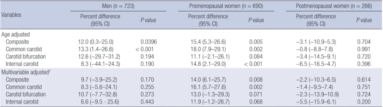
Table 2. Association* between CIMT and BMD at lumbar spine Variables
Men (n = 723) Premenopausal women (n = 690) Postmenopausal women (n = 266) Percent difference
(95% CI) P value Percent difference
(95% CI) P value Percent difference
(95% CI) P value
Age adjusted Composite Common carotid Carotid bifurcation Internal carotid
12.0 (0.3–25.0) 13.3 (1.4–26.6) 12.6 (−29.7–31.2)
8.3 (−44.1–24.3)
0.0396
< 0.001 0.194 0.190
15.4 (5.3–26.6) 18.0 (7.9–29.1) 11.1 (−2.1–26.1) 14.8 (2.1–29.0)
0.005 0.002 0.084
< 0.001
−3.1 (−10.9–5.3)
−0.8 (−8.8–7.8)
−3.4 (−14.5–9.1)
−6.5 (−16.5–4.7)
0.704 0.991 0.720 0.396 Multivariable adjusted†
Composite Common carotid Carotid bifurcation Internal carotid
9.7 (−3.9–25.2) 8.3 (−5.6–24.1) 10.7 (−7.7–32.8) 6.6 (−9.5 - 25.6)
0.170 0.255 0.273 0.443
14.0 (6.1–25.7) 16.1 (5.7–27.6) 13.0 (−1.3–29.3) 11.9 (−1.2–26.7)
0.008 0.002 0.071 0.068
−2.2 (−10.3–6.5)
−1.4 (−9.5–7.4)
−2.3 (−13.9–10.9)
−5.5 (−15.9–6.1)
0.614 0.751 0.724 0.200 CIMT = carotid intima-media thickness, BMD = bone mineral density, CI = confidence interval, BMI = body mass index.
*β coefficients (95% CI) for log-transformed carotid intima media thickness per 1 g/cm2 increase in BMD were assessed by linear mixed model. Then, percent difference of ca- rotid intima media thickness was calculated by multiplying 100 to the value of (exponentiated β coefficient −1). In all analytic models, household and twin pair was adjusted as the random effects; †In the multivariable-adjusted model, age, height, BMI, hypertension, diabetes, thyroid stimulating hormone, low-density lipoprotein cholesterol, high-density lipoprotein cholesterol, triglyceride, lipid lowering medication, smoking habit, physical exercise, calcium supplement and estrogen replacement therapy (for postmenopausal women) were additionally adjusted as fixed effects.
Table 3. Percent difference (95% CI)* of CIMT according to BMI†
Sites of IMT measure
Men Premenopausal women Postmenopausal women
Low BMI
(n = 361) High BMI
(n = 362)
P interac-
tion
Low BMI
(n = 536) High BMI (n = 154)
P interac-
tion
Low BMI
(n = 141) High BMI (n = 125)
P interac-
tion Composite −9.6 (−25.6–9.9) 24.5 (5.2–47.5)‡ 0.014 14.5 (2.7–27.7)‡ 16.1 (−4.9–41.6) 0.976 −4.2 (−18.2–12.2) 6.3 (−9.2–24.5) 0.437 Common carotid −5.7 (−23.8–16.6) 24.1 (4.1–48.0)‡ 0.062 13.5 (2.3–25.9)‡ 30.0 (−4.6–61.4) 0.233 −1.4 (−5.0–14.2) 4.4 (−10.7–21.9) 0.794 Carotid bifurcation −14.7 (−34.6–11.3) 24.0 (−2.3–57.4) 0.007 12.5 (−3.0–30.5) 8.1 (−18.1–42.6) 0.812 −8.5 (−28.6–17.4) 13.5 (−9.4–42.2) 0.246 Internal carotid −13.5 (−31.7–9.4) 12.0 (−9.0–37.7) 0.047 13.8 (−0.5–30.2) 15.5 (−12.0–51.4) 0.948 −1.5 (−22.7–25.4) −0.8 (−16.9–18.5) 0.986 CI = confidence interval, CIMT = carotid intima-media thickness, BMI = body mass index.
*β coefficients (95% CI) for log-transformed carotid intima media thickness per 1 g/cm2 increase in lumbar BMD according to two BMI groups were assessed by linear mixed model. Then, percent thickness difference of carotid intima media thickness was calculated by multiplying 100 to the value of (exponentiated β coefficient −1). In the model, household and twin pair was adjusted as the random effects. Age, height, hypertension, diabetes, thyroid stimulating hormone, low-density lipoprotein cholesterol, high-density lipoprotein cholesterol, triglyceride, lipid lowering medication, smoking habit, physical exercise, calcium replacement therapy and estrogen replacement therapy (for postmeno- pausal women) were additionally adjusted as fixed effects; †BMI was divided as two-group ( < 25 or ≥ 25 kg/m2); ‡P < 0.05.
exercise status did not differ between men and postmenopaus- al women. The prevalence of thyroid disease did not significant- ly differ between premenopausal women and postmenopausal women. Similarly, the level of thyroid-stimulating hormone and the prevalence of lipid-lowering medication were not sig- nificantly different between men and premenopausal women.
The study variable relationships for men, premenopausal wom- en, and postmenopausal women with respect to composite CIMT tertile distribution are shown in Table 1. In all three groups, sub- jects with a higher composite CIMT tended to be older. With in- creasing composite CIMT, the lumbar BMD gradually increased in premenopausal women (P for trend = 0.026), whereas no specific trend was observed in postmenopausal women or in men. As the composite CIMT increased, height decreased in all three groups, whereas BMI increased only in premenopausal women. Moreover, as the composite CIMT increased, total cho- lesterol and LDL-cholesterol levels increased in both men and premenopausal women, while the HDL-cholesterol level de- creased in both men and postmenopausal women. The preva- lence of hypertension gradually increased as composite CIMT
increased in all three groups. The prevalence of diabetes increa- sed as composite CIMT increased; however, this trend was ob- served only in men. In premenopausal women, calcium sup- plementation was more prevalent among women with higher composite CIMT.
The relationship between lumbar BMD and CIMT is shown in Table 2. After adjusting for age, positive associations were observed between BMD and composite IMT and CCA-IMT in men and between BMD and composite CIMT, CCA-IMT, and ICA-IMT in premenopausal women. After adjusting for covari- ates, this significant association persisted for composite CIMT and CCA-IMT. Borderline significant associations were also ob- served between lumbar BMD and BIF-IMT and between lum- bar BMD and ICA-IMT in premenopausal women. However, the association in men between CIMT and lumbar BMD did not persist after this adjustment. In postmenopausal women, no association was observed between CIMT and BMD. We re- peated the multivariable adjusted analysis after stratifying male subjects into two groups by the age cut-off level (≥ 50 years, < 50 years) and found no difference in the association between BMD
and CIMT depending on the age level in men (data not shown).
The findings from stratified analysis regarding the association between lumbar BMD and CIMT according to the BMI level are shown in Table 3. In premenopausal women, a positive associ- ation was observed, while statistically significant positive asso- ciation with BMD was confirmed only with composite CIMT and CCA-IMT in low BMI women. In premenopausal women, no significant association was observed between BMI and lum- bar BMD. Lumbar BMD tended to be positively associated with CIMT in men with high BMI, while an inverse but statistically insignificant relationship was observed in men with low BMI.
The interaction between BMI and lumbar BMD on the associa- tion with CIMT was statistically significant in men. In postmeno- pausal women, no association between BMD and CIMT was observed at any site, regardless of BMI level.
DISCUSSION
In this Korean study, we observed a positive association between lumbar BMD and CIMT, although this association was restrict- ed to premenopausal women and obese men, and mainly to composite CIMT and CCA-IMT. The positive associations be- tween lumbar BMD, composite CIMT, and CCA-IMT in preme- nopausal women are consistent with the findings of a previous study in Mexican American young women (mean age = 26.7 years) in which the BMDs at the hip, radius, and spine were found to be positively associated with CCA-IMT (8). Cecelja et al. (12) also observed positive associations between hip and lumbar BMD and CIMT in British women (mean age 57.7 years, stan- dard deviation 8.9 years). However, these associations were not evaluated according to menopausal status in the British study.
Estrogen may play a role in the positive association between BMD and atherosclerosis observed in premenopausal women, given the biological relationships that have been described be- tween BMD and CIMT. Specifically, both BMD and CIMT have been shown to be regulated by estrogen receptor-alpha gene polymorphisms (23,24); mitogen-activated protein (MAP) ki- nase, a serine/threonine kinase that mediates tumor necrosis factor-α signaling, and various interleukins that are associated positively with CIMT have been shown to be regulated by estro- gen (25,26); single nucleotide polymorphism variants of the es- trogen receptor have been reported to be associated with CIMT in Taiwanese women, but not in men (24); and the vitamin D receptor polymorphism was reported to affect BMD by com- bining the estrogen receptor polymorphism (27). These biologi- cal mechanisms may underlie the association between BMD and CIMT and strongly suggest that future studies on BMD and atherosclerosis in women should consider menopausal status.
Estrogen levels are known to decrease after menopause, which results in reduced estrogen-mediated protection against ath- erosclerosis and osteoporosis in postmenopausal women (7).
Postmenopausal women also experience changes in their body composition such as increased fat distribution and decreased lean mass (28) that are associated with both BMD and athero- sclerosis (29,30). Moreover, increasing calcium supplementation during the menopausal period may accelerate vascular athero- genesis (31). Therefore, an inverse association between BMD and atherosclerosis seems plausible in postmenopausal wom- en. Some studies found an inverse association between BMD and CIMT (8,13,32). However, other studies in Morocco, the USA, Finland, China, Japan, and Korea (including the present study) did not find any association between lumbar BMD and CIMT, or any other surrogate marker of atherosclerosis, in postmeno- pausal women (10,11,14,16,20).
These discrepancies have a number of potential explanations.
Variation regarding the site of BMD measurement is one possi- ble reason. In support of this explanation, one study found that femur BMD was inversely associated with CIMT, whereas lum- bar BMD was not (10). However, since even the findings regard- ing the association of lumbar spine BMD with CIMT vary be- tween studies (10,11,32), variation regarding the site of BMD measurement cannot fully explain the discrepancies between these studies. Second, different age distributions between stud- ies may have resulted in these discrepancies. The age distribu- tion of postmenopausal women in our study (mean age = 59.5 years) was somewhat younger than that in another study that found an inverse association (mean age = 69.2 years) (8). How- ever, another study of older women (mean age = 73.6 years) did not find any association, which is consistent with our findings.
To clarify this issue, the association needs to be studied accord- ing to age or time after menopause. Third, study-to-study varia- tion of covariates such as osteoporosis treatment and hormonal replacement therapy may also explain the different findings.
However, we did not observe any significant influence by co- variates after adjustment in the present study. Forth, differences in ethnicity/race might be another reason (13,20). Finally, the association between BMD and CIMT might not be shown dis- tinctly due to the effect modification by other factors such as BMI level on the association. In our study, stratified analysis by BMD level in postmenopausal women has revealed that BMD may be associated with CIMT inversely in lower BMI group but positively in higher BMI group. Although the estimates lacked statistical significance, this finding seems compatible with the findings observed in men. In our study the sample size of post- menopausal women may not be enough to do a subgroup anal- ysis to examine the effect modifying role of BMI. We think fur- ther study with larger sample size of postmenopausal women would be needed to clarify this issue.
Few studies have been conducted in men regarding the rela- tionship between BMD and CIMT compared with women. Al- though two studies in Chinese population did not identify sig- nificant association (16,33), the direction was consistent with
our study. However, another study in multiethnic population reported an inverse association (20). In our study, age-adjusted analysis revealed a positive relationship between lumbar BMD and CIMT, but this relationship did not persist after adjusting for covariates. All other studies also considered known con- founding variables such as smoking status, BMI, lipid level, hy- pertension status, and diabetes status. Thus, these discrepan- cies do not seem to be due to different covariate adjustments.
Obesity is known to be positively associated with BMD, since a high BMI can increase the mechanical load on the bones (29, 34). Moreover, a high BMI increases the risk of atherosclerosis through mechanisms related to chronic inflammation and in- sulin resistance (35). The known relationships between obesity and both BMD and atherosclerosis prompted us to carefully in- vestigate the effects of BMI via stratified analysis. Although our results need to be confirmed in future studies, interestingly, we found that BMD was positively associated with CIMT in men with high BMI but not in men with low BMI. This finding seems to suggest an effect modification by BMI in men. We assume that obesity related factors such as increased mechanical load, chronic inflammation and insulin resistance may explain the effect modifying role of BMI on the association between CIMT and BMD. However, we were unable to examine the biological mechanism. Further study is needed to investigate and to ex- plain the association between BMD and CIMT in men.
The present study has a number of strengths. First, a large number of subjects were included, which allowed us to consid- er both sex and menopausal status. Second, a wide range of co- variates including thyroid-stimulating hormone status, estro- gen replacement therapy status, and calcium supplementation status could be considered. Therefore, we believe that the influ- ence of most confounding factors on our results was minimized.
However, the present study has some limitations. First, we measured lumbar BMD using whole body dual-energy X-ray absorptiometry, which does not provide BMD at each segment of the lumbar spine. Thus, measurement error could have af- fected our results. Second, we could not take into consideration of the probable influence by aortic calcification or osteophytes of spine on the measurement of lumbar BMD because we could not obtain those information. Third, we were unable to evalu- ate the extent of association of CIMT with femoral BMD, which was a measurement that is commonly used in the clinical set- ting along with lumbar BMD, because whole body dual-energy X-ray absorptiometry does not provide femoral BMD informa- tion. Fourth, the sample size of postmenopausal women might be relatively limited to disclose the association between CIMT and BMD with enough power.
We found that lumbar BMD has a positive association with CIMT in premenopausal women and men with high BMI. This finding suggests that the association between lumbar BMD and subclinical atherosclerosis may differ according to sex and meno-
pausal status.
DISCLOSURE
The authors have no potential conflicts of interest to disclose.
AUTHOR CONTRIBUTION
Participating in the conception and design: Sung J. Analysis and interpretation of data: Shin J, Park JH, Song YM, Lee K, Sung J.
Drafting the article or critically revising: Shin J, Park JH, Song YM, Lee K, Sung J. Approving the final version submitted: all authors.
ORCID
Jinyoung Shin http://orcid.org/0000-0001-9558-1853 Joo-Hyun Park http://orcid.org/0000-0002-4358-4208 Yun-Mi Song http://orcid.org/0000-0001-9232-5563 Kayoung Lee http://orcid.org/0000-0002-2816-554X Joohon Sung http://orcid.org/0000-0001-9948-0160 REFERENCES
1. Sarkisian CA, Liu H, Gutierrez PR, Seeley DG, Cummings SR, Mangione CM. Modifiable risk factors predict functional decline among older wom- en: a prospectively validated clinical prediction tool. The study of osteo- porotic fractures research group. J Am Geriatr Soc 2000; 48: 170-8.
2. Bliuc D, Nguyen ND, Alarkawi D, Nguyen TV, Eisman JA, Center JR. Ac- celerated bone loss and increased post-fracture mortality in elderly wom- en and men. Osteoporos Int 2015; 26: 1331-9.
3. Tabas I, García-Cardeña G, Owens GK. Recent insights into the cellular biology of atherosclerosis. J Cell Biol 2015; 209: 13-22.
4. Hofbauer LC, Brueck CC, Shanahan CM, Schoppet M, Dobnig H. Vascu- lar calcification and osteoporosis--from clinical observation towards mo- lecular understanding. Osteoporos Int 2007; 18: 251-9.
5. Demer LL. Vascular calcification and osteoporosis: inflammatory respons- es to oxidized lipids. Int J Epidemiol 2002; 31: 737-41.
6. Stevenson JC, Lees B, Devenport M, Cust MP, Ganger KF. Determinants of bone density in normal women: risk factors for future osteoporosis?
BMJ 1989; 298: 924-8.
7. Losordo DW, Kearney M, Kim EA, Jekanowski J, Isner JM. Variable expres- sion of the estrogen receptor in normal and atherosclerotic coronary ar- teries of premenopausal women. Circulation 1994; 89: 1501-10.
8. Kammerer CM, Dualan AA, Samollow PB, Périssé AR, Bauer RL, MacClu- er JW, O’Leary DH, Mitchell BD. Bone mineral density, carotid artery in- timal medial thickness, and the vitamin D receptor BsmI polymorphism in Mexican American women. Calcif Tissue Int 2004; 75: 292-8.
9. Anagnostis P, Karagiannis A, Kakafika AI, Tziomalos K, Athyros VG, Mikha- ilidis DP. Atherosclerosis and osteoporosis: age-dependent degenerative processes or related entities? Osteoporos Int 2009; 20: 197-207.
10. Hmamouchi I, Allali F, Khazzani H, Bennani L, El Mansouri L, Ichchou L, Cherkaoui M, Abouqal R, Hajjaj-Hassouni N. Low bone mineral density
is related to atherosclerosis in postmenopausal Moroccan women. BMC Public Health 2009; 9: 388.
11. Frost ML, Grella R, Millasseau SC, Jiang BY, Hampson G, Fogelman I, Cho- wienczyk PJ. Relationship of calcification of atherosclerotic plaque and arterial stiffness to bone mineral density and osteoprotegerin in postmeno- pausal women referred for osteoporosis screening. Calcif Tissue Int 2008;
83: 112-20.
12. Cecelja M, Jiang B, Bevan L, Frost ML, Spector TD, Chowienczyk PJ. Arte- rial stiffening relates to arterial calcification but not to noncalcified ather- oma in women. A twin study. J Am Coll Cardiol 2011; 57: 1480-6.
13. Shaffer JR, Kammerer CM, Rainwater DL, O’Leary DH, Bruder JM, Bauer RL, Mitchell BD. Decreased bone mineral density is correlated with in- creased subclinical atherosclerosis in older, but not younger, Mexican American women and men: the San Antonio Family osteoporosis study.
Calcif Tissue Int 2007; 81: 430-41.
14. Yamada S, Inaba M, Goto H, Nagata M, Ueda M, Nakatuka K, Tahara H, Yokoyama H, Emoto M, Shoji T, et al. Significance of intima-media thick- ness in femoral artery in the determination of calcaneus osteo-sono in- dex but not of lumbar spine bone mass in healthy Japanese people. Os- teoporos Int 2005; 16: 64-70.
15. Lorenz MW, Markus HS, Bots ML, Rosvall M, Sitzer M. Prediction of clini- cal cardiovascular events with carotid intima-media thickness: a system- atic review and meta-analysis. Circulation 2007; 115: 459-67.
16. Liang DK, Bai XJ, Wu B, Han LL, Wang XN, Yang J, Chen XM. Associations between bone mineral density and subclinical atherosclerosis: a cross- sectional study of a Chinese population. J Clin Endocrinol Metab 2014;
99: 469-77.
17. El Khoudary SR, Wildman RP, Matthews K, Thurston RC, Bromberger JT, Sutton-Tyrrell K. Progression rates of carotid intima-media thickness and adventitial diameter during the menopausal transition. Menopause 2013;
20: 8-14.
18. Iki M, Dohi Y, Nishino H, Kajita E, Kusaka Y, Tsuchida C, Yamamoto K, Ishii Y. Relative contributions of age and menopause to the vertebral bone density of healthy Japanese women. Bone 1996; 18: 617-20.
19. Underhill HR, Yuan C, Terry JG, Chen H, Espeland MA, Hatsukami TS, Saam T, Chu B, Yu W, Oikawa M, et al. Differences in carotid arterial mor- phology and composition between individuals with and without obstruc- tive coronary artery disease: a cardiovascular magnetic resonance study.
J Cardiovasc Magn Reson 2008; 10: 31.
20. Hyder JA, Allison MA, Barrett-Connor E, Detrano R, Wong ND, Sirlin C, Gapstur SM, Ouyang P, Carr JJ, Criqui MH. Bone mineral density and ath- erosclerosis: the multi-ethnic study of atherosclerosis, abdominal aortic calcium study. Atherosclerosis 2010; 209: 283-9.
21. Sung J, Cho SI, Lee K, Ha M, Choi EY, Choi JS, Kim H, Kim J, Hong KS, Kim Y, et al. Healthy twin: a twin-family study of Korea--protocols and current status. Twin Res Hum Genet 2006; 9: 844-8.
22. Gombojav B, Song YM, Lee K, Yang S, Kho M, Hwang YC, Ko G, Sung J.
The healthy twin study, Korea updates: resources for omics and genome epidemiology studies. Twin Res Hum Genet 2013; 16: 241-5.
23. Nam HS, Shin MH, Kweon SS, Park KS, Sohn SJ, Rhee JA, Choi JS, Son MH.
Association of estrogen receptor-alpha gene polymorphisms with bone mineral density in postmenopausal Korean women. J Bone Miner Metab 2005; 23: 84-9.
24. Wu MM, Hsieh YC, Lien LM, Chen WH, Bai CH, Chiu HC, Chen HH, Chung WT, Lee YC, Hsu CY, et al. Association of estrogen receptor {alpha} geno- types/ haplotypes with carotid intima-media thickness in Taiwanese wom- en. Angiology 2010; 61: 275-82.
25. Miller VM, Petterson TM, Jeavons EN, Lnu AS, Rider DN, Heit JA, Cun- ningham JM, Huggins GS, Hodis HN, Budoff MJ, et al. Genetic polymor- phisms associated with carotid artery intima-media thickness and coro- nary artery calcification in women of the Kronos early estrogen preven- tion study. Physiol Genomics 2013; 45: 79-88.
26. Mahmoodzadeh S, Dworatzek E, Fritschka S, Pham TH, Regitz-Zagrosek V. 17beta-Estradiol inhibits matrix metalloproteinase-2 transcription via MAP kinase in fibroblasts. Cardiovasc Res 2010; 85: 719-28.
27. Kim JG, Lim KS, Kim EK, Choi YM, Lee JY. Association of vitamin D re- ceptor and estrogen receptor gene polymorphisms with bone mass in postmenopausal Korean women. Menopause 2001; 8: 222-8.
28. Ley CJ, Lees B, Stevenson JC. Sex- and menopause-associated changes in body-fat distribution. Am J Clin Nutr 1992; 55: 950-4.
29. Park JH, Song YM, Sung J, Lee K, Kim YS, Kim T, Cho SI. The association between fat and lean mass and bone mineral density: the healthy twin study. Bone 2012; 50: 1006-11.
30. Song Y, Lee K, Sung J, Lee D, Lee MK, Lee JY. Genetic and environmental relationships between Framingham risk score and adiposity measures in Koreans: the healthy twin study. Nutr Metab Cardiovasc Dis 2012; 22: 503-9.
31. Reid IR, Bolland MJ, Avenell A, Grey A. Cardiovascular effects of calcium supplementation. Osteoporos Int 2011; 22: 1649-58.
32. Fodor D, Bondor C, Albu A, Muntean L, Simon SP, Poanta L, Craciun A.
Relation between intima-media thickness and bone mineral density in postmenopausal women: a cross-sectional study. Sao Paulo Med J 2011;
129: 139-45.
33. Wang YQ, Yang PT, Yuan H, Cao X, Zhu XL, Xu G, Mo ZH, Chen ZH. Low bone mineral density is associated with increased arterial stiffness in par- ticipants of a health records based study. J Thorac Dis 2015; 7: 790-8.
34. Skerry TM, Suva LJ. Investigation of the regulation of bone mass by me- chanical loading: from quantitative cytochemistry to gene array. Cell Bio- chem Funct 2003; 21: 223-9.
35. Reinehr T, Kiess W, de Sousa G, Stoffel-Wagner B, Wunsch R. Intima me- dia thickness in childhood obesity: relations to inflammatory marker, glu- cose metabolism, and blood pressure. Metabolism 2006; 55: 113-8.