Time Dependent Bladder Apoptosis Induced by Acute Bladder Outlet Obstruction and Subsequent Emptying is Associated with Decreased MnSOD Expression and Bcl-2/Bax Ratio
Ischemia/reperfusion (I/R) injury-induced oxidative stress plays an important role in the functional impairment of the bladder following acute bladder outlet obstruction (BOO) via induction of apoptosis. The purpose of this study was to investigate the time course of the bladder apoptosis, and apoptosis related molecular changes in the early stage of acute BOO. Twelve-week-old male Sprague Dawley rats were divided into control, acute BOO only (I), and acute BOO plus subsequent emptying (I/R) for 30, 60, 120 min, 3 days and 2 weeks.
We examined the extent of bladder apoptosis, expression of Mn-superoxide dismutase (Mn-SOD), Bcl-2, Bax, caspase 3 and poly (ADP-ribose) (PAR) in the bladder. Bladder apoptosis was significantly increased in the I/R group at 30, 60, and 120 min following bladder emptying. BOO plus subsequent emptying for 30, 60, 120 min showed significant decrease in MnSOD and Bcl-2 expression, and significant increase in caspase 3, Bax expression, and amounts of PAR. These results indicate that bladder apoptosis, induced by acute BOO and subsequent emptying, is associated with decreased MnSOD expression, increased PARP activity and imbalance in apoptosis pathways.
Key Words: Urinary Bladder Neck Obstruction; Apoptosis; Oxidative Stress; Poly (ADP- ribose) Polymerases
Wen Ji Li1, Mi-Kyung Shin2, and Seung-June Oh1
Department of Urology1, Seoul National University Hospital, and Neuroscience Research Institute, Medical Research Center, Seoul National University, Seoul; Department of Pharmacology2, Center for Advanced Medical Education, Inha University College of Medicine, BK-21 Project, MTRC, Inha Research Institute for Medical Science, Inha University, Incheon, Korea
Received: 7 February 2010 Accepted: 11 June 2010 Address for Correspondence:
Seung-June Oh, M.D.
Department of Urology, Seoul National University Hospital, Seoul National University College of Medicine, 101 Daehang-no, Jongno-gu, Seoul 110-744, Korea
Tel: +82.2-2072-2406, Fax: +82.2-742-4665 E-mail: [email protected]
This study was supported by grant 06-2008-1670 from the Handok Pharmaceuticals.
DOI: 10.3346/jkms.2010.25.11.1652 • J Korean Med Sci 2010; 25: 1652-1656 Urology
INTRODUCTION
Bladder outlet obstruction (BOO) is a common clinical prob- lem and can occurs in several urologic diseases, including be- nign prostatic hyperplasia, urethral stricture disease, and con- genital anomalies of the posterior urethral valve. BOO induces prolonged bladder overdistension, which produces damage of the detrusor muscle, progressive denervation of the bladder, detrusor receptor upregulation, and decrease in bladder wall blood flow, leading to bladder dysfunction and structural alter- ations (1). Currently, investigation of the primary cause of BOO and long-term partial BOO-induced changes of the bladder are well established (2, 3). However, acute complete outlet obstruc- tion-induced early changes in the bladder have not been well studied. Several recent reports have indicated that acute urinary retention (AUR) induced not only bladder damage, but also func- tional and structural impairment of the heart, liver and kidney (4-6). Therefore, it is necessary to elucidate the molecular chang- es and the underlying mechanisms in acute BOO.
Increasing evidence has suggested that ischemia/reperfusion
(I/R) injury plays an important role in the progression of blad- der dysfunction induced by BOO. Bladder overdistension causes increase in intravesical pressure and subsequent blood vessel compression, which induces relative decrease in blood flow to the bladder tissue, and results in ischaemia and hypoxia. Blood supply recovers after bladder emptying and allows reperfusion (7). A previous study has indicated that reperfusion following ischemia resulted in more severe bladder injury than ischemia itself (8). Reperfusion and re-oxygenation of ischemic bladder tissue increase generation of reactive oxygen species (ROS), which cause oxidative damage and cell apoptosis (9-11). How- ever, apoptosis related molecular changes in the early stage of acute BOO have not been investigated.
In the present study, we investigated the time dependent changes in apoptosis related proteins in rat bladder following acute BOO. In addition, we also examined the activity of poly (ADP-ribose) polymerase (PARP), because recent studies have demonstrated that increase in ROS following I/R injury increas- es PARP activity and causes damage in several organs, such as brain, kidney and the heart (12-14).
MATERIALS AND METHODS Animal preparation
Twelve-week-old male Sprague Dawley rats were randomly di- vided into three groups: control group (n=5), ischemia only group (I, n=5) and ischemia/reperfusion group (I/R, n=25). Acute BOO followed by bladder distension (ischemia) and drainage (reper- fusion) was induced as described elsewhere (15) with slight mod- ifications. Briefly, the rats were anesthetized by an intraperito- neal injection of 50 mg/kg zoletil (tiletamine/zolazepam combi- nation), and a 3-0 a silk ligature was placed around the prepuce of the rat penis. To promote bladder overdistension, 12 mL/kg Ringer solution was given intraperitoneally with intramuscular administration of furosemide (12 mg/kg) in I and I/R groups. A capacity of 3±0.5 mL was reached 15 min after this application.
After the bladder was kept overdistended for 60 min, it was ex- posed via a lower midline abdominal incision and drained by a syringe with 18 G needle, until the rat woke up from anesthesia.
Five rats in the I/R group was allowed to reperfuse for 30 min, 60 min, 120 min, 3 days and 2 weeks, respectively (n=5 in each I/R subgroup). The control group was anesthetized for 60 min by zoletil without administration of Ringer solution and furose- mide, and the I group was anesthetized and overdistended for 60 min without reperfusion. After this, the rats were sacrificed and the bladders were extirpated. The bladder dome was main- tained overnight in 10% formaldehyde solution and embedded in paraffin for histological studies. The remaining bladder tissue was rapidly frozen in liquid nitrogen and stored at -80°C until processing. The animal experiment was approved by the Insti- tutional Animal Care and Use Committee (IACUC) of the Clini- cal Research Institute at the Seoul National University Hospital (AAALAC accredited facility) (IACUC number: 08-0259).
Detection of apoptosis
For detection of apoptosis, the terminal deoxynucleotidyl trans- ferase-medited dUTP-biotin nick end labeling (TUNEL) method was used. Briefly, 5 μm thick bladder tissue sections were de- paraffinized and re-hydrated in graded series of xylene and eth- anol. Then, the slides were pretreated with 20 μg/mL proteinase K and quenched in 3% hydrogen peroxidase in PBS at room tem- perature for 5 min. After the equilibration, all sections were in- cubated in a humidified chamber at 37°C for 1 hr with a termi- nal deoxynucleotidyl transferase enzyme. The reaction stopped and then the slides were incubated with an anti-digoxignenin- peroxidase conjugate at room temperature for 30 min. After stained by 0.05% diaminobenzidine in staining buffer, the color development was monitored under the microscope. When the dark brown apoptotic bodies were detected, the slides were counterstained in 0.5% methyl green for 10 min. The slides were then washed with 100% N-butanol, and mounted. In each slide, three high-power (×400) fields were randomly selected and the
apoptotic index was expressed as the percentage of apoptotic cells relative to the number of total cells in a given area (non- apoptotic nuclei plus apoptotic cells).
Western blot analysis
After bladder tissues were homogenized in an ice-cold lysis buf- fer, whole cell homogenates were centrifuged at 14,000 g, 4°C for 20 min and the supernatants were collected. After quantita- tion of protein concentration, equal amounts of proteins (20-50 µg) were electrophoresed on SDS-polyacrylamide gel, and trans- ferred onto polyvinyl difluoride. The membranes were blocked with 5% nonfat dry milk in TBST for 1 hr at room temperature, and incubated overnight at 4°C with primary antibodies (poly [ADP-ribose] PAR, Trevigen, Gaithersburg, MD, USA; caspase 3, Cell Signaling Technology, Beverly, MA, USA; mitochondrial manganese superoxide dismutase (MnSOD), Stressgen, Ann Arbor, MI, USA; Bcl-2 and Bax, Santa Cruz Biotechnology, Santa Cruz, CA, USA, diluted 1:1,000, respectively). Membranes were then incubated with appropriate horseradish peroxidase-linked secondary antibodies (Pierce, Rockford, IL, USA) for 2 hr at room temperature, and visualized using the ECL detection system (Pierce, Rockford, IL, USA). The results of western blots were quantified by densitometry using Bio-Rad imaging software Quantity One 4.6.2 (Bio-Rad Laboratories, Hercules, CA, USA).
Statistical analysis
All results are expressed as means±SD. Because of the modest sample size, the nonparametric Kruskal-Wallis test was per- formed to analyze the total groups, and the Mann–Whitney U- test was conducted to compare between two groups. A P value
<0.05 was considered statistically significant.
RESULTS
Detection of apoptosis
At 30 min, 60 min, and 120 min following bladder emptying, tissue sections from the I/R group exhibited a significant in- crease in the number of TUNEL positive cells than the controls.
However, compared to the control group, there was no statisti- cal difference in the number of TUNEL positive cells in the I group, I/R groups at 3 days and 2 weeks after bladder emptying (Fig. 1).
Western blot analysis
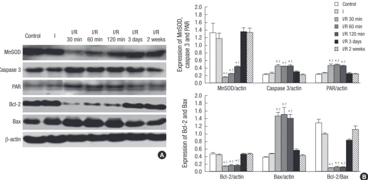
Western blot analysis of the expression of MnSOD, caspase 3, Bcl-2 and Bax and amounts of PAR showed time-dependent changes. Expression of MnSOD significantly decreased in the I/
R group at 30 min, 60 min, and 120 min after bladder emptying compare to the control group, and began to recover thereafter.
Expression of caspase 3 significantly increased in the I/R group at 30 min, 60 min, and 120 min after bladder emptying compared
to the control group, and significantly reduced to the control level at 3 days and 2 weeks after bladder emptying. When com- pared to the control group, the I/R group showed significant decrease in Bcl-2 expression and significant increase in the Bax expression at 30 min, 60 min, and 120 min after bladder empty- ing. As a result, the ratio of Bcl-2/Bax was significantly reduced.
The expression of Bcl-2 and Bax, and the ratio of Bcl-2/Bax re- covered to the control level at 3 days and 2 weeks after bladder emptying. On the other hand, the amounts of PAR (a marker of PARP activity) significantly increased at 30 min, 60 min, and 120 min after bladder emptying compared to the control group. Three days and 2 weeks after bladder emptying in the I/R group, there were no statistical difference in the expression of MnSOD, capase 3, Bcl-2 and Bax and the amounts of PAR between the I group and the control group (Fig. 2).
DISCUSSION
Apoptosis is an important element in maintaining the structur- al integrity and homeostasis of organisms. Although bladder apoptosis is frequently associated with AUR, the time course of the bladder apoptosis and the underlying mechanism of apop- tosis have not been well established. In the present study, we found that bladder apoptosis was significantly increased in ear- lier time points following bladder overdistension and subse- quent emptying. Another important finding was that bladder apoptosis induced by AUR after acute BOO and subsequent bladder emptying was associated with decreased MnSOD ex- pression, increased PARP activity and imbalance between pro- and anti-apoptotic factors.
AUR secondary to acute BOO is a clinical and experimental syndrome characterized by bladder dysfunction and apoptotic injury of the bladder (11, 16). Ischemia/reperfusion injury and
A B C
D
G
E F
Fig. 1. Detection of apoptosis: Representative micrographs show TUNEL positive cells as black-brown (magnification ×400), that are indicated by using arrows. Bar graphs show quantitative image analysis. The apoptotic index represents the percent of apoptotic cells within the total number of cells in a given area. *P<0.01 vs control group;
†P<0.01 vs ischemia only group. (A): Control group; (B): Overdistension without reperfusion (ischemia only group, I); (C)-(G): Overdistension with reperfusion for 30 min, 60 min, 120 min, 3 days and 2 weeks, respectively (ischemia/reperfusion group, I/R).
Mean apoptotic index (%)
Control I I/R 30 min I/R 60 min I/R 120 min I/R 3 days I/R 2 weeks 40
35 30 25 20 15 10 5 0
*,†
*,† *,†
subsequent oxidative stress are believed to play a central role in AUR induced bladder dysfunction, in addition to several other factors such as bladder overdistension and ischemia (8, 16).
Oxidative stress is often defined as imbalance between free radical productions and antioxidant capability. It has been dem- onstrated that nitric oxide and superoxide generated during the process of I/R, which can react to form the powerful nitrating agent peroxynitrite (17). Peroxynitrite as a potent oxidizing and nitrating agent has been shown to cause numerous injurious events, such as lipid peroxidation, reduction in antioxidant de- fenses and inactivation of enzymes that may adversely affect their function and signal transduction processes (18, 19). Impaired response to oxidative stress antioxidative proteins, include Mn- SOD, heat shock proteins (HSPs), and IGF-1 (20). MnSOD is the major antioxidative enzyme in the mitochondria, which cata- lyzes superoxide.
Numerous studies have shown that MnSOD was essential for life, because homozygous MnSOD knockout mice exhibited myocardial and liver injury and endothelial dysfunction (21-23), whereas overexpression of MnSOD reduced I/R-related dam- age (24, 25). Therefore a loss of MnSOD activity during I/R ap- pears to be related to tissue injury. On the other hand, MnSOD is a major mitochondrial antioxidant enzyme involved in the suppression of apoptosis, via regulation of the Bcl-2 family pro- teins such as Bcl-2 and Bax (26, 27). In our study, western blot analysis showed decreased MnSOD expression was parallel to bladder apoptosis. In addition, western blot analysis also showed decreased Bcl-2 expression and increased Bax and caspase-3 expression. These results indicate that downregulation of Mn-
SOD expression and imbalance of Bcl-2 family proteins expres- sion play a critical role in the development of bladder apoptosis following AUR and subsequent reperfusion.
Recent studies have demonstrated that increase ROS genera- tion following I/R injury increased PARP activity and caused damage in several organs, such as brain, kidney and the heart (12-14). Therefore we examined whether activity of PARP is in- creased in the bladder after AUR and subsequent reperfusion.
Western blot analysis showed a significant increase in activation of PARP in earlier time points after bladder overdistension and subsequent emptying. The present study indicates that activation of PARP may participate in the development of bladder apopto- sis following AUR and subsequent reperfusion. However, how PARP activation induces bladder apoptosis remains unclear.
Further studies are needed to elucidate the exact mechanism.
We did not investigate whether inhibition of PARP could at- tenuate bladder apoptosis following bladder I/R injury. This is the main limitations of our study. Since PARP inhibition counter- acts several organs damage caused by I/R injury, such as brain, kidney and the heart, further studies are required to determine whether inhibition of PARP could attenuate bladder apoptosis.
In conclusion, the results of the present study provide evi- dence that bladder apoptosis induced by AUR after acute BOO and subsequent bladder emptying is associated with decreased MnSOD expression, increased PARP activity and imbalance between pro- and anti-apoptotic factors. These results add to our understanding of the pathophysiology and molecular mech- anisms involved in bladder damage induced by I/R injury after acute BOO and subsequent bladder emptying.
*,†*,†
*,†
*,† *,† *,†
*,†
*,†
*,†*,†*,†
*,†
*,†
*,†*,†*,†
*,† *,†
MnSOD
Control I/R
30 min
I I/R
60 min I/R 120 min
I/R 3 days
I/R 2 weeks
Caspase 3 PAR Bcl-2 Bax
β-actin
A
B
Expression of Bcl-2 and Bax
Bcl-2/actin Bax/actin Bcl-2/Bax
2.0 1.8 1.6 1.4 1.2 1.0 0.8 0.6 0.4 0.2 0.0 Expression of MnSOD, caspase 3 and PAR
MnSOD/actin Caspase 3/actin PAR/actin 2.0
1.8 1.6 1.4 1.2 1.0 0.8 0.6 0.4 0.2 0.0
Control I I/R 30 min I/R 60 min I/R 120 min I/R 3 days I/R 2 weeks
Fig. 2. Western blot analysis: Representative western blot shows expression of MnSOD, caspase 3, PAR, Bcl-2 and Bax. Bar graphs depict quantitative analysis for each protein expression in the bladder. The expression of each protein was normalized to β-actin. *P<0.01 vs control group; †P<0.01 vs ischemia only group. I, overdistension without reperfusion (ischemia only group); I/R 30 min-2 weeks, overdistension with reperfusion for 30 min, 60 min, 120 min, 3 days and 2 weeks, respectively.
REFERENCES
1. Park JS, Lee CY, Lee W, Lee JZ. The effect of terazosin on the histological changes of rat bladder after partial outlet obstruction. J Korean Conti- nence Soc 2006; 10: 106-15.
2. Emberton M, Fitzpatrick JM. The Reten-World survey of the management of acute urinary retention: preliminary results. BJU Int 2008; 101 Suppl 3: 27-32.
3. Kaplan SA, Wein AJ, Staskin DR, Roehrborn CG, Steers WD. Urinary re- tention and post-void residual urine in men: separating truth from tra- dition. J Urol 2008; 180: 47-54.
4. Lee TM, Su SF, Suo WY, Lee CY, Chen MF, Lee YT, Tsai CH. Distension of urinary bladder induces exaggerated coronary constriction in smok- ers with early atherosclerosis. Am J Physiol Heart Circ Physiol 2000; 279:
H2838-45.
5. Yu HJ, Lin BR, Lee HS, Shun CT, Yang CC, Lai TY, Chien CT, Hsu SM.
Sympathetic vesicovascular reflex induced by acute urinary retention evokes proinflammatory and proapoptotic injury in rat liver. Am J Physi- ol Renal Physiol 2005; 288: F1005-14.
6. Chen SS, Chen WC, Hayakawa S, Li PC, Chien CT. Acute urinary blad- der distension triggers ICAM-1-mediated renal oxidative injury via the norepinephrine-renin-angiotensin II system in rats. J Formos Med Assoc 2009; 108: 627-35.
7. Shimizu S, Saito M, Kinoshita Y, Kazuyama E, Tamamura M, Satoh I, Satoh K. Acute urinary retention and subsequent catheterization cause lipid peroxidation and oxidative DNA damage in the bladder: preven- tive effect of edaravone, a free-radical scavenger. BJU Int 2009; 104: 713-7.
8. Bratslavsky G, Kogan BA, Matsumoto S, Aslan AR, Levin RM. Reperfu- sion injury of the rat bladder is worse than ischemia. J Urol 2003; 170:
2086-90.
9. Ohnishi N, Liu SP, Horan P, Levin RM. Effect of repetitive stimulation on the contractile response of rabbit urinary bladder subjected to in vitro hypoxia or in vitro ischemia followed by reoxygenation. Pharmacology 1998; 57: 139-47.
10. Juan YS, Chuang SM, Kogan BA, Mannikarottu A, Huang CH, Leggett RE, Schuler C, Levin RM. Effect of ischemia/reperfusion on bladder nerve and detrusor cell damage. Int Urol Nephrol 2009; 41: 513-21.
11. Hong SK, Son H, Kim SW, Oh SJ, Choi H. Effect of glycine on recovery of bladder smooth muscle contractility after acute urinary retention in rats.
BJU Int 2005; 96: 1403-8.
12. Liu R, Gao M, Yang ZH, Du GH. Pinocembrin protects rat brain against oxidation and apoptosis induced by ischemia-reperfusion both in vivo and in vitro. Brain Res 2008; 1216: 104-15.
13. Wu HH, Hsiao TY, Chien CT, Lai MK. Ischemic conditioning by short periods of reperfusion attenuates renal ischemia/reperfusion induced apoptosis and autophagy in the rat. J Biomed Sci 2009; 16: 19.
14. Sodhi RK, Singh M, Singh N, Jaggi AS. Protective effects of caspase-9 and
poly(ADP-ribose) polymerase inhibitors on ischemia-reperfusion-in- duced myocardial injury. Arch Pharm Res 2009; 32: 1037-43.
15. Yildirim A, Onol FF, Haklar G, Tarcan T. The role of free radicals and ni- tric oxide in the ischemia-reperfusion injury mediated by acute bladder outlet obstruction. Int Urol Nephrol 2008; 40: 71-7.
16. Yu HJ, Chien CT, Lai YJ, Lai MK, Chen CF, Levin RM, Hsu SM. Hypoxia preconditioning attenuates bladder overdistension-induced oxidative injury by up-regulation of Bcl-2 in the rat. J Physiol 2004; 554: 815-28.
17. Mladenka P, Simunek T, Hubl M, Hrdina R. The role of reactive oxygen and nitrogen species in cellular iron metabolism. Free Radic Res 2006;
40: 263-72.
18. Dohare P, Varma S, Ray M. Curcuma oil modulates the nitric oxide sys- tem response to cerebral ischemia/reperfusion injury. Nitric Oxide 2008;
19: 1-11.
19. Gandhi C, Zalawadia R, Balaraman R. Nebivolol reduces experimentally induced warm renal ischemia reperfusion injury in rats. Ren Fail 2008;
30: 921-30.
20. Lawler JM, Kwak HB, Kim JH, Suk MH. Exercise training inducibility of MnSOD protein expression and activity is retained while reducing pro- oxidant signaling in the heart of senescent rats. Am J Physiol Regul Integr Comp Physiol 2009; 296: R1496-502.
21. Van Remmen H, Qi W, Sabia M, Freeman G, Estlack L, Yang H, Mao Guo Z, Huang TT, Strong R, Lee S, Epstein CJ, Richardson A. Multiple deficiencies in antioxidant enzymes in mice result in a compound increase in sensitivity to oxidative stress. Free Radic Biol Med 2004; 36: 1625-34.
22. Ohashi M, Runge MS, Faraci FM, Heistad DD. MnSOD deficiency in- creases endothelial dysfunction in ApoE-deficient mice. Arterioscler Thromb Vasc Biol 2006; 26: 2331-6.
23. Kessova IG, Ho YS, Thung S, Cederbaum AI. Alcohol-induced liver inju- ry in mice lacking Cu, Zn-superoxide dismutase. Hepatology 2003; 38:
1136-45.
24. Saba H, Batinic-Haberle I, Munusamy S, Mitchell T, Lichti C, Megyesi J, MacMillan-Crow LA. Manganese porphyrin reduces renal injury and mitochondrial damage during ischemia/reperfusion. Free Radic Biol Med 2007; 42: 1571-8.
25. Jones SP, Hoffmeyer MR, Sharp BR, Ho YS, Lefer DJ. Role of intracellu- lar antioxidant enzymes after in vivo myocardial ischemia and reperfu- sion. Am J Physiol Heart Circ Physiol 2003; 284: H277-82.
26. Sharma DR, Sunkaria A, Bal A, Bhutia YD, Vijayaraghavan R, Flora SJ, Gill KD. Neurobehavioral impairments, generation of oxidative stress and release of pro-apoptotic factors after chronic exposure to sulphur mustard in mouse brain. Toxicol Appl Pharmacol 2009; 240: 208-18.
27. Xu Z, Lin S, Wu W, Tan H, Wang Z, Cheng C, Lu L, Zhang X. Ghrelin prevents doxorubicin-induced cardiotoxicity through TNF-alpha/NF- kappaB pathways and mitochondrial protective mechanisms. Toxicolo- gy 2008; 247: 133-8.