ISSN 1225-6552, eISSN 2287-7630 https://doi.org/10.7853/kjvs.2017.40.4.287
< Case Report >
Veterinary Service
Available online at http://kjves.org
*Corresponding author: Chul Park, Tel. +82-63-850-0962, Fax. +82-63-850-0972, E-mail. [email protected]
#
The first co-authors are equally contributed to this study.
Prognosis evaluation of a great dane dog with dilated cardiomyopathy
Yun-Hye Kim 1# , Jiung Na 1# , Kyung-Min So 2 , Chul Park 1 *
1
BK21 Plus Team and College of Veterinary Medicine, Chonbuk National University, Iksan 54596, Korea
2
Animal Nutrition and Physiology Team, National Institute of Animal Science, Rural Development Administration, Wanju 55365, Korea
(Received 16 November 2017; revised 23 December 2017; accepted 23 December 2017)
Abstract
A five-year-old, male, Great Dane weighing 107 kg was presented with anorexia, abdominal distension, and dyspnea for 5 days. Physical examination, blood works, radiography, electrocardiography (ECG), and echocardiography were performed. Based on severely low fractional shortening (FS) and marked four chamber enlargement in echocardiography, continuous atrial fibrillation and occasional ventricular premature complex (VPC) on ECG, the dog was diagnosed as dilated cardiomyopathy (DCM) con- current with congestive heart failure. Pleural effusion and ascites were modified transudate. In accord- ance with DCM scoring system recommended by European Society of Veterinary Cardiology (ESVC), DCM score was 13/15 in this case. Concentrations of cTnI and NT-pro-BNP were 1.0 ng/mL and 693 pmol/L, respectively. Since the former and the latter were remarkably high values, it was certain that the patient had grave prognosis. Intensive care was performed for the dog and the clinical signs as well as the radiographic abnormalities were resolved. However, when he presented serious dyspnea again at 25 days post therapy, the dog was dead. In case of canine DCM, the scoring system for the diagnosis and cardiac biomarkers including NT-pro-BNP and cTnI could be useful to advise owners on the status and prognosis of their dog with DCM.
Key words : Dilated cardiomyopathy, Dog, NT-pro-BNP, cTnI, Great dane
INTRODUCTION
Dilated cardiomyopathy (DCM) is a myocardial dis- ease characterized by atrial and ventricular enlargement, and myocardial (predominantly but not solely systolic) dysfunction with congestive heart failure (CHF) often developing at some stage without other cardiovascular diseases (Wynne and Braunwald, 2001). In the dog, spe- cific breed predispositions exist and the Doberman pinscher, Great Dane, Scottish deerhound, and Irish wolfhound appear to be over represented. For etiology of DCM, a lot of theories have been proposed, such as taurine (Kittleson et al, 1997) and/or carnitine (Keene et
al, 1991) deficiency, antibodies against beta-receptors (Limas et al, 1989) or the adenine nucleotide trans- locator (Schultheiss and Bolte, 1985), mutations of in the cardiac actin gene (Olson et al, 1998), toxic factors (Sisson et al, 2000) and possibly pregnancy (Sandusky and Cho, 1984). Recently DCM was reported the poten- tial for immunologic (Limas et al, 1989; Wynne and Braunwald, 2001) or viral etiology (Wynne and Braun- wald, 2001). Typical findings include increased left ven- tricular end-systolic dimension (ESD) and end-diastolic dimension (EDD), decreased fractional shortening (FS), and increased E-point septal separation (EPSS) which are the major criteria for the diagnosis of canine DCM.
Usually, M-mode and B-mode echocardiographic param-
eters are the most reliable methods of diagnosing DCM
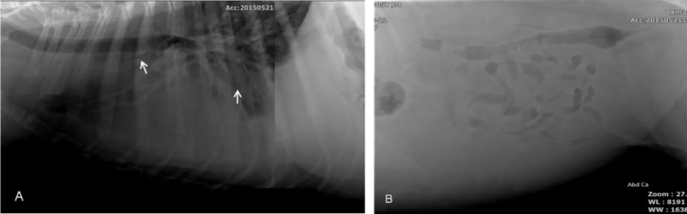
Fig. 1. Right lateral thoracic (A) and abdominal (B) radiographs in the patient. (A) The whole lung field is alveolar pattern and lungs lobe margins are disappeared (arrows). (B) Loss of intra-abdominal contrast and increased intra-abdominal fluid opacity were found due to abdominal fluid.
Great Danes also are known to be predisposed to DCM, and this is one of the most common breeds iden- tified in retrospective analyses (Borgarelli et al, 2006;
Martin et al, 2009; Monnet et al, 1995). Great Dane with DCM often also have atrial fibrillation, an elec- trical defect of the heart (Martin et al, 2009; Menaut et al, 2005; Meurs et al, 2001). And this dysrhythmias may cause or contribute to congestive heart failure oc- curring and reduce survival time in affected dogs.
Recent studies in United Kingdom suggest that Great Dane have shorter median survival times than other breeds (Martin et al, 2010).
In this case, we evaluated the prognosis of a Great Dane dog with DCM based on clinical, cardiac bio- markers, electrocardiography and echocardiographic study using scoring system recommended by European Society of Veterinary Cardiology (ESVC).
CASE
A five-year-old, male, Great Dane dog weighing 107 kg was presented to the Chonbuk National University Animal Medical Center with anorexia, abdominal dis- tension and dyspnea for 5 days.
On the physical examination, the dog had severe ta- chycardia, lethargy and approximately 5% dehydration.
Respiratory sound was determined to rapid, shallow breathing and expiratory labored breathing. Heart rate
breathings per minute, respectively. Heart sound was gallop rhythms with third heart sound and lung sound was crackled on auscultation.
The whole lung field on thoracic radiography was al- veolar pattern and lungs lobe margins were disappeared.
On abdominal radiography, loss of serosal detail was evident (Fig. 1).
No significant findings were detected except for mild- ly increased BUN value on complete blood count and serum chemistry profiles. The plasma concentration of NT-proBNP and cTnI were evaluated in a IDEXX labo- ratory (Seoungnam, Republic of Korea), resulting in 693 pmol/L and 1.0 ng/mL, respectively.
The result of ECG (MAC 5000, GE, Milwaukee, USA) showed no P waves, supraventricular QRS com- plexes and irregularly irregular R-R intervals with fast rhythms indicating clearly atrial fibrillation and inter- mittently ventricular premature complexes were recorded (Fig. 2).
Conventional echocardiographic images (HD 15, Phillips Ultrasound, Bothell, Washington, USA) were taken from the right parasternal and left apical views.
Based on B-mode echocardiographic examination, four chambers enlargement of the heart was detected (Fig.
3). The M-mode echocardiographic images taken from right parasternal short axis view at papillary muscles level showed marked dilation of left ventricle (LVIDd 72.6 mm, reference range of Great Dane: 47∼54.8 mm;
LVIDs 66.6 mm, reference range of Great Dane: 35.0∼
Fig. 2. Electrocardiography in the dog (paper speed: 50 mm/s, Sensi- tivity: 10 mm/mV). The strip rep- resents no P waves, supraventri- cular QRS complexes and irregu- larly irregular R-R intervals with fast rhythms, which indicate atrial fibrillation (A). Intermittently ven- tricular premature complexes (ar- rows) were shown (B).
Fig. 3. On the echocardiography from the right parasternal long axis 4 chambers view of the dog, all chambers of the heart are enlarged.
Fig. 4. M-mode echocardiography (right parasternal short axis view at the level of papillary muscles) shows marked dilation of left ven- tricle and severely decreased fractional shortening (8.2%).
38.2 mm) and severely decreased fractional shortening (8.2%, reference range of Great Dane: 22.3∼33.3%) (Fig. 4). From right parasternal short axis view at the level of mitral valve, Increased E-point septal separation (EPSS) was shown (4.15 mm, reference range of Great Dane: 0.3∼0.86 mm).
The dog was diagnosed as overt dilated cardiomyop- athy on the basis of history, physical examination, blood works, radiography, ECG, and echocardiography. In ac- cordance with DCM scoring system recommended by ESVC, the score was 13/15 in this case.
Thoracocentesis and abdominocentesis were performed to remove a large volume of effusion from the pleural and abdominal space to alleviate respiratory distress.
Pleural effusion and ascites were modified transudate.
Oxygen was supplied by an oxygen cage and fol-
lowed by 2 times of furosemide (Lasix
Ⓡ, Handok phar-
ma Co, Chungbuk, Korea, 4 mg/kg, IV) and nitro-
glycerine patch (Angiderm
Ⓡpatch, Samyangsa, Daejeon,
Korea, 0.5 mg/kg) administration to manage the pulmo-
nary edema. As respiratory distress was improved and
the dog could swallow food or water without difficulty,
furosemide (2 mg/kg, PO, q 12 hrs), pimobendan
(Vetmedin
Ⓡ, Boehringer Ingelheim, North Ryde NSW,
0.25 mg/kg, PO, q12 hrs), and enalapril (Enalapril,
CMG, Korea, 0.5 mg/kg, PO, q12 hr) were given for
management of the CHF caused by DCM. For manage-
ment of the atrial fibrillation, we administrated diltiazem
q12 hrs).
On 10th day after initial therapy, the clinical signs as well as the radiographic abnormalities were resolved.
On 25th day, however, he was presented again with difficulty breathing. Although intensive care was per- formed, the dog died eventually.
DISCUSSION
DCM is one of the most common heart diseases in large and giant breed dogs (Tidholm et al, 2001). Great Danes are included among the predisposed breeds (Martin et al, 2009). Dogs with DCM usually have a long occult phase in which dogs may have no detectable echocardiographic or electrical abnormalities (Calvert and Wall, 2001; Wess et al, 2010). An ESVC taskforce suggested guideline for the diagnosis of canine DCM, which helps for early as well as reliable identification.
They proposed a scoring system based on M-mode left ventricular dimensions, LV geometry, LV systolic func- tion, and suggested the dog get a total score of SIX or more should diagnose the DCM (Dukes-McEwan et al, 2003). In this case, the dog got score 13/15 by in- creased left ventricular dimensions and sphericity, de- creased FS, the presence of atrial fibrillation, increased EPSS, and LA enlargement. Especially, FS was severely low, 8.2%. A previous study reported that Great Dane with DCM had systolic dysfunction more than other giant breeds (Koch et al, 1996).
Prognosis for dogs with DCM varies ranging from days to years, depending on many variables (Tidholm, 2006), while, the survival time of Great Danes was re- ported shorter compared to other breed dogs (Martin et al, 2010). Survival time is known to be associated with cardiac-related clinical signs, echocardiographic or elec- trical abnormalities (Tidholm, 2006). Recently, cardiac biomarkers have been described as being advantageous in that blood sample-based biomarkers are easy to assay and less expensive than methods which require ex- pensive equipment in dogs with DCM (O'Sullivan et al, 2007; Oyama et al, 2007).
NT-pro-BNP is a neuroendocrine hormone that is syn-
stretch caused by pressure or volume overloads. High concentration of circulating NT-pro-BNP was reported in dogs and humans with many cardiovascular diseases including cardiomyopathies, myxomatous mitral valve degeneration, and congenital heart diseases (Fox et al, 2009; Oyama et al, 2008; Tarnow et al, 2009). It is considered as a useful indicator to distinguish cardiac from non-cardiac causes in dyspneic dogs. A recent study suggested that plasma concentration of NT-pro-BNP
>445 pmol/l distinguished dogs with cardiovascular disea- ses from healthy ones (Oyama et al, 2008). DCM might be detected early by measuring the concentration of cir- culating NT-pro-BNP. The level of NT-pro-BNP was re- ported to be more sensitive compared to 24-hour ambu- latory ECG recording (Holter) or ECG, and echocardio- graphy especially in the preclinical stage (Boswood et al, 2008; Oyama et al, 2013; Wess et al, 2011). In a study of 328 Doberman Pinschers with DCM, plasma NT-pro-BNP concentration was increased before echo- cardiographic morphologic or electrical changes. At the level of NT-pro-BNP >550 pmol/L, the dogs were like- ly to have echocardiographically detectable DCM, whereas less likely at the level of NT-pro-BNP <400 pmol/L (Wess et al, 2011).
Furthermore, According to previous studies, plasma NT-pro-BNP concentration was closely correlated with heart size and mortality in dogs with DCM. DCM dogs with higher concentration of NT-proBNP had shorter median survival time, indicating that a high level of NT-pro-BNP is a negative prognosis factor (Noszc- zyk-Nowak, 2011).
In this case, plasma NT-pro-BNP concentration of the dog was 693 pmol/L. According to previous studies, prognosis of the dog was predicted poor. The dog had significant clinical signs related CHF by DCM. Also, echocardiographic and electrical abnormalities were ob- vious, including chamber enlargement, systolic dysfunc- tion, and atrial fibrillation. Although we performed ap- propriate management for the dog, he died 25 days after initiating therapy.
The level of cTnI is very low or below the detectable
level in normal dogs. cTnI blood concentration increases
in the case of myocardial cellular damage leading to
cTnI release into the circulation (Sleeper et al, 2001).
Increased cTnI concentration has been determined in dogs with various cardiac diseases (Oyama and Sisson, 2004). Moreover, elevation of cTnI > 0.22 ng/mL could indicate any form of cardiomyopathy, with sensi- tivity of 79.5% and a specificity of 84.4% (Wess et al, 2010). A Correlation between concentration of cTnI and mortality in dog with DCM was reported. Higher plas- ma cTnI concentration was related to shorter survival time in human and animals (Oyama and Sisson, 2004).
In a study of 120 dogs with cardiac diseases, increased cTnI concentration (>1.0 ng/mL) indicated poor prognosis. Additionally, the same study suggested that cTnI concentration of a single sample was not sufficient as a prognostic factor. Rather than, persistently in- creased or reduced concentration of cTnI upon repeat sampling had more correlation with survival time (Fonfara et al, 2010). In this case, plasma cTnI concen- tration was highly increased, with the value of 1.0 ng/mL. The value could indicate poor prognosis as well as presence of cardiomyopathy.
CONCLUSION
In accordance with DCM scoring system recom- mended by European Society of Veterinary Cardiology, DCM score was 13/15 in this case. And cTnI and NT-pro-BNP, specific biomarker of heart failure, could be good indicators for evaluation of progression and prognosis in patient with DCM. Since the former and the latter were remarkably high values, it was certain that the patient had grave prognosis. So, in case of ca- nine DCM, scoring system for the diagnosis of DCM and hematological biomarkers including NT-pro-BNP and cTnI may be useful to advice to owners on the sta- tus and prognosis of their dog with DCM.
ACKNOWLEDGEMENTS
This work was carried out with the support of
“Cooperative Research Program for Agriculture Science
& Technology Development (Project title: Functional
feed material development and efficacy evaluation for detector dog’s ability improvement, Project No: PJ011989)”
Rural Development Administration, Republic of Korea.
REFERENCES
Borgarelli M, Santilli RA, Chiavegato D, D’Agnolo G, Zanatta R, Mannelli A, Tarducci A. 2006. Prognostic indicators for dogs with dilated cardiomyopathy. J Vet Intern Med 20:
104-110.
Boswood A, Dukes‐McEwan J, Loureiro J, James R, Martin M, Stafford‐Johnson M, Smith P, Little C, Attree S. 2008.
The diagnostic accuracy of different natriuretic peptides in the investigation of canine cardiac disease. J Small Anim Pract 49: 26-32.
Calvert CA, Pickus CW, Jacobs GJ, Brown J. 1997. Signalment, Survival, and Prognostic Factors in Doberman Pinschers With End‐Stage Cardiomyopathy. J Vet Intern Med 11:
323-326.
Calvert CA, Wall M. 2001. Results of ambulatory electro- cardiography in overtly healthy Doberman Pinschers with equivocal echocardiographic evidence of dilated cardiomyopathy. J Am Vet Med Assoc 219: 782-784.
Dukes-McEwan J, Borgarelli M, Tidholm A, Vollmar AC, Häggström J, The ESVC Taskforce for Canine Dilated Cardiomyopathy. 2003. Proposed guidelines for the diag- nosis of canine idiopathic dilated cardiomyopathy. J Vet Cardiol 5: 7-19.
Fonfara S, Loureiro J, Swift S, James R, Cripps P, Dukes- McEwan J. 2010. Cardiac troponin I as a marker for se- verity and prognosis of cardiac disease in dogs. Vet J 184: 334-339.
Fox PR, Oyama MA, Reynolds C, Rush JE, DeFrancesco TC, Keene BW, Atkins CE, MacDonald KA, Schober KE, Bonagura JD, Stepien RL, Kellihan HB, Nguyenba TP, Lehmkuhl LB, Lefbom BK, Moise NS, Hogan DF. 2009.
Utility of plasma N-terminal pro-brain natriuretic peptide (NT-proBNP) to distinguish between congestive heart failure and non-cardiac causes of acute dyspnea in cats.
J Vet Cardiol 11: S51-S61.
Keene B, Panciera D, Atkins C, Regitz V, Schmidt M, Shug A.
1991. Myocardial L-carnitine deficiency in a family of dogs with dilated cardiomyopathy. J Am Vet Med Assoc 198: 647-650.
Kittleson MD, Keene B, Pion PD, Loyer CG. 1997. Results of the Multicenter Spaniel Trial (MUST): Taurine‐and Carnitine‐Responsive Dilated Cardiomyopathy in American Cocker Spaniels With Decreased Plasma Taurine Con- centration. J Vet Intern Med 11: 204-211.
Koch J, Pedersen H, Jensen AL, Flagstad A. 1996. M‐mode Echocardiographic Diagnosis of Dilated Cardiomyopathy in Giant Breed Dogs. J Vet Intern Med Series A 43:
Limas C, Goldenberg I, Limas C. 1989. Autoantibodies against beta-adrenoceptors in human idiopathic dilated cardio- myopathy. Circ Res 64: 97.
Martin M, Stafford Johnson M, Celona B. 2009. Canine dilated cardiomyopathy: a retrospective study of signalment, presentation and clinical findings in 369 cases. J Small Anim Pract 50: 23-29.
Martin M, Stafford Johnson M, Strehlau G, King J. 2010. Canine dilated cardiomyopathy: a retrospective study of prog- nostic findings in 367 clinical cases. J Small Anim Pract 51: 428-436.
Menaut P, Bélanger MC, Beauchamp G, Ponzio NM, Moïse NS.
2005. Atrial fibrillation in dogs with and without struc- tural or functional cardiac disease: a retrospective study of 109 cases. J Vet Cardiol 7: 75-83.
Meurs KM, Miller MW, Wright NA. 2001. Clinical features of dilated cardiomyopathy in Great Danes and results of a pedigree analysis: 17 cases (1990-2000). J Am Vet Med Assoc 218: 729-732.
Monnet E, Orton EC, Salman M, Boon J. 1995. Idiopathic dilated cardiomyopathy in dogs: survival and prognostic indicators.
J Vet Intern Med 9: 12-17.
Noszczyk-Nowak A. 2011. NT-pro-BNP and troponin I as pre- dictors of mortality in dogs with heart failure. Pol J Vet Sci 14: 551-556.
O'Sullivan ML, O'Grady MR, Minors SL. 2007. Plasma Big Endothelin‐1, Atrial Natriuretic Peptide, Aldosterone, and Norepinephrine Concentrations in Normal Doberman Pinschers and Doberman Pinschers with Dilated Cardiomyopathy. J Vet Intern Med 21: 92-99.
Olson TM, Michels VV, Thibodeau SN, Tai Y-S, Keating MT.
1998. Actin mutations in dilated cardiomyopathy, a her- itable form of heart failure. Science 280: 750-752.
Oyama MA, Boswood A, Connolly DJ, Ettinger SJ, Fox PR, Gordon SG,Rush JE, Sisson DD, Stepien RL, Wess G, Zannad F. 2013. Clinical usefulness of an assay for measurement of circulating N-terminal pro-B-type natriu- retic peptide concentration in dogs and cats with heart disease. J Am Vet Med Assoc 243: 71-82.
Oyama MA, Fox PR, Rush JE, Rozanski EA, Lesser M. 2008.
Clinical utility of serum N-terminal pro-B-type natriu- retic peptide concentration for identifying cardiac disease in dogs and assessing disease severity. J Am Vet Med Assoc 232: 1496-1503.
for occult cardiomyopathy in dogs by measurement of plasma atrial natriuretic peptide, B-type natriuretic pep- tide, and cardiac troponin-I concentrations. Am J Vet Res 68: 42-47.
Oyama MA, Sisson DD. 2004. Cardiac Troponin‐I Concentration in Dogs with Cardiac Disease. J Vet Intern Med 18:
831-839
Sandusky GE, Cho D. 1984. Congestive cardiomyopathy in a dog associated with pregnancy. Cornell Vet 74: 60-64.
Schultheiss H, Bolte H. 1985. Immunological analysis of au- to-antibodies against the adenine nucleotide translocator in dilated cardiomyopathy. J Mol Cell Cardiol 17: 603- 617.
Sisson DD, Thomas WP, Keene BW. Primary myocardial disease in the dog. In: Ettinger SJ, Feldman EC, editors.
Textbook of veterinary internal medicine 5th edition Philadelphia. WB Saunders 2000: 874-895.
Sleeper, MM, Clifford, CA, Laster, LL. 2001. Cardiac troponin I in the normal dog and cat. J Vet Intern Med 15: 501- 503
Tarnow I, Olsen LH, Kvart C, Hoglund K, Moesgaard SG, Kamstrup TS,Pedersen HD, Häggström J. 2009.
Predictive value of natriuretic peptides in dogs with mi- tral valve disease. Vet J 180: 195-201.
Tidholm A, Häggström J, Borgarelli M, Tarducci A. 2001.
Canine idiopathic dilated cardiomyopathy. Part I: aetiol- ogy, clinical characteristics, epidemiology and pathology.
Vet J 162: 92-107.
Tidholm A. 2006. Survival in dogs with dilated cardiomyopathy and congestive heart failure treated with digoxin, furo- semide and propranolol: A retrospective study of 62 dogs. J Vet Cardiol 8: 41-47.
Wess G, Butz V, Mahling M, Hartmann K. 2011. Evaluation of N-terminal pro-B-type natriuretic peptide as a diagnostic marker of various stages of cardiomyopathy in Doberman Pinschers. Am J Vet Res 72: 642-649.
Wess G, Schulze A, Butz V, Simak J, Killich M, Keller L, Maeurer J, Hartmann K. 2010. Prevalence of dilated car- diomyopathy in Doberman Pinschers in various age groups. J Vet Intern Med 24: 533-538.
Wynne J, Braunwald E. The cardiomyopathies and myocardities.
In: Heart disease: a textbook of cardiovascular medicine.
6th ed. Philadelphia (Pa): WB Saunders. 2001: 1751- 1806.