서 론
유방염은 젖소에서 가장 흔하게 발생하는 질병 중 하나로 우 유 생산량의 감소, 유질 저하, 치료 비용 및 환축 도태 등의 이유 로 낙농 산업에 큰 경제적 손실을 초래하는 질병이다(Zhao과 Lacasse 2008; Jeong 등, 2017). 유방염으로 인한 연간 경제
적 손실은 전세계적으로 350억 달러, 한국은 108억 원, 미국의 경우 약 20억 달러에 이르는 것으로 보고되고 있다(Leitner 등, 2003; Park 등, 2011). 유방염은 발생 양상에 의해서 전염성과 환경성으로 분류되며, 전염성 유방염 원인균으로는 Staphylo- coccus aureus, Streptococcus dysgalactia, Streptococcus agalactia 등이 있고 환경성 유방염 원인균으로 Escherichia
KJVS
Korean Journal of Veterinary Service
봉독의 젖소 유방염 유래 그람 양성 및 음성 세균별 항균효과 분석
정숙한
1
ㆍ오상익1
ㆍ이한규1
ㆍ정영훈1
ㆍ허태영1
ㆍ한상미2
ㆍ백귀정3
ㆍ조아라1
*농촌진흥청 국립축산과학원 가축질병방역과1, 농촌진흥청 국립농업과학원 잠사양봉소재과2, 전라북도 동물위생시험소3
Antibacterial effect of bee venom against Gram-positive and negative bacteria isolated from mastitis in dairy cattle
Sukhan Jung
1, Sang-Ik Oh
1, Han-Gyu Lee
1, Young-Hun Jung
1, Tai-Young Hur
1, Sangmi Han
2, Kui-Jeong Baek
3, Ara Cho
1*
1Division of Animal Diseases and Health, National Institute of Animal Science, Rural Development Administration, Wanju 55365, Korea
2Department of Agricultural Biology, National Institute of Agricultural Science, Rural Development Administration, Wanju 55365, Korea
3Jeollabuk-do Institute of Livestock & Veterinary Research, Jangsu 55632, Korea
Received September 19, 2021 Revised September 29, 2021 Accepted September 29, 2021
Corresponding author:
Ara Cho
E-mail: [email protected] https://orcid.org/0000-0001-5309-7721
Mastitis is an inflammatory condition of the mammary gland, most often caused by bacterial infec- tions, resulting in significant economic losses to the dairy industry. Antimicrobial resistance has been of great concern because of the extensive clinical use of antibiotics. For this reason, the development of new compounds as an alternative treatment to bovine mastitis is needed. Bee venom has been widely used as an oriental treatment for several inflammatory diseases and bacterial infections. The aim of the present study was to evaluate the antimicrobial activity of bee venom on bacteria isolated from bovine mastitis. A total of 107 isolates from bovine mastitic milk samples collected in 2019 and 2020 in Jeonbuk province. All bacterial isolates were tested for susceptibility to bee venom of the honey bee (Apis mellifera). In order to obtain comprehensive antibacterial activities of the bee venom, we measured the minimal inhibitory concentration (MIC) of the bee venom against bacte- rial strains. Bee venom showed significant inhibition of bacterial growth of Gram-negative bacteria Citrobacter spp., Escherchia coli, Klebsiella spp., Pseudomonas spp., Serratia spp. and Raoultella with MIC values of 96, 81, 72, 230, and 85 μg/mL, respectively, and Gram-positive bacterial Enterococcus spp., Staphylococcus spp. and Streptococcus spp. with MIC values of 29, 21 and 16 μg/mL, respec- tively. The results indicated that the MIC values were different depending on the bacterial strains, and those of Gram-positive bacteria were lower than those of Gram-negative bacteria for bee venom.
These findings suggested that bee venom could be an effective antimicrobial treatment for bovine mastitis; however, further research is necessary to evaluate the mechanism underlying the antimicro- bial action, its effectiveness/safety in vivo and effective application for therapeutic use.
Key Words:
Mastitis, Bee venom, Antimicrobial effect, Minimal inhibitory concentration (MIC), Dairy cattleCopyright ⓒ The Korean Society of Veterinary Service.
This is an Open Access article distributed under the terms of the Creative Commons Attribution Non-Commercial License (http://creativecommons.org/licenses/
https://doi.org/10.7853/kjvs.2021.44.3.169 pISSN: 1225-6552 eISSN: 2287-7630
Korean J Vet Serv 2021;44(3):169-174
SHORT COMMUNICATION
coli와 Streptococcus uberis 등이 있다(Lee 등, 2007; Kim 등, 2017). 전염성 유방염 원인균은 유방의 유선에 감염되어 착 유 과정을 통해 우군에 전파되어 준임상형 및 만성 유방염을 일 으키는 것으로 알려져 있다. 한편 환경성 유방염 원인균은 토양, 분변 및 생식기 분비물, 깔짚, 유방 및 유두 그리고 착유기 세척 에 사용되는 오염된 물 등의 주변 환경을 통해 유선 조직에 침 입하여 기회 감염을 일으켜 일시적으로 임상형 유방염을 일으 키는 것으로 보고되고 있다(Watts 1988; Lee 등, 2007). 최근 국내 젖소 유방염에서 분리되는 원인균의 균종별 분리 빈도는 Staphylococcus 종이 50% 이상을 차지하며, Streptococcus 종이 6.8%, Enterococcus 종이 4.8%, Escherichia coli 종이 4.5% 차지하는 것으로 보고되어 있다(Kim 등, 2017). 대표적인 유방염 유발 세균인 Staphylococcus aureus는 숙주 탐식 세포 내 기생함으로 항생제에 의해 잘 박멸되지 않으며, 재발 확률이 높고 착유 시 한 개체에서 또 다른 개체로 전염이 잘되는 특징이 있다. 그리고 Escherichia coli의 경우 유관을 통해 감염되어 유선 세포를 파괴하거나 내독소를 분비하여 염증을 일으킨다고 보고 되어있다(Eberhart 등, 1979; Erskine 등. 1991; Kim 등, 2011).
지난 수십년 동안, 항생제는 젖소 유방염의 예방과 치료를 위 해 사용되어 왔다. 항생제의 사용이 가축의 생산성 증진에 크게 기여하였으나 이에 따른 항생제 내성균의 출현으로 인해 치료율 이 저하되는 부작용이 나타나고 있다(Han 등, 2014). 뿐만 아니 라 항생제 내성균으로 인한 감염을 치료하기 위하여 과량의 항 생제 사용, 치료 기간이 장기화 됨에 따라 유제품 내 항생제 잔 류 등의 문제로 사람의 건강에 영향을 미치게 되어 사회적인 관 심과 그 중요성이 점차 커지고 있다(Kim 등, 1997; 남, 2010;
Han 등, 2015). 또한 유방염은 발병 메커니즘 및 발생요인이 매 우 복잡하고 그 원인균이 다양하여 특정 몇 종의 항생제로는 치 료에 많은 어려움이 있어 안전하고 효과적인 젖소 유방염 치료 제 개발이 절실히 요구되는 실정이다(Kang 등, 2001; Han 등, 2007). 이러한 문제를 해결하기 위해 천연물질을 이용한 유방염 치료제 연구가 활발히 진행되고 있다(Han 등, 2014).
봉독은 벌의 독낭세포에서 분비되는 액체로 독낭에 저장되 었다가 외부의 자극이나 공격으로부터 종족을 보호하기 위한 방어 물질이다(Park 등, 2016). 봉독은 phospholipase A2와 hyaluronidase 등의 효소 및 melittin, apamin, adolapin, mast cell degranulating peptide와 같은 peptide 성분과 그 외 nonpeptide 성분 등 40가지의 구성성분으로 이루어져 있으 며 그 중 50% 이상을 차지하는 melittin은 세포막을 파괴하고 세포막 수용체에 결합하여 용혈 작용, 항염증 작용 및 항균 작용
등의 역할을 한다고 알려져 있다(Habermann과 Reiz, 1965;
Rudenko과 Nipot, 1996; Lee 등, 2015). 최근 꿀벌에서 미세 전류를 이용한 봉독 채집 장치를 이용하여 봉독을 정제하는 방 법을 통하여 대량 생산되어 사람의 치료 및 화장품으로 사용되 고 있다(Kim 등 2002; Lee 등 2015).
축산 분야에 있어 봉독은 가축의 성장촉진, 면역기능 증가 및 각종 질병 치료에 유효하다고 알려져 있다(Han 등, 2009; Oh 등, 2011). 젖소에서 봉독은 분만 소요시간 단축과 후산 정체율 감소 등 분만 효율을 개선하고 젖소 유방염의 경우 봉독 투여로 체세포수가 크게 감소하였으며 유방염에 걸린 젖소로부터 채취 한 원유에서 분리한 균에 대한 항균력이 우수한 것으로 보고된 바 있다(Han 등 2007). 젖소 유방염 질환 치료에는 10 mg/두 이하 농도의 봉독이 주사제로 이용되고 있으며(Han 등, 2009).
젖소에 봉독을 투여한 후 우유에서 봉독 성분이 검출되지 않아 천연물질인 봉독은 축산물 내에 잔류하지 않는 것으로 확인되었 다(Han 등 2015). 또한 가축에 사용되는 봉독의 용량은 극소량 으로 증류수와 생리식염수로 희석할 경우 균질성의 정확성과 정 밀성이 모두 확보되었으며 실온에서 7일간 보관하더라도 봉독 성분의 안정성이 유지된다고 보고하였다(Han 등 2016).
본 연구에서는 젖소 유방염 주요 원인균 9종 Staphylococ- cus spp., Streptococcus spp., Enterococcus spp., Citro- bacter spp., Escherichia spp., Klebsiella spp., Proteus spp., Serratia spp., Raoultella spp. 등에 대하여 봉독의 농도 에 따른 항균성을 조사하고 향후 젖소 유방염 치료를 위한 기초 자료를 제공하고자 한다.
재료 및 방법
시험 물질인 봉독은 전기 채집 장치를 사용하여 서양종 꿀벌 (Apis mellifera)로부터 채취하였다. 이후 국립농업과학원 잠사 양봉소재과에서 봉독 정제를 마친 동결건조 상태의 봉독을 받아 실험에 사용하였다. 봉독은 멸균 증류수로 희석한 후 syringe filter(0.45μm)를 이용하여 무균 여과하였다.
본 연구에서 사용한 균주는 2019년부터 2020년까지 전북 젖소목장에서 유방염으로 진단된 원유 시료로부터 분리, 동정 한 균주를 전라북도 동물위생시험소에서 분양 받아 사용하였 으며, 그람 양성균으로는 Staphylococcus spp., Enterococ- cus spp., Streptococcus spp. 등 3종 52개 균주와 그람 음 성균 Citrobacter spp,, Escherichia spp., Klebsiella spp., Proteus spp., Serratia spp., Raoultella spp. 등 6종 55개 균 주 총 9종 107개 균주를 Blood agar (Kisan Biotech, Seoul,
정숙한ㆍ오상익ㆍ이한규ㆍ정영훈ㆍ허태영ㆍ한상미ㆍ백귀정ㆍ조아라
KJVS
Korea) 배지에서 37℃, 호기조건으로 배양하였다.
각 균주에 대한 봉독의 항균 효과를 파악하기 위하여 액체 배 지 희석법을 사용하여 최저 성장 억제 농도를 구하였다. 측정은 96-well plate에 균 배양액 Muller-Hinton broth 90 μL와 농 도 별로 희석한 봉독 시료 10 μL를 차례대로 첨가하여 최종 농 도를 0, 0.5, 1, 2, 4, 8, 16, 32, 64, 128, 256, 512 μg/mL이 되도록 설정하였다. 37℃ 18시간 배양 후 육안으로 균의 성장을 확인하여 증식이 일어나지 않은 농도를 최소발육저지농도로 나 타내었다. 같은 종의 균주(strain) 중 최소발육저지농도에 차이 가 있는 경우 최소값(Min)과 최대값(Max)를 확인하였다.
유방염 균주 최소발육저지농도 측정 결과에 대한 통계적 처 리는 RStudio (RStudio Version 2.3.5033, USA)의 one-way analysis of variance (ANOVA), Duncan’s multiple range test를 이용하여 처리군 간의 유의성을 검정하였으며(P<0.05), 그 결과는 평균+표준편차(mean±STD)로 표시하였다.
결과 및 고찰
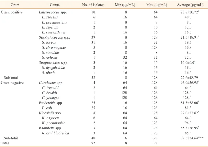
전북 젖소 농장에서 수집한 유방염 분리균에서 최소발육저지 농도를 그람 음성균과 그람 양성균으로 나누어 Table 1, 2에 각 각 나타내었다.
봉독의 최소 발육 저지 농도는 그람 양성균에서 평균 22.6 μg/
mL, 그람 음성균에서 평균 97.8 μg/mL로 그람 음성균에서 더 높은 MIC 수치를 확인하였다. 그람 양성균에서 균주별 비교 결 과 Enterococcus spp.와 Staphylococcus spp., Strepto- coccus spp. 균에서 봉독의 최소 발육 저지 농도는 각각 29 μg/
mL와, 21 μg/mL, 16 μg/mL이었으며, 그람 음성균에서 균주 별 비교 결과 Citrobacter spp., Escherichia, Klebsiella spp., Serratia spp,, Raoultella 균에서 봉독의 최소 발육 저지 농도 는 96 μg/mL, 81 μg/mL, 72 μg/mL, 230 μg/mL, 85 μg/mL 로 그람 음성균과 비교하였을 때 그람 양성균이 더 낮은 농도에
Table 1. Minimum inhibitory concentration (MIC) of the Bee Venom on bacterial mastitis pathogens isolates from dairy cows in Korea
Gram Genus No. of isolates Min (μg/mL) Max (μg/mL) Average (μg/mL)Gram positive Enterococcus spp. 10 8 64 28.8±20.72a
E. faecalis 6 16 64 40.0
E. pseudoavium 1 8 8 8.0
E. faecium 2 8 16 12.0
E. casseliflavus 1 16 16 16.0
Staphylococcus spp. 39 8 128 21.5±18.91a
S. aureus 31 16 32 19.6
S. chromogenes 5 8 128 36.8
S. simulans 2 8 8 8.0
S. xylosus 1 32 32 32.0
Streptococcus spp. 3 16 16 16.0±0.0a
S. dysgalactiae 2 16 16 16.0
S. uberis 1 16 16 16.0
Sub-total 52 8 128 22.6±18.79
Gram negative Citrobacter spp. 4 64 128 96.0±36.95b
C. freundii 2 64 64 64.0
C. braakii 1 128 128 128.0
C. youngae 1 128 128 128.0
Escherchia spp. 25 16 128 81.3±38.06b
E. coli 25 16 128 81.3
Klebsiella spp. 8 64 128 72.0±22.62b
K. oxytoca 6 64 64 64.0
K. pneumoniae 2 64 128 96.0
Raoultella spp. 3 64 128 85.3±36.95b
R. ornithinolytica 3 64 128 85.3
Sub-total 40 16 128 97.8±34.64***
Total 92 8 128
Average data are presented as mean±STD per group. Differences between multiple groups were compared using one-way analysis of variance (ANOVA) while individual comparisons were obtained using a Duncan’s Multiple Range Test (Different letters indicate signi- ficant differences, P<0.05). Differences between Gram positive and negative group was compared using student’s t-test (***P<0.001).
봉독의 젖소 유방염 유래 그람 양성 및 음성 세균별 항균효과 분석
KJVS
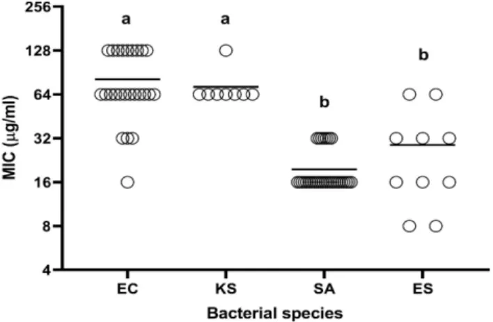
서 균의 성장이 억제되어 봉독의 더 높은 항균 효과를 확인 할 수 있었다. 그람 양성 및 음성 중 각 2종 세균의 MIC 값을 산점 도(Scatter dot plot)으로 나타낸 Fig. 1에서 그 차이를 추가적 으로 확인할 수 있다. 이러한 결과는 기존 연구에서 봉독이 그람 음성균에서 보다는 그람 양성균에서의 항균 효과가 뛰어나다는 보고 결과와 일치한다(Han 등, 2007).
또한 Proteus vulgaris는 4.8% 빈도로 유방염을 유발하는 원 인균 중 하나로 보고 되어있다(Kim 등, 2017). 유방염 젖소 환 축으로부터 분리된 Proteus spp. 종에서 봉독의 항균효과를 검 증하는 연구는 아직 보고된 바가 없었으나 본 연구에서는 그람 음성균인 Proteus spp. 균주가 본 실험에서 적용된 최고 농도 인 512 μg/mL에서도 균이 자라 봉독의 항균 효과가 없음을 확 인하였다. Steiner 등(1981)은 봉독의 항균 작용은 그람 양성균 과 일부 그람 음성균에 작용한다고 보고하였다.
봉독 중 melittin 성분은 세균의 세포막 지질에 친화력이 높 아 세포막의 구멍을 통하여 전위하여 세포막을 파괴하면서 항 균작용을 나타낸다고 보고되어 있다(Lee 등, 2015). 그람 음성 균보다 그람 양성균에서 항균 활성이 높게 나타난 것은 Naka- mura 등(1991)도 보고한 바와 같이 그람 양성균의 세포벽은 peptidoglycan이 표면에 노출되어 항균 활성 물질의 공격을 받기 쉽지만, 그람 음성균은 lipopolysaccharide를 주성분으 로 하는 외막이 peptidoglycan을 보호하는 작용을 하기 때문인 것으로 사료된다. Han 등(2007)의 보고에서도 황색포도상구균 을 포함한 그람 양성균에서 대장균 등의 그람 음성균에 비해 유 방염 치료효과가 크게 나타나는 것을 확인한 바 있다.
또한 봉독은 기존 항생제인 penicillin과 비교하였을 때 약 1,000~1,200배나 강력하며 penicillin에 저항성이 있는 포도 상구균의 변종균 Staphylococcus aureus 균에도 효과가 있 다고 보고되어 있다(Kim 등, 2006). 이 외 다재 내성을 보이는 Acinetobacter baumannii에 봉독을 투여하였을 때 항생제 내
성과 관련 없이 항균효과를 보이는 것을 확인하였다.
결 론
유방염으로 진단된 원유 시료로부터 분리 동정한 Staphy- lococcus spp., Enterococcus spp., Streptococcus spp., Citrobacter spp., Escherichia, Klebsiella spp., Proteus spp., Serratia spp., Raoultella 총 9종 107개 균주에 대한 봉 독의 항균효과를 검정한 결과 Proteus 균주를 제외한 모든 균주 에서 항균력을 나타내었으며 최소 발육 저지 농도는 그람 양성 균에서 평균 22.6 μg/mL, 그람 음성균에서 평균 97.8 μg/mL 로 그람 음성균과 비교하였을 때 그람 양성균이 더 낮은 농도에 서 균의 성장이 억제되어 봉독의 더 높은 항균 효과를 확인할 수 있었다.
본 연구 결과를 고려하여 항생제의 오남용이나 부작용으로 인 해 항생제 사용이 어려운 축산 농가에서 세균의 특성에 따른 봉 독의 항균 효과에 대한 기초 자료를 제공하여 추후 젖소 유방염 에 적용 가능성을 확인하였으며 안전성 측면에서의 연구와 유즙 으로부터 체세포수 검사를 통하여 유방염에 대한 치료효과를 확 인하는 연구가 추가적으로 필요할 것으로 사료된다.
감사의 글
본 논문은 농촌진흥청 연구사업(세부과제명: 가축 유래 세
Table 2. Minimum inhibitory concentration (MIC) of the Bee
Venom on Proteus spp. and Serratia spp. isolates from dairy cows in Korea
Gram Number Min
(μg/mL) Max (μg/mL) Gram negative
Proteus spp. 5 >512 -
P. vulgaris 4 >512 - P. mirabilis 1 >512 - Serratia spp. 10 128 >512 S. liquefaciens 7 128 >512 S. marcescens 3 512 >512
Fig. 1. Scatter dot plot with individual MICs of bee venom in
bacterial species. Line represent mean of all BV against the test strains. EC, E. coli (n=25); KS, Klebsiella spp. (n =8); SA, Staphylococcus aureus (n=31); ES, Enterococcus spp. (n=10).Differences between multiple groups were compared using one- way analysis of variance (ANOVA) while individual comparisons were obtained using a Duncan’s Multiple Range Test (Different letters indicate significant differences, P<0.05.
정숙한ㆍ오상익ㆍ이한규ㆍ정영훈ㆍ허태영ㆍ한상미ㆍ백귀정ㆍ조아라
KJVS
포주에서 봉독의 안전성 및 작용 효과 분석, 세부과제번호:
PJ014298022021)의 지원 및 2021년도 농촌진흥청(국립축산 과학원) 전문연구원 과정 지원사업에 의해 이루어진 것임.
CONFLICT OF INTEREST
No potential conflict of interest relevant to this article was reported.
ORCID
Sukhan Jung, https://orcid.org/0000-0002-5821-9027 Sang-Ik Oh, https://orcid.org/0000-0003-0877-9170 Han-Gyu Lee, https://orcid.org/0000-0002-3531-1971 Young-Hun Jung, https://orcid.org/0000-0002-8094-0304 Tai-Young Hur, https://orcid.org/0000-0003-3129-2942 Sangmi Han, https://orcid.org/0000-0002-6840-4509 Kui-Jeong Baek, https://orcid.org/0000-0002-1915-9439 Ara Cho, https://orcid.org/0000-0001-5309-7721
REFERENCES
남향미. 2010. 젖소 유방염에 대한 항생제 사용 지침. 대한수의 사회지. 46(7): 638-644.
Eberhart RJ, Natzke RP, Newbould FSH. 1979. Coliform mastitis-a review. J Dairy Sci 62(1): 1-22.
Erskine RJ, Tyler TW, Riddler MG. 1991. Theory use and realities of efficacy and food safety of antimicro- bial treatment of acute coliform mastitis. J Am Vet Med Assoc 198(6): 980-984.
Habermann, E. and Reiz, K. G. 1965. On the biochem- istry of bee venom peptides, melittin and apamine.
Biochemistry 343(2): 192-203.
Han SM, Hong IP, Woo SO, Kim SG, Jang HR. 2015.
Analysis of bee venom residues in milks of dairy cattle using UHPLC with newly developed pre- processing method. Korean J Vet Serv 38(1): 25-30.
Han SM, Kim JM, Yeo JH, Hong IP, Woo SO, Lee KG, Kweon HY. 2014. Origin and effective ingredient standards of honeybee venom as natural antibiotic ingredients. Korean J Vet Serv 37(2): 123-129.
Han SM, Kim SG. Hong IP, Woo SO, Jang HR, Lee KW.
2016. Antibacterial effects of purified bee venom against some pathogenic bacteria isolated from dead chickens. Korean J Vet Serv 39(3): 159-166.
Han SM, Lee KG, Yeo JH, Hwang SJ, Jang CH, Che- noweth PJ, Pak SC. 2009. Effects of bee venom treatment on growth performance of young pigs.
Am J Chin Med 37(2): 253-260.
Han SM, Lee KG, Yeo JH, Kweon HY, Kim BS, Kim JM, Baek HJ, Kim ST. 2007. Antibacterial activity of the honey bee venom aganist bacterial mastitis patho- gens infecting dairy cows. Int J Indust Entomol 14(2): 137-142.
Han SM, Lee KG, Yeo JH, Kweon HY, Woo SO, Oh BY, Lee YG, Kim BS, Baek HJ, Kim ST. 2007. Therapeu- tic effects of honeybee (Apis Mellifera L.) venom injection on bovine mastitis. Korean J Vet Serv 30(1): 115-123.
Han SM, Woo SO, Kim SG, Jang HR, Lee KW. 2018. An- tibacterial effects of purified bee venom against Bacillus cereus, Streptococcus agalactiae, and Pseudomonas aeruginosa. Journal of Apiculture 33(1): 9-16.
Jeong CH, Cheng WN, Bae H, Lee KW, Han SM, Petriel- lo M C, Lee HG, Seo HG, Han SG. 2017. Bee venom decreases LPS-induced inflammatory responses in bovine mammary epithelial cells. J Microbiol Bio- technol 27(10): 1827-1836.
Kang HJ, Kim IC, Kim JH, Son WG, Lee DS. 2001 Iden- tification and antimicrobial susceptibility of mi- croorganisms isolated from bovine mastitic milk.
Korean J Vet Serv 41(4): 511-521.
Kim DI, Kim EK, Seong WJ, Ro YH, Ko DS, Kim NH, Kim JH, Kwon HJ. 2017. Identification of microbi- ome with 16S rRNA gene pyrosequencing and an- timicrobial effect of egg white in bovine mastitis.
Korean J Vet Res 57(2): 117-126.
Kim J, Ochoa, MT, Krutzik, SR, Takeuchi, O, Uematsu S, Legaspi AJ, Brightbill HD, Holland D, Cunliffe WJ, Akira S, Sieling PA, Godowski PJ, Modlin RL.
2002. Activation of toll-like receptor 2 in acne trig-
봉독의 젖소 유방염 유래 그람 양성 및 음성 세균별 항균효과 분석
KJVS
gers inflammatory cytokine responses. J. Immunol 169(3):1535-41.
Kim JH, Kim BM, Ham JS, Oh MH. 2017. Detection and Characteristics of Coagulase-Negative Staphylo- coccus sp. isolated from Dairy Cattle Milk. J Milk Sci Biotechnol 35(3): 162-168.
Kim SE, Hah DY, Jang EH, Kwon HN, Jo SS, Kwon YT, Park DY, Lee KC, Kim JS. 2011. Survey of mastitis management and incidence of mastitis in high so- matic cell count of bulk milk at dairy farms in the Gyeongnam. Korean J Vet Serv 34(4): 379-388.
Kim ST, Hwang JY, Sung MS, Je SY, Bae DR, Han SM, Lee SH. 2006. The minimum inhibitory concentra- tion (MIC) of bee venom against bacteria isolated from pigs and chickens. Korean J Vet Serv 29(1):19- 26.
Kim ST, Kim S, Kim SY, Son JK. 1997. Comparison of fatty acid composition of Staphylococcus sp. iso- lated from bovine mastitis milk. Korean J Vet Serv 20(1): 37-45.
Lee DG, Shin HH. 2008. Pharmacokinetics and phar- macodynamics of antibiotics: General concepts and recent advances. Infect Chemother 40(3): 140- 147.
Lee ES, Kang HM, Chung CI, Moon JS. 2007. Antimicro- bial susceptibility and prevalence of gram-negative bacteria isolated from bovine mastitis. Korean J Vet Res 47(4): 67-75.
Lee G, Kang HM, Chung CI, Moon JS. 2007. Antimicro- bial susceptibility and genetic characteristics of Streptococcus uberis isolated from bovine mastitis milk. Korean J Vet Res 47(1): 33-41.
Lee SJ, Kim KH, Lee WR, Kim JY, An HJ, Park KK. 2015.
Anti-bacterial and anti-inflammatory effect of melittin on propionibacterium acnes-induced in-
flammatory skin disease in vivo. Journal of Apicul- ture 30(2): 95-101.
Leitner Gabriel, Lubashevsky, Evgenia, Glickman Anita, Winkler Marta, Saran Arthur, Trainin Zeev. 2003.
Development of a Staphylococcus aureus vaccine against mastitis in dairy cows. Vet Immunol. Im- munopathol 93(1-2): 31-38.
Nakamura S, Kato AM, Kobayashi K. 1991. New anti- microbial characteristics of lysozyme-dextran con- jugate. J Agric Food Chem 39:647-650.
Oh BY, Han SM, Oh YI, Kim ST. 2011. Effects of the blood chemistry of honeybee (Apis mellifera L.) venom on the Hanwoo calves. Korean J Vet Serv 34(1): 87-93.
Park JK, Shen CZ, Kim CG, Kim IK. 2016. The stability comparison of purified bee venom and bee venom melittin in aqueous solution. Anal. Sci. Technol 29(4): 194-201.
Park YJ, Yang DK, Han HY. 2011. Efficacy of a herd specific mastitis vaccine against Staphylococcus aureus in dairy cows. Journal of preventive veteri- nary medicine 35(2): 81-89.
Rudenko SV, Nipot EE. 1996. Modulation of melittin- induced hemolysis of erythrocytes. Biokhimiia 61(12), 2116-2124.
Steiner, H., D. Hultmark, A. Engstrom, H. Bennich and H.G. Boman. 1981. Sequence and specificity of two antibacterial proteins involved in insect immunity.
Nature 292(5820): 246-248.
Watts, J. 1988. Etiological agents of bovine mastitis. Vet Microbiol 16(1): 41-66.
Zhao X, Lacasse P. 2008. Mammary tissue damage dur- ing bovine mastitis: causes and control. J Anim Sci 86(13): 57-65.
정숙한ㆍ오상익ㆍ이한규ㆍ정영훈ㆍ허태영ㆍ한상미ㆍ백귀정ㆍ조아라