판상형 산화아연의 합성 및 응용에 관한 연구 동향
글 _ 장의순 금오공과대학교 응용화학과 반도체용 세라믹스
특 집 특 집 반도체용 세라믹스
CERAMIST
ABSTRACT
As one of the most versatile semiconductors, zinc oxide (ZnO) with one-dimensional (1-D) nanostructures has been significantly developed for the application of ultraviolet (UV) lasers, photochemical sensors, photocatalysts, and so on. Such 1-D nanowires could be easily achieved due to the anisotropic growth rate along the [0001] direction. However, such typical growth habit leads to decrease the surface area of the (0001) plane, which plays a central role in not only UV lasing action but also photocatalytic reaction. This fact lead us to develop ZnO crystal with enhanced polar surface area through crystal growth control.
The purpose of this review is to provide readers a simple route to plate-type ZnO crystal with highly enhanced polar surfaces and their applications for UV-laser, photocatalyst, and antibacterial agents. In addition, we will highlight the recent study on pilot-scale synthesis of plate-type ZnO crystal for industrial applications.
Keywords : Plate-type ZnO, Polar planes, UV-laser, Photocatalyst, Antibacterial mechanism
1. Introduction
Zinc oxide (ZnO)는 2012년 기준 global market에서 1년 생산량이 33,400 톤에 이를 만큼 photocatalysts, optoelectronic devices, cosmetics, antibacterial agents, food supplement additives, animal feeds, textiles, pigments 등 다양한 산업분야에 걸쳐 활용되 고 있는 물질이다.
1),2)특히, 2001년 Peidong Yang 그룹 에서 gold를 catalyst로 한 vapor-liquid-solid (VLS) crystal growth 방법을 통해 sapphire (Al
2O
3single crystal) substrate 위에 gold가 코팅된 부위에만 선택적 으로 ZnO nanorod의 crystal growth가 가능함을 보여 줌으로써
3)photo-lithography에 의존하였던 기존 IT
산업의 Top-down 공정이 갖는 기술적 한계를 Bottom- up 공정으로 해결할 수 있다는 점이 부각되면서 ZnO 반 도체에 대한 학술적, 산업적 관심은 크게 증가하였다.
Wurzite structure를 갖는 ZnO는 3.37eV의 direct
wide band-gap을 가지고 있으며 Exciton binding
energy가 60 meV로써 room-temperature의 thermal
energy인 26 meV보다 크기 때문에 polariton lasing이
가능하다.
4-6)따라서 기존 laser가 갖는 복잡한 multi-
stacking quantum-well structure가 없어도 ZnO 자
체로 UV-laser로 활용될 수 있기 때문에 2000년대 초반
UV-laser 기반의 Blue-lay disc의 핵심기술로 주목 받
게 되었다. 그러나 UV-laser는 IR-laser와 비교하여
bandgap이 큰 물질을 사용해야 하기 때문에 laser
특 집 장의순
CERAMIST
emission을 위한 threshold power density 값이 매우 크다는 단점이 있으며 이로 인해 optoelectronic device 에서 열이 많이 발생하게 된다. laser threshold power density 값을 낮추기 위해서는 cavity length를 늘리거 나 reflecting mirror planes 의 reflectance를 증가시켜 야 한다.
7),8)그러나 ZnO의 crystal 특성상 [0001]
direction으로 빠르게 자라나려는 anisotropic crystal growth rate (V[0001] >> V[0110] > V[0001])으로
9)인 해 crystal이 rod형태로 자라날수록 reflecting mirror planes으로 작용하는(0001)면의 면적이 줄어들게 되어 laser threshold power density 값이 증가하게 된다. 실 제로 P. Yang 그룹이 만든 ZnO nanorod 기반의 UV light-laser는 threshold power density 값이 40 kW/
cm
2로 일반적인 적외선 laser의 threshold power density 값 ~ 0.1 kW/cm
2보다 400배나 크며 cavity length를 약 500 µm로 증가시킨 ZnO nanofiber 조차 threshold power density 값이 8 kW/cm
2에 이른다.
3),8)한편, ZnO crystal에서(0001)면은 optoelectronic device의 reflecting mirror planes으로 작용할 뿐만 아 니라 많은 atomic defect sites를 가지고 있어 화학적 반 응성도 우수하다. 이에 대한 논의는 다음에 다루고자 한 다. 따라서 ZnO의 물리, 화학적 특성을 높이기 위해서는
crystal growth rate control을 통해 넓은(0001)면을 갖 는 plate-type ZnO의 합성이 요구되며 본 리뷰 논문에 서는 plate-type ZnO의 합성 방법, UV-lasing 특성, photocatalytic 특성, dark conditions에서의 ZnO nanoparticles의 antibacterial mechanism, 및 plate- type ZnO의 pilot-scale 합성 방법에 관하여 최근 연구 동향을 기술하고자 한다.
2. Synthesis of ZnO nanorod array by VLS method and its drawbacks
VLS 합성법은 1964년 R. S. Wagner와 W. C. Ellis에 의해 Si-wafer 위에 코팅된 gold thin-film을 이용하여 silicon whiskers를 제조하는 합성법
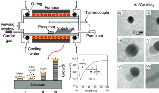
9)으로 개발되었으 며 이후 다양한 semiconducting nanorods를 합성하는 데 이용되었다. Fig. 1은 VLS 방법을 이용해 Ge nanorod 를 합성하는 원리와 성장과정을 transmission electron microscopy (TEM) 사진을 통해 직접적으로 보여주고 있다.
11),12)먼저 Precursor인 Ge powder를 furnace에서 온도가 가장 높은 영역에서 melting point 이상으로 가열하여 vaporization 시키면 carrier gas에 의해 gold가 코팅된
Fig. 1. Preparation of Ge nanorod via VLS method and its direct TEM observation.
12)CERAMIST
판상형 산화아연의 합성 및 응용에 관한 연구 동향
substrate로 이동하게 되고 liquid gold와 alloy가 형성 되면서 crystal growth가 이루어지게 된다. Fig. 2는 VLS 방법으로 합성된 ZnO의 nanorod array의 Scanning electron microscopy (SEM) 사진을 보여주 고 있으며 substrate 위에 catalyst로 작용하는 gold가 코팅된 부위에만 매우 균일하게 ZnO nanorod들의 crystal growth가 이루어진 것을 확인할 수 있다.
3)ZnO crystal이 위와 같이 육각기둥 형태로 자라나는 것은 Fig. 3과 같이 {0001}과 같은 polar planes들이 nonpolar planes들인 (1010)과 (1120)면들 보다 surface energy가 높아 [0001] direction으로의 crystal growth rate이 다른 direction보다 빠르기 때문이다.
13),14)이러한 이유로 ZnO의 crystal growth가 지속되면 Fig. 4와 같 이 (0001)면의 면적이 줄어든 hexagonal pencil 형태가 되며 이때 외부에서 빛이 조사되면 (0001)면을 통해 ZnO crystal 내부로 들어오게 되고 reflecting mirror planes 인 (0001)과 (0001)면 사이에서 polariton lasing을 통해 생성된 stimulated emission이 증폭되면서 (0001)면을 통해 외부로 발진하게 된다. 즉, (0001)면은 외부 빛이 들 어오는 window이자 생성된 stimulated emission의 reflecting mirror planes 역할을 하기 때문에 anisotropic crystal growth 속도로 인해 줄어든(0001) 면의 면적은 ZnO의 threshold power density 값을 높 이는 요인으로 작용하게 된다.
따라서 ZnO의 crystal growth rate control을 통하여
Fig. 2. SEM images of ZnO nanorod array by VLS method.
3)Fig. 4. Schematic representation of crystal growth of ZnO nanorod and its polartiton lasing mechanism.
5),15)Fig. 3. Theoretically calculated surface energy (g) of polar and
nonpolar planes for ZnO crystal.
14)특 집 장의순
CERAMIST
(0001)면이 넓은 plate-type ZnO를 합성할 수 있다면 기존의 nanorod structure와 비교하여 UV-lasing 특 성이 개선될 수 있을 것이라 기대할 수 있다.
3. Soft-solution route to ZnO nanowall array and its UV-lasing properties
2000년대 초반 UV-laser 물질로 ZnO가 주목 받으면 서 VLS 방법을 이용하여 tetra-pod,
16),17)nano-belt,
18),19)nano-ring,
20),21)등 다양한 ZnO nanostructures가 경쟁 적으로 발표되었으나 합성 온도가 매우 높아 사용할 수 있는 substrate의 종류가 sapphire와 같이 melting
temperature가 높은 물질로 한정되어 있었다. 본 연구 실에서는 2003년 약 3 ~ 4nm 크기의 ZnO nanoparticles 들을 nanorod crystal growth를 위한 seed이자 Si- wafer와 ZnO nanorod 간의 lattice mismatch를 줄이 기 위한 buffer layer로 사용하여 95°C 저온에서 hydrothermal reaction을 통해 Fig. 5와 같이 Si- wafer 표면에 균일한 ZnO nanorod array 제조가 가능 함을 최초로 보고 하였다.
22)이후 gold nanoparticles이 결합된 ZnO nanorod array를 합성하고자 하는 시도에서 우연히 Fig. 6과 같 은 plate-type ZnO nanowall array가 합성 되었다.
Fig. 5. Schematic illustration of seed-mediated synthesis of ZnO nanorod array and its electronic microscopy images; (a) TEM images of ZnO seed crystal, (b-c) FE-SEM images for top surface and cross section of ZnO nanorod array, (d) high-resolution TEM image of ZnO nanorod.
22)Fig. 7. (a) EDS and (b) AES spectra for plate-type ZnO nanowall array
Fig. 6. FE-SEM and TEM images of plate-type ZnO nanowall
array(NWA); (a, b) SEM and high-resolution TEM images
for top surface of NWA, (b, d) SEM image and high-reso-
lution TEM image including selected area electron diffrac-
tion pattern (inset) for cross section of NWA, (e) simulated
image (inset) is well matched with high-resolution TEM
one for the (0001) plane of nanowall.
23)CERAMIST
판상형 산화아연의 합성 및 응용에 관한 연구 동향
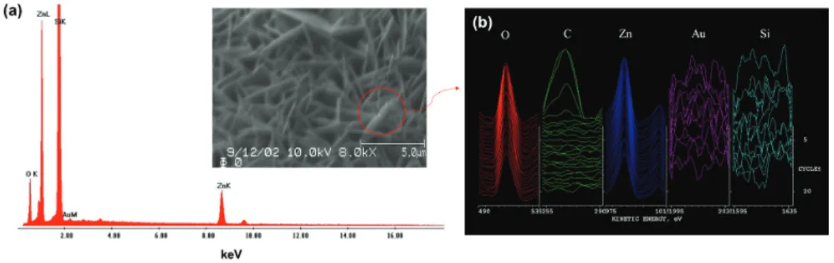
Si-wafer 표면에 생성된 plate-type ZnO nanowall들 은 nanorod structure와 달리 [0110] direction으로 성 장한 것을 확인할 수 있었으며 이로 인해 넓은 (0001)면 을 갖게 되었다. Gold nanoparticles이 plate-type ZnO의 생성에 미치는 영향을 확인하고자 Fig. 7과 같이 energy dispersive X-ray spectroscopy(EDS)와 auger electron spectroscopy(AES)를 이용하여 분석하 였다. 그러나 gold nanoparticles의 존재를 확인할 수 없 었다.
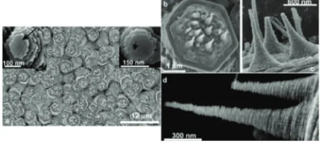
한편, Z. R. Tian 그룹이 sodium citrate 첨가에 의해 Fig. 8과 같이 ZnO nanorod 표면이 spiral structure로 변형된다는 연구결과를 보고하였다.
24)이로부터 우리는 sodium citrate를 reduction agent로 하여 합성한 gold nanoparticles로부터 sodium citrate가 hydrothermal reaction 중 우연히 releasing 되면서 Fig. 9와 같이 Si- wafer 표면에 코팅된 ZnO seed의 (0001)면 Zn
2+sites 에 결합되어 [0001] direction으로의 crystal growth가 억제되면서 ZnO nanowall array가 합성 되었음을 추측 할 수 있었다. 실제로 growth solution에 gold nanoparticles을 넣지 않고 sodium citrate만을 첨가하 여 동일한 plate-type ZnO nanowall array를 제조할 수 있었다.
23)Plate-type ZnO nanowall array의 UV-lasing 특 성을 확인한 결과 Fig. 10과 같이 low-temperature- hydrothermal reaction procedure로 제조한 ZnO nanorod array의 UV-lasing 특성보다 threshold power density 값이 70 kW/cm
2에서 25 kW/cm
2로 약
Fig. 8. SEM images of spiral ZnO nanorods on oriented ZnO crystals. (a) large arrays of ZnO nanorods on top of base ZnO rods, (b) precisely aligned ZnO nanorods on the (0001) surface of one ZnO rod, (c, d) high-magnification SEM images of spiral nanorods.
24)Fig. 10. (a, c) Excitation irradiation (Iex)-dependent PL spectra of ZnO nanorod and nanowall array dispersed with 150 g/
mm grating. The Iex of each spectrum is given inside.
The fitted solid line of the inset shows the PL intensity increases non-linearly above the threshold (Ith) of 70 and 25 kW/cm
2, respectively. (b, d) PL emission spectrum, dispersed with 1200 g/mm grating, of ZnO nanorod and nanowall array at 200 kW/cm
2for nanorod array and 20
~ 50 kW/cm
2for nanowall array.
22),23)Fig. 11. (a) Randomly oriented ZnO nanowalls on the Si-wafer cause even the reflected excitation light to be almost completely absorbed via WGM-type modes, (b) sche- matic view of the ZnO nanowall, (c) whereas nonreflected incident excitation light is totally absorbed to excite the medium, emission light stimulates emission extremely ef- ficiently via total internal reflection, (d) PL emission intensity variation upon incident angle (q) of pumping laser against c-axis of Si-substrate. Excitation light was fixed as 10 kW/cm
2.
28)Fig. 9. Schematic representation for crystal growth of plate-type
ZnO nanostructure.
특 집 장의순
CERAMIST
1/3로 감소되었음을 확인 하였다.
22),23)이는 plate-type ZnO nanowall이 넓은 (0001)면을 가지고 있어 nanorod structure보다 외부에서 조사되는 빛이 좀 더 쉽게 plate-type ZnO crystal 안으로 입사되며 reflecting mirror planes인 (0001)면이 넓기 때문에 stimulated emission의 reflection이 용이해지기 때문일 것으로 판 단된다. 또한, silicon substrate 위에 random하게 배열 된 house of card structure가 조사된 빛이 Fig. 11과 같 이 plate-type ZnO의 (0001)면들에 의해 반사되면서 쉽 게 외부로 빠져나가지 못하도록 빛의 vortex flow 현상 을 유도할 수 있다는 점도 plate-type ZnO의 threshold
power density 값을 낮추어 주는 요인이 될 수 있다.
23)이렇게 refractive index가 큰 물질들의 구조적 특성에 의해 빛을 가주어 두는 효과를 whispering-gallery mode(WGM)라고 한다.
25-27)WGM은 영국 St. Paul’s cathedral의 돔 주위로 그림을 전시한 gallery에서 옆 사 람과 속삭인 작은 소리가 돔 벽면을 따라 멀리까지 퍼지 는 것에서 유래되었다.
한편, plate-type ZnO의 random한 배열에 의한 외 부 빛의 WGM 현상 뿐만 아니라 crystal 내부로 입사된 빛들도 WGM 현상을 발생시키는 것으로 확인되었다.
28)Fig. 12는 단일 plate-type ZnO nanoparticles로부터
Fig. 12. (a) Room temperature cathodoluminescence(CL) spectra of ZnO nanoplate measured, (b) blowup spectra from (a). Arrows mark the calculated wavelengths in WGMs. The contribution from WGM-like-enhanced luminescence were approximated with six Gaussians. Note that the total intensity was assumed to consist of two parts; spontaneous emission (two broad Gaussians) and the WGM-like-enhanced emission (relatively narrow six Gaussians). (c) The monochromatic CL image of ZnO nanoplate.
28)Fig. 13. Schematic illustration of the growth mechanism of ZnO nanowall array. (a) a digital photograph of ZnO product on Al substrate, (b,
c) low- and high-magnification SEM images of ZnO nanowall array, (e, d) the EDS and XRD patterns of ZnO nanowall array.
29)CERAMIST
판상형 산화아연의 합성 및 응용에 관한 연구 동향
발생한 cathodoluminescence(CL) spectrum을 보여주 고 있으며 378 nm wavelength에서 나타난 주요 peak 를 분석한 결과 2개의 spontaneous emission과 6개의 WGM emission이 약 75%:25% 비율로 존재함을 확인할 수 있다.
28)이렇듯 plate-type ZnO의 random한 배열 에서 오는 WGM 현상과 crystal 자체의 WGM 현상이 UV-lasing을 발생시키는데 필요한 threshold power density 값을 낮추는데 효과적으로 기여한 것으로 판단 된다.
한편, 최근 W. Li 그룹의 연구결과에 따르면 aluminum (Al)-foil을 0.02 mol zinc nitrate(Zn (NO
3)
2·6H
2O) 와 8mL 25%(v/v) ammonia (NH
3·H
2O) solution에 담근 결과 Fig. 13과 같이 plate-type ZnO nanowall structure가 형성됨을 보고 하였다.
29)이들은 자세한 crystal growth mechanism을 제시하지는 않았지만 다 음 화학식과 같이 ammonia solution에 의해 생성된 Al(OH)
4-ion이 Zn(NH
3)
42+ion으로부터 생성된 ZnO seed의 (0001)면에 선택적으로 결합하여 plate-type ZnO nanowall structure가 형성되는 것으로 보고하였 다. 아직 정확한 crystal growth mechanism이 밝혀지 지 않았지만 이러한 방법은 flexible한 Al-foil에 plate- type ZnO nanowall structure를 room-temperature 에서 구현함으로써 상업적으로 좀 더 이용 가치가 높을 것이라 판단된다.
2Al+2OH
-+6H
2O → 2Al(OH)
4-+ 3H
2(1)
Zn
2++2OH
-→ Zn(OH)
2(2)
Zn(OH)
2+4NH
3·H
2O → Zn(NH
3)
42++4H
2O + 2OH
-(3) Zn(NH
3)
42++2OH
-→ ZnO + 4NH
3+ H
2O (4)
4. Photocatalytic properties of plate-type ZnO nanostructure
앞서 설명한 바와 같이 ZnO는 c-축을 따라 Zn
2+ion 으로 끝나 있는 (0001)면과 O
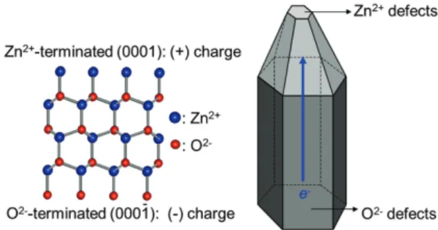
2-ion으로 끝나 있는 (0001) 면이 존재한다. 그러나 이러한 서로 다른 polarity를 띄 는 crystal planes의 존재는 현대 surface science 측면 에서 고려하면 crystal 내부의 dipole moment와 electrostatic potential 값을 높이는 요인으로 작용하기 때문에 crystal을 불안정하게 만드는 원인으로 작용한다.
따라서 ZnO crystal은 Fig. 14와 같이 다음 2가지 방법 으로 polar planes들의 surface charge를 낮춰 crystal 을 안정화 시킨다고 알려져 있다.
13),31),32)(1) (0001)면에서 (0001)면으로 electrons의 이동 (2) (0001)면에서 Zn
2+원자의 제거 또는 (0001)면에서
O
2-원자의 제거
또한, 앞서 Fig. 3에서 언급한 바와 같이 ZnO crystal 에서 polar planes인 {0001}의 surface energy는 3.92 J/m
2으로 (1010)면과 같은 nonpolar planes들의 surface energy인 2.01 J/m
2보다 높은 것으로 잘 알려 져 있다.
14)이러한 이론적 고찰들을 바탕으로 polar planes인 {0001}의 화학적 반응성이 다른 nonpolar planes들 보다 높다는 것을 쉽게 짐작할 수 있으며 photocatalytic reaction이 주로 polar planes에서 발생 할 것이라는 점을 예상할 수 있다.
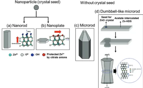
이러한 이론적 가정을 입증하고자 Fig. 15와 같이 4가 지 ZnO crystal(nanorod, nanoplate, microrod, 및 dumbbell-like microrod)을 합성하였다.
33)nanorods 와 nanoplates는 앞서 설명한 바와 같이 ZnO seed로부 터 합성하였으며 microrod와 dumbbell-like microrod 는 seed crystal 없이 합성하였다.
Fig. 14. Schematic representation for stabilization mechanism of
polar ZnO crystal with the {0001}.
특 집 장의순
CERAMIST
특히, dumbbell-like microrod는 수열합성 과정에서 pH 변화에 의해 ZnO crystal이 dissolution 되면서 acetate anion이 intercalation된 zinc hydroxy double salt(Zn-HDS)가 생성되고 다시 Zn-HDS 표면에서 ZnO crystal이 형성됨에 따라 Zn-HDS에 intercalation 된 acetate anions 주위로 (0001)면이 형성되어있어 (0001)면이 외부로 노출되는 것이 차단되어 있다.
33),34)Fig. 16은 서로 다른 4가지 ZnO crystal의 SEM 사진을 보여주고 있으며 각각의 입자 크기 및 이론적으로 계산된 전체 surface area와 (0001)면의 surface area을 보여주 고 있다. 즉, (0001)면의 surface area가 nanoplate >>
nanorod > microrod > dumbbell-like microrod 순서 로 감소함을 알 수 있다.
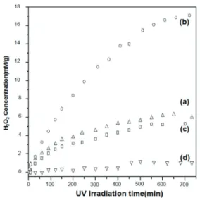
Photocatalyst 실험은 2mM acetic acid solution을
Fig. 15. Schematic illustration of the growth models for (a) nanorods, (b) nanoplates, (c) microrods, and (d) dumbbell-like microrods of zinc oxide.
33)Fig. 16. SEM images of ZnO (a) nanorods, (b) nanoplates, (c) microrods, 및 (d) dumbbell-like microrods. Table shows estimated areas
of total and the (0001) faces of the four different ZnO nanocrystals.
33)CERAMIST
판상형 산화아연의 합성 및 응용에 관한 연구 동향
hole scavenger로 사용하여 4가지 ZnO crystal이 각각 0.01g씩 담긴 solution에 UV light을 조사하면서 생성된 hydrogen peroxide(H
2O
2) 양을 iodometric titration으 로 측정하였다.
33)ZnO crystal의 photocatalytic effect 에 의한 hydrogen peroxide 생성에 대한 mechanism은 다음과 같다.
ZnO+hv → e
-cb+h
+vb(1)
O
2+e-e
-cb→ O
2·-(2)
O
2·-+2H+ → H
2O
2+O
2(3) CH
3CO
2-+h
+vb→ CH
3·+CO
2(4) H
2O
2+3I
-+2H
+→I
3-(λ
abs≈350 nm)+2H
2O (5)
위 식에서 hv 는 photon energy, h
+vb는 valence band에 존재하는 holes, e
-cb는 conduction band에 존 재하는 electrons을 의미하며 ZnO의 photocatalytic effect로 생성된 hydrogen peroxide는 iodide anion을 triiodide anion으로 산화 시키게 되고 absorption spectrum을 통해서 생성된 triiodide anion (λ
abs≈3 350 nm)의 농도로부터 hydrogen peroxide의 농도를 측정할 수 있다.
Fig. 17은 UV light 조사 시간에 따른 ZnO nanorod, nanoplate, microrod, 및 dumbbell-like microrod로 부터 발생한 hydrogen peroxide의 농도 변화를 보여주 고 있으며 hydrogen peroxide 생성양이 (0001)면의 surface area 증가 순서인 nanoplate >> nanorod >
microrod > 아령형 microrod의 순서로 증가하고 있음을 확인할 수 있다. 즉, polar planes인 {0001}면의 surface arear가 증가할수록 photocatalyst 특성 및 화학적 반응 성이 증가된다는 것을 의미하며 이는 앞서 언급한 polar planes들에 대한 이론적 고찰과도 정확히 일치하는 결과 이다. 본 연구실에서 이러한 사실을 최초로 보고한 이후 에 많은 유사한 연구결과들이 보고 되었으며 ZnO crystal을 이용한 photocatalyst, solar cell, UV-laser 등의 응용연구에서 효율을 높이기 위한 이론적 근거가 되 고 있다.
35-40)5. Antibacterial mechanism of ZnO nanostructures under dark conditions
최근 organic antibacterial agents의 무분별한 사용 으로 인해 항균제에 대한 내성을 보이는 super-bacteria 의 출현이 빈번해지면서 인류 건강에 큰 위협요인이 되고 있다. 또한, 한국에서는 가습기 살균제로 사용된 polyhexamethyleneguanidine phosphate(PHMG)와 oligo-(2-(2-ethoxy)-ethoxyethyl)-guanidinium chloride(PGH)와 같은 organic antibacterial agents 사용으로 인해 급속한 폐손상에 따른 사망사건이 발생하 면서 organic antibacterial agents를 대체할 수 있는 inorganic antibacterial agents에 대한 관심이 커지고 있다.
41-43)다양한 inorganic antibacterial agents 중 ZnO는 생체독성이 적으면서 Gram-positive bacteria 와 Grapm-negative bacteria에 걸쳐 넓은 항균력을 갖 는다는 장점이 있다.
44-51)빛을 조사하였을 때 ZnO의 antibacterial mechanism은 photocatalytic effect에 의해 발생한 reactive oxygen species(ROS)가 살균효과 를 나타낸다는 사실은 잘 알려져 있다.
52),53)그러나 우리 가 사용하는 대부분의 생활용품들이 photocatalytic
Fig. 17. Time profiles of the evolution of H
2O
2in UV-illuminated
(l > 300 nm) suspensions of ZnO (a) nanorods, (b) nano-
plates, (c) microrods, 및 (d) dumbbell-like microrods.
33)특 집 장의순
CERAMIST
effect를 유발시킬 수 있을 정도로 강한 빛을 사용하는 조 건들이 아니기 때문에 photocatalytic effect에 의존하는 inorganic antibacterial agents의 활용범위는 제한되 어 있다는 단점이 있다. 그러나 최근에 dark conditions 에서도 ZnO가 antibacterial effect를 나타낸다는 보고 가 나오면서 생활용품에 ZnO nanoparticles을 사용하 려는 시도가 많아지고 있다.
54),55)아직까지 이에 대한 명 확한 antibacterial mechanism이 규명되어 있지 않지 만 다음과 같이 크게 세가지 원인들이 제시되고 있다.
① polar planes에서 oxygen defect sites에 의한
ROS 발생; 앞서 Fig. 14에서 설명한 바와 같이 ZnO crystal의 안정을 위해 (0001)면에서 (0001)면 으로 electrons들이 이동하게 되는데 만일 Fig. 18 과 같이 (0001)면에 oxygen defect sites가 있다면 electrons들이 trap되게 되고 이들에 의해 H
2O molecules들이 oxidation-reduction 되면서 hydrogen peroxide와 같은 ROS가 생성되어 antibacterial effect가 나타날 수 있다는 mechanism으로 electron-spin resonance(ESR) spectroscopy 데이터를 근거로 제시하고 있다.
56-58)그러나 ZnO nanoparticles로부터 발생한 ROS에 대한 직접적인 증거는 아직까지 제시된바 없다.
② ZnO로부터 releasing된 Zn
2+ion에 의한 antibacterial effect; solution 상에서 ZnO nanoparticles로부터 releasing된 Zn
2+ion이 antibacterial effect를 나타낸다는 mechanism이 제안되었으나 일부 논문들에서 releasing된 Zn
2+ion의 농도가 antibacterial effect를 발휘하기에는 너무 낮다고 반박하고 있다.
56),59-61)③ Electrostatic interaction에 의한 ZnO nanoparticles 의 흡착; (+) charge를 띄는 ZnO nanoparticles이
Fig. 18. Schematic representation of oxygen defect sites-induced antibacterial mechanism of ZnO nanoparticles under dark conditions.
57)Fig. 19. TEM images of ZnO (a) nanoplate (NP), (b) nano-assembly (NA), and (c) conventional nanoparticles (CN). (d) Room-temperature
PL emission spectra for NPs, NAs, and CNs. (e) Schematic energy band diagram of ZnO crystals. Zni and Ov indicate interstitial
zinc and oxygen vacancy levels, respectively.
64)CERAMIST
판상형 산화아연의 합성 및 응용에 관한 연구 동향
(-) charge를 띄는 bacteria의 cell wall에 결합되어 membrane fluidity를 방해하고 cell wall을 destruction하여 균의 사멸을 유도한다는 mecha- nism이 제시되고 있으나 명확한 mechanism은 밝혀 지지 않고 있다.
62),63)이상과 같이 dark conditions에서 ZnO nanoparticles 의 antibacterial effect에 대한 mechanism은 아직까지 는 불분명한 상태이지만 oxygen defect sites에 의한 antibacterial mechanism이 ESR data를 근거로 주요 원인으로 자리 잡혀가고 있다.
한편, plate-type ZnO nanoparticles는 넓은 (0001) 면을 가지고 있으며 이로 인해 많은 oxygen defect sites 가 존재한다. 따라서 dark conditions에서 ZnO nanoparticles의 antibacterial mechanism이 oxygen defect sites에서 생성된 ROS에 의한 것인가를 규명하는 데 최적의 물질이라고 할 수 있다. 이에 대한 연구를 위해 Fig. 19와 같이 ZnO nanoplate(NP), nano-assembly (NA), 및 conventional nanoparticle(CN, Sumitomo Osaka Cement Co., Ltd. (ZnO-310))을 이용하였으며 room-temperature PL spectrum을 확인한 결과 bandgap transition에 의한 free excitonic emission (FEE)의 intensity는 모든 ZnO nanocrystal에서 유사 하였으나 l ≈ 600 nm에서 관찰된 conduction band 또 는 zinc interstitial level(Zn
i)에서 oxygen vacancy level(O
v)로의 transition에 의한 deep level emission (DLE)은 nanoplate에서 월등히 증가하였음을 확인할 수 있다.
64)ZnO crystal에서 DLE는 oxygen defect sites와 밀접 한 관련이 있으며 Fig. 19의 PL 결과는 (0001)면이 넓은 nanoplate에서 많은 oxygen defect sites가 존재함을 의미한다.
65)따라서 앞서 설명한 dark conditions에서 oxygen defect sites에 의한 antibacterial mechanism 이 옳다면 위 3가지 ZnO nanostructures에서 nano- plate가 가장 antibacterial effect가 높게 나타날 것이라 고 예측할 수 있다.
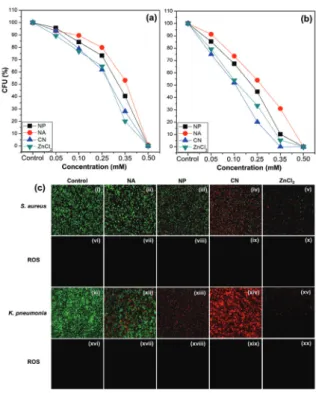
그러나 Gram-positive bacteria인 Staphylococcus
aureus와 Gram-negative bacteria인 Klebsiella pneumoniae을 대상으로 dark conditions에서 ZnO NP, NA, 및 CN의 antibacterial effect을 비교한 결과 Fig. 20과 같이 CN이 가장 antibacterial effect가 높은 것으로 나타났으며 ZnCl
2solution을 처리했을 때와 유 사한 antibacterial effect를 보였다. 이러한 사실은 dual immunofluorescence 기법을 통해서도 확인할 수 있었 으며 특이한 사실은 CN을 처리하였을 때는 단위 면적당 관찰된 bacteria의 수가 많은 반면 ZnCl
2를 처리하였을 때는 그 수가 상대적으로 적은 것을 알 수 있었다. 이는 dark conditions에서 ZnO nanoparticles의 antibac- terial effect가 ZnCl
2의 antibacterial effect 보다 더 늦 게 발현되기 때문인 것으로 판단된다. ROS staining을 통해 관찰한 결과 모든 샘플을 처리한 bacteria에서 ROS 에 의한 green fluorescence는 관찰되지 않았다. 이는 ZnO의 oxygen defect sites에 의한 ROS 발생이 거의
Fig. 20. Relative CFU (%) variation of (a)
S. aureusand (b)
K.pneumoniae
in increasing concentrations of ZnO NA, NP,
CN, and ZnCl
2solutions under dark conditions. (c) Dual
immunofluorescence and ROS staining images for
S. au- reus(i–x) and
K. pneumoniae(xi–xx) treated with NAs,
NPs, CNs, and ZnCl
2(0.35 mM) under dark conditions.
64)특 집 장의순
CERAMIST
없다는 것을 뒷받침해준다. 즉, dark conditions에서 ZnO nanoparticles의 antibacterial effect는 crystal에 존재하는 oxygen defect sites의 양과는 무관하며 Zn
2+ion releasing에 의한 antibacterial effect일 가능성이 높다는 것을 의미한다. 실제로 Fig. 21과 같이 0.35 mM 의 ZnO nanoparticles 및 ZnCl
2solution을 넣은 Staphylococcus aureus과 Klebsiella pneumoniae에 선택적 Zn
2+ion 결합 dye인 AZn2(red fluorescence)를 처리한 후 two-photon fluorescence microscopy을 통 해 관찰한 결과 ZnO nanoparticles들을 처리한 모든 균 내부에서 ZnCl
2을 처리한 균과 유사한 Zn
2+ion 농도가
존재함을 관찰할 수 있다.
다음으로는 ZnO nanoparticles들이 electrostatic interaction에 의해 bacteria와 결합하여 cell wall을 파 괴하는가를 살펴보기 위하여 Fig. 22와 같이 0.35 mM의 CN을 처리한 후 Staphylococcus aureus의 TEM 분석 을 실시하였다. Control의 TEM 사진은 정상적인 Staphylococcus aureus의 특징을 잘 보여주고 있으며 ZnO CN을 처리한 후의 TEM 사진을 보면 Staphylococcus aureus의 cell wall은 유지되고 있으나 내부의 일부분이 파괴되어 사라진 것을 볼 수 있다. 그러 나 cell wall에 붙어 있는 ZnO CN은 관찰되지 않았다.
일부 보이는 heavy metal agglomerates들은 EDS 분석 결과 ZnO가 아닌 것으로 확인 되었으며 Staphylococcus aureus의 TEM 시편 준비과정에서 contrast agents로 첨가하는 OsO
4와 같은 heavy metal agents에 의해 나 타난 것으로 보인다. 그러나 Fig. 23의 confocal microscopy 사진을 보면 Staphylococcus aureus의 표 면에 결합된 ZnO nanoparticles들로부터 DLE emission에 의한 green fluorescence가 명확히 관찰되 고 있으며 죽은 bacteria를 staining한 PI fluorescence (red)와 위치가 동일한 것을 알 수 있다. 이러한 사실들을 종합해 보면 ZnO nanoparticles들은 bacteria와
Fig. 21. Two-photon fluorescence microscopy and bright-field images of AZn2-labeled (a–j)
S. aureusand (k–f)
K. pneumoniae. Both species were treated with ZnO nanoparticles (NA, NP, and CN) and ZnCl
2of 0.35 mM.
64)Fig. 22. TEM images of (a)
S. aureus(control) and (b) CN (0.35
mM)-treated
S. aureus.Arrows indicate bacterial cell
wall (CW), plasma membrane (PM), nucleoid (DNA), and
heavy metal (HM) aggregates
.64)CERAMIST
판상형 산화아연의 합성 및 응용에 관한 연구 동향
electrostatic interaction을 통해 결합은 하고 있지만 cell wall을 파괴할 만큼 강한 결합이 아니기 때문에 TEM 시편 전처리 과정에서 bacteria 표면에 결합된 ZnO nanoparticles들이 분리되어 TEM 관찰에서는 ZnO nanoparticles들을 관찰할 수 없었던 것이 아닌가
판단된다.
따라서 dark conditions에서 ZnO nanoparticles에 의한 antibacterial mechanism은 Fig. 24와 같이 ZnO nanoparticles들이 electrostatic interaction에 의해 cell wall에 붙어 있으면서 천천히 releasing된 Zn
2+ion
Fig. 23. (a–e) bright-field (BF), (f–j) DLE, (k–o) PI staining, and (p–t) merged images (MI) for
S. aureustreated with NA, NP, CN, and ZnCl
2solutions (0.35 mM) under dark conditions (scale bar = 20 µm). DLE images were achieved by excitation at a wavelength of 488 nm.
64)Fig. 24. Schematic illustration of ZnO nanoparticle attachment to the outer cell wall of Gram (+) and Gram (-) bacteria.
64)특 집 장의순
CERAMIST
들이 cell wall의 특이 specific metalloprotein을 통해 균 내부로 들어가게 되고 bacteria가 견딜 수 있는 Zn
2+ion 농도의 critical concentration을 넘어서면서 사멸하 게 된다. 이러한 antibacterial mechanism을 명확히 밝 힐 수 있었던 것은 (0001)면이 넓은 plate-type ZnO nanoparticles의 합성이 가능하였기 때문이다.
6. Pilot-scale synthesis of plate-type ZnO
앞서 기술한 바와 같이 plate-type ZnO nanoparticles 의 경우 많은 장점을 가지고 있음에도 불구하고 학술적 연구를 위한 소용량 합성법만이 보고되었다. plate-type
ZnO의 산업적 이용가치를 높이기 위해서는 pilot-scale 합성기술이 반드시 필요하다. plate-type ZnO의 합성 방법은 약 3~4 nm 크기의 ZnO seed로 이용하여 합성 하게 되는데 안타깝게도 이러한 seed가 없으면 합성이 잘 되지 않는 것으로 확인되었다. 본 연구실에서 ZnO seed를 합성하는데 있어서 L. Sphanhel 방법을 이용하 였으나 이 방법은 복잡할 뿐만 아니라 시간이 오래 소요 된다.
66)이에 반해 P. S. Hale에 의해 제안된 ZnO seed 합성방법은 간단하면서도 합성시간이 한 시간 이내로 짧 다는 장점이 있다.
67)Table 1은 3L 용량에서 plate-type ZnO를 합성하기 위해 seed 합성방법 및 crystal growth solution의 조건
Fig. 25. SEM images for as-prepared products ((a) to (h)) from conditions ((1) to (8)) summarized in Table 1.
68)Table 1. Variation of seed and growth solution for large-scale (3L) preparation of plate-type ZnO. a),b)ZnO seed was synthesized by L.
Spanhel and P. Hall method, respectively, c)Sodium citrate was dissolved in seed solution and then added into growth solution, d)NaOH solution was slowly dropped to growth solution for 30min.
68)조건 seed
(mL)
Zn(OAc)
2·2H
2O (M)
Trisodium citrate (mM)
Citric acid (M)
Acetic acid
(M) NaOH (M) growth so-
lution의 pH 수득양 (g)
(1) 30 a) 0.1 0.28 - - 0.05 7.0 5.9
(2) 60 a) 0.2 7.6 - - 1 7.0 50.7
(3) 300 b) 1 2.8 - - 1 7.0 114.3
(4) 600 b) 1 5.6 - - 1 6.9 113.4
(5) 300 b) 1 2.8 0.2 - 1.5 6.9 297.9
(6) 300 b) 1 2.8 - 0.12 1.5 7.0 151.8
(7) 300 b) 1 5.6 c) - 0.12 1.5 7.0 176.48
(8) 300 b) 1 5.6 c) - 0.12 1.5 d) 7.0 181.28
CERAMIST
판상형 산화아연의 합성 및 응용에 관한 연구 동향
들을 변화시키면서 측정한 수득양 결과를 보여주고 있으 며 각각의 SEM 사진은 Fig. 25에서 보여주고 있다.
68)growth solution의 pH 조건이 산성일 경우 ZnO crystal이 용해되면서 Zn-HDS와 같은 불순물이 생성 되며 염기성일 경우 rod 형태의 ZnO crystal이 생성되 기 때문에 모든 조건에서 growth solution의 pH는 중성 을 유지 시켰다. 먼저, Table 1의 조건(1) 방법은 기존의 소용량(100mL)으로 합성할 때 사용한 방법으로써 3L 용 량으로 합성하였을 때는 Fig. 25(a)와 같이 입자크기가 약간 증가하였지만 균일한 plate-type ZnO가 합성되었 음을 확인할 수 있었다. 그러나 수득양이 약 5.9g으로 매 우 낮다.
조건(2)에서는 수득양을 높이고자 NaOH의 양을 20배 증가시켰으며 pH 증가로 인한 rod 형태의 ZnO 생성을 억제하고자 trisodium citrate의 양을 늘려 growth solution의 pH를 7.0으로 조절하였다. 또한, acidic condition 또는 basic condition에서 seed crystal의 dissolution 또는 crystal growth가 일어나는 것을 방지 하고자 중성 조건의 growth solution을 만든 후 seed solution은 마지막에 첨가하였다. 이로부터 얻어진 생성 물의 SEM 사진 (Fig. 25(b))을 살펴보면 불균일한 크기 의 plate-type ZnO가 생성되었으며 rod 형태가 다수 발 견 된 것을 알 수 있다. 그러나 수득양은 growth solution 에 첨가한 NaOH 양이 증가하면서 약 10배로 증가한 것 을 알 수 있다. 조건 (3-8)에서는 Hale 방법으로 합성된
ZnO seed를 이용하였다. 조건 (3)과 (4)에서는 조건 (2) 와 동일하게 NaOH의 양은 1M로 유지시키고 growth solution의 pH를 중성으로 유지시키고자 아세트산아연 이수화물(Zn(OAc)
2·2H
2O)과 trisodium citrate의 양 을 조절하였다. 이에 대한 생성물들은 rod 형태의 ZnO 는 거의 사라졌으나 plate-type ZnO의 입자크기가 여전 히 불균일한 것을 알 수 있다(Fig. 25(c)와 (d)). 그러나 수득양은 약 110g으로 크게 향상되었다. 조건 (5)에서는 NaOH 양을 1.5M로 증가시키고 pH를 중성으로 맞추고 자 citric acid를 growth solution에 첨가하였다. 이로부 터 얻어진 생성물의 수득량은 약 300g으로 가장 많은 수 득양을 나타내었으나 amorphous crystal로 보이는 다 량의 impurity들이 확인되었다 (Fig. 25(e)). 이는 citric acid가 생성된 ZnO를 dissolution하면서 Zn-HDS가 생성되는 것으로 확인되었다.
34)조건 (6)에서는 growth solution의 pH를 조절하기 위하여 acetic acid를 사용하 였으며 생성물은 Fig. 25(f)와 같이 불규칙한 입자크기를 갖는 plate-type ZnO가 생성되었고 수득양은 약 152g 으로 약간 감소하였다. 그러나 citric acid를 첨가하였을 때 생성된 Zn-HDS는 관찰되지 않았다. 조건 (7)에서는 조건 (6)과 같이 동일하게 acetic acid를 사용하여 pH를 조절하였으며 seed의 (0001)면을 효과적으로 citrate molecules로 protection 시키기 위해 trisodium citrate 를 seed solution에 넣어 반응시킨 후 growth solution 에 첨가하였다. 이로부터 Fig. 25(g)와 같이 균일한 크기
Fig. 26. Pilot-scale (100L) synthesis of plate-type ZnO crystals via high capacity reactor (Easthill inc.) and SEM images of the resulting
products.
특 집 장의순
CERAMIST
의 plate-type ZnO가 생성된 것을 확인 할 수 있었으며 수득양은 약 177g으로 조건 (1)의 수득양과 비교하면 수 득률이 약 30배 향상된 것을 알 수 있다. 조건 (8)은 조건 (7)과 반응조건이 같으나 growth solution 제조시 NaOH solution을 약 30분에 걸쳐 천천히 넣어주면서 합 성하였다. 그 결과 Fig. 25(h)와 같이 균일한 plate-type ZnO crystal이 제조되었으나 입자크기가 약 2배 증가한 것으로 확인되었다. 이는 NaOH를 천천히 투여하면서 OH- ion이 Zn(OAc)
2·2H
2O와 반응이 충분히 이루어 져 ZnO의 precursor인 Zn(OH)
42-생성이 원활히 이루 어지기 때문인 것으로 판단된다. 따라서 균일한 plate- type ZnO crystal을 수득양이 높게 획득하기 위한 합성 방법은 조건 (7)이 가장 적합하였다. 이와 같은 조건을 바 탕으로 Fig. 26과 같이 500L급 반응기에서 100L 용량으 로 hydrothermal reaction 하였을 때 균일한 plate- type ZnO를 수득량 약 6.7kg으로 제조할 수 있었다. 그 러나 산업적 이용가치를 더 높이기 위해서는 더 높은 수 득률이 필요하며 pilot-scale으로 합성하였을 때 입자크 기가 증가하는 문제와 seed 없이 합성하는 방법 등에 대 한 해결방안을 향후 탐색해야 할 것으로 판단된다.
7. Conclusion
seed의 crystal growth 조절을 통해 합성한 plate- type ZnO는 넓은 (0001)면으로 인해 rod 형태의 ZnO crystal과 비교하여 우수한 UV-lasing 특성을 보였으며 (0001)면에 존재하는 많은 atomic defect sites에 의해 photocatalytic property가 뛰어난 것으로 확인되었다.
또한, 기존에 명확히 밝히기 어려웠던 dark conditions 에서 ZnO nanoparticles의 antibacterial mechanism 을 plate-type ZnO의 결정학적 특성을 이용하여 규명 할 수 있었다. plate-type ZnO의 pilot-scale 합성은 crystal growth solution과 seed 합성방법을 변화시켜 가능하였으나 산업적 이용가치를 높이기 위한 pilot- scale 합성법이 더욱 개량되어야 할 것으로 판단된다.
plate-type ZnO는 photo-electronic device, solar cell, photocatalyst, suncare, antibacterial agents
등 다양한 산업분야에 걸쳐 활용도가 높은 물질이며 향후 활발한 학술연구와 산업화 개발을 통해 지금까지 알려지 지 않은 더 많은 특성과 활용분야를 개척해 나가야 할 것 이다.
8. Acknowledgment
This research was supported by the MSIT(Ministry of Science and ICT), Korea, under the ITRC(Information Technology Research Center) support program (IITP- 2017-2014-0-00639) supervised by the IITP(Institute for Information & communications Technology Promotion).
참고문헌
1. G. A. O. Oprea, E. Andronescu, D. Ficai, A. Ficai, F.
N. Oktar, and M. Yetmez, “ZnO Applications and Challenges,”Curr. Org. Chem., 18 [2] 192-203 (2014).
2. O. Bondarenko, K. Juganson, A. Ivask, K. Kasemets, M. Mortimer, and A. Kahru, “Toxicity of Ag, CuO and ZnO Nanoparticles to Selected Environmentally Relevant Test Organisms and Mammalian Cells In Vitro: a Critical Review,” Arch. Toxicol., 87 [7] 1181-
1200 (2013).
3. M. H. Huang, S. Mao, H. Feick, H. Yan, Y. Wu, H.
Kind, E. Weber, R. Russo, and P. Yang, “Room- Temperature Ultraviolet Nanowire Nanolasers,”
Science, 292 [8] 1897-99 (2001).
4. D. C. Reynolds, D. C. Look, B. Jogai, and T. C.
Collins, “Polarition and Free-Excition-Like Photoluminescence in ZnO”, Appl. Phys. Lett., 79 [23]
3794-96 (2001).
5. H. Deng, G. Weihs, C. Santori, J. Bloch, and Y.
Yamamoto, “Condensation of Semiconductor Microcavity Exciton Polaritons”, Science, 298 [4] 199- 202 (2002).
6. M. Zamfirescu, A. Kavokin, B. Gil, and G. Malpuech,
“ZnO as a Material Mostly Adapted for Realization of Room-Temperature Polariton Lasers”, Phys. Stat.
Sol. (a), 195 [3] 563-67 (2003).
7. D. M. Bagnall, Y. F. Chen, Z. Zhu, T. Yao, S. Koyama,
M. Y. Shen, and T. Goto, “Optically Pumped Lasing
of ZnO at Room Temperature”, Appl. Phys. Lett., 79
[23] 3794-96 (2001).
CERAMIST
판상형 산화아연의 합성 및 응용에 관한 연구 동향
8. J.–H. Choy, E.–S. Jang, J.–H. Won, J.–H.
Chung, D.–J. Jang, Y.–W. Kim, “Hydrothermal Route to ZnO Nanocoral Reefs and Nanofibers”, Appl.
Phys. Lett., 84 [2] 287-89 (2004).
9. R. A. Laudise, and A. A. Ballman, “Hydrothermal Synthesis of Zinc Oxide and Zinc Sulfide,” J. Phys.
Chem., 64 [5] 688-91 (1960).
10. R. S. Wagner, and W. C. Ellis, “Vapor-Liquid-Solid Mechanism of Single Crystal Growth,” Appl. Phys.
Lett., 70 [17] 2230-32 (1997).
11. Y. Wu, and P. Yang, “Direct Observation of Vapor- Liquid-Solid Nanowire Growth”, J. Am. Chem. Soc., 123 [13] 3165-66 (2001).
12. Y. Wu, P. Yang, Y. Sun, Y. Wu, B. Mayers, B. Gates, Y. Yin, F. Kim, and H. Yan, “One-Dimensional Nanostructures: Synthesis, Characterization, and Applications”, Adv. Mater., 15 [5] 353-89 (2003).
13. A. Wander, F. Schedin, P. Steadman, A. Norris, R.
McGrath, T. S. Turner, G. Thornton, and N. M.
Harrison, “Stability of Polar Oxide Surfaces”, Phys.
Rev. Lett., 86 3811-14 (2001).
14. J. I. Sohn, W. –K. Hong, S. Lee, S. Lee, J. Ku, Y. J.
Park, J. Hong, S. Hwang, K. H. Park, J. H. Warner, S. Cha, and J. M. Kim, “Surface Energy-Mediated Construction of Anisotropic Semiconductor Wires with Selective Crystallographic Polarity”, Scientific Reports, 4 5680 (2014).
15. M. M. Versteegh, D. Vanmaekelbergh, and J. I.
Dijkhuis, “Room-Temperature Laser Emission of ZnO Nanowires Explained by Many-Body Theory”, Phys. Rev. Lett., 108 [15] 157402 (2012).
16. Y. Dai, Y. Zhang, Q. K. Li, and C. W. Nan, “Synthesis and optical properties of tetrapod-like zinc oxide nanorods”, Chem. Phys. Lett., 358 [1-2] 83-6 (2002).
17. Z. Chen, Z. Shan, M. S. Cao, L. Lu, and S. X. Mao,
“Zinc Oxide Nanotetrapods”, Nanotechnology, 15 [3]
365 (2004).
18. Z. W. Pan, Z. R. Dai, and Z. L. Wang, “Nanobelts of Semiconducting Oxides”, Science, 291 [9] 1947-49 (2001).
19. Z. R. Dai, Z. W. Pan, and Z. L. Wang, “Novel Nanostructures of Functional Oxides Synthesized by Thermal Evaporation”, Adv. Funct. Mater., 13 [1]
9-24 (2003).
20. X. Y. Kong, and Z. L. Wang, “Spontaneous Polarization-Induced Nanohelixes, Nanosprings, and Nanorings of Piezoelectric Nanobelts”, Nano Lett., 3
[12] 1625-31 (2003).
21. X. Y. Kong, Y. Ding, R. Yang, and Z. L. Wang,
“Single-Crystal Nanorings Formed by Epitaxial Self- Coiling of Polar Nanobelts”, Science, 303 [27] 1348-51 (2004).
22. J. –H. Choy, E. –S. Jang, J. –H. Won, J. –H.
Chung, D. –J. Jang, and Y. –W. Kim, “Soft Solution Route to Directionally Grown ZnO Nanorod Arrays on Si Wafer; Room-Temperature Ultraviolet Laser”, Adv. Mater., 15 [22] 1911-14 (2003).
23. E. –S. Jang, X. Chen, J. –H. Won, J. –H. Chung, D. –J. Jang, Y. –W. Kim, and J. –H. Choy, “Soft- Solution Route to ZnO Nanowall Array with Low Threshold Power Density”, Appl. Phys. Lett., 97 043109 (2010).
24. Z. R. Tian, J. A. Voigt, J. Liu, B. Mckenzie, and M.
J. Mcdermott, “Biomimetic Arrays of Oriented Helical ZnO Nanorods and Columns”, J. Am. Chem. Soc., 124 12954-55 (2002).
25. S. L. McCall, A. F. J. Levi, R. E. Slusher, S. J.
Pearton, and R. A. Logan, “Whispering-Gallery Mode Microdisk Lasers”, Appl. Phys. Lett., 60 [3]
289-91 (1992).
26. R. G. Nazmitdinov, K. N. Pichugin, I. Rotter, and P.
Seba, Phys. Rev. E, 64 056214 (2001).
27. A. C. Tamboli, E. D. Haberer, R. Sharma, K. H. Lee, S. Nakamura, and E. L. Hu, “Room-Temperature Continuous-Wave Lasing in GaN/InGaN Microdisks”, Nature Photonics, 1 61-64 (2007).
28. C. Kim, Y. –J. Kim, E. –S. Jang, G. –C. Yi, and H.
H. Kim, “Whispering-Gallery-Modelike-Enhanced Emission from ZnO Nanodisk”, Appl. Phys. Lett., 88 093104 (2006).
29. W. Li, S. Gao, L. Li, S. Jiao, Q. Yu, H. Li, J. Wang, Q. Yu, Y. Zhang, and D. Wang, “A Facile Solution Synthesis of ZnO Nanoplates on Al Substrate at Room Temperature”, Mater. Lett., 185 161-64 (2016).
30. P. W. Tasker, “The Stability of Ionic Crystal Surfaces”, J. Phys. C: Solid State Phys., 12 4977-84 (1979).
31. O. Dulub, U. Kiebold, and G. Kresse, “Novel Stabilization Mechanism on Polar Surfaces:
ZnO(0001)-Zn”, Phys. Rev. Lett., 90 [1] 016102 (2003).
32. V. Staemmler, K. Fink, B. Meyer, D. Marx, M.
Kunat, S. G. Girol, U. Burghaus, and C. Woll,
“Stabilization of Polar ZnO Surfaces: Validating
Microscopic Models by Using CO as a Probe Molecule”,
Phys. Rev. Lett., 90 [10] 106102 (2003).
특 집 장의순
CERAMIST
33. E.–S. Jang, J.–H. Won, S.–J. Hwang, and J.–H.
Choy, “Fine Tuning of the Face Orientation of ZnO Crystals to Optimize Their Photocatalytic Activity”, Adv. Mater., 18 3309-12 (2006).
34. E.–S. Jang, J.–H. Won, Y.–W. Kim, Z. Cheng, and J.–H. Choy, “Dynamic Transition between Zn-HDS and ZnO; Growth and Dissolving Mechanism of Dumbbell-like ZnO Bipod Crystal”, CrystEngComm, 13 546-52 (2011).
35. J. H. Zeng, B. B. Jin, and Y. F. Wang, “Facet Enhanced Photocatalytic Effect with Uniform Single- Crystalline Zinc Oxide Nanodisks”, Chem. Phys.
Lett., 472 90-95 (2009).
36. R. Boppella, K. Anjaneyulu, P. Basak, and S. V.
Manorama, “Facile Synthesis of Face Oriented ZnO Crystals: Tunable Polar Facets and Shape Induced Enhanced Photocatalytic Performance”, J. Phys.
Chem. C, 117 4597-4605 (2013).
37. M. Huang, Y. Yan, W. Feng, S. Weng, Z. Zheng, X.
Fu, and P. Liu, “Controllable Tuning Various Ratios of ZnO Polar Facets by Crystal Seed-Assisted Growth and Their Photocatalytic Activity”, Cryst. Grwoth Des., 14 2179-86 (2014).
38. G. Tang, S. Tian, Z. Zhou, Y. Wen, A. Pang, Y.
Zhang, D. Zeng, H. Li, B. Shan, and C. Xie, “ZnO Micro/Nanocrystals with Tunable Exposed (0001) Facets for Enhanced Catalytic Activity on the Thermal Decomposition of Ammonium Perchlorate”, J. Phys.
Chem. C, 118 11833-41 (2014).
39. Y. Chen, H. Zhao, B. Liu, and H. Yang, “Charge Separation between Wurtzite ZnO Polar {0001}
Surfaces and Their Enhanced Photocatalytic Activity”, Appl. Catalysis B: Environmental, 163 189-97 (2015).
40. Y. Zhang, C. Liu, F. Gong, B. Jiu, and F. Li, “Large Scale Synthesis of Hexagonal Simonkolleit Nanosheets for ZnO Gas Sensors with Enhanced Performances”, Mater. Lett., 186 7-11 (2017).
41. E. D. Brown, and G. D. Wright, “New Targets and Screening Approaches in Antimicrobial Drug Discovery”, Chem. Rev., 105 759-74 (2005).
42. H.–J. Yang, H.–J. Kim, J. Yu, E. Lee, Y.–H.
Jung, H.–Y. Kim, J.–H. Seo, G.–Y. Kwon, J.–H.
Park, J. Gwack, S.–K. Youn, J.–W. Kwon, B.–Y.
Jun, K. W. Kim, K. Ahn, S.–Y. Lee, J.–D. Park, J.
–W. Kwon, B.–J. Kim, M.–S. Lee, K.–H. Do, S.
–J. Jang, B.–Y. Pyun, and S. J. Hong, “Inhalation Toxicity of Humidifier Disinfectants as a Risk Factor
of Children’s Interstitial Lung Disease in Korea: A Case-Control Study”, PLOS ONE, 8 [6] e64430 (2013).
43. H. R. Kim, K. Lee, C. W. Park, J. A. Song, D. Y.
Shin, Y. J. Park, and K. H. Chung, “Polyhexamethylene guanidine phosphate aerosol particles induce pulmonary inflammatory and fibrotic responses”, Archives of Toxicology, 90 [3] 617-32 (2016).
44. R. Brayner, R. F-Iliou, N. Brivois, S. Djediat, M.F.
Benedetti, and G. Fiévet, “Toxicological Impact Studies Based on Escherichia coli Bacteria in Ultrafine ZnO Nanoparticles Colloidal Medium”, Nano Lett., 6 [4] 866-70 (2006).
45. G. Applerot, A. Lipovsky, R. Dror, N. Perkas, Y.
Nitzan, R. Lubart, and A. Gedanken, “Enhanced Antibacterial Activity of Nanocrystalline ZnO Due to Increased ROS-Mediated Cell Injury” Adv. Funct.
Mater., 19 [6] 842-52 (2009).
46. K. Ali, S. Dwivedi, A. Azam, Q. Saquib, M. S.
Al-Said, A. A. Alkhedhairy, and J. Musarrat, “Aloe Vera Extract Functionalized Zinc Oxide Nanoparticles as Nanoantibiotics Against Multi-Drug Resistant Clinical Bacterial Isolates”, J Colloid Interface Sci., 472 [15] 145-56 (2016).
48. S. Dwivedi, R. Wahab, F. Khan, Y. K. Mishra, J.
Musarrat, and A. A. Al-Khedhairy, “Reactive Oxygen Species Mediated Bacterial Biofilm Inhibition via Zinc Oxide Nanoparticles and Their Statistical Determination”, PLOS ONE, 9 e111289 (2014).
49. A. Azam, A. S. Ahmed, M. Oves, M. S. Khan, S. S.
Habib, and A. Memic, “Antimicrobial Activity of Metal Oxide Nanoparticles against Gram-Positive and Gram-Negative Bacteria: a Comparative Study”, Int.
J. Nanomed., 7 6003-09 (2012).
50. S. A. Ansari, Q. Husain, S. Qayyum, and A. Azam,
“Designing and Surface Modification of Zinc Oxide Nanoparticles for Biomedical Applications”, Food &
Chem. Toxicology, 49 [9] 2107-15 (2011).
51. M. A. Ansari, H. M. Khan, A. A. Khan, A. Sultan, and A.
Azam, “Synthesis and Characterization of the Antibacterial Potential of ZnO Nanoparticles against Extended- Spectrum β-Lactamases-Producing Escherichia coli and Klebsiella pneumoniae Isolated from a Tertiary Care Hospital of North India”, Appl. Microbiol & Biotech., 94 [2]
467-77 (2012).
52. Y. Li, W. Zhang, J. Niu, and Y. Chen, “Mechanism of
Photogenerated Reactive Oxygen Species and
CERAMIST
판상형 산화아연의 합성 및 응용에 관한 연구 동향
Correlation with the Antibacterial Properties of Engineered Metal-Oxide Nanoparticles”, ACS Nano, 6 [6] 5164-73 (2012).
53. N. Padmavathy, and R. Vijayaraghavan, “Enhanced Bioactivity of ZnO Nanoparticles—an Antimicrobial Study”, Sci. Technol. Adv. Mater., 9 [3] 035004 (2008).
54. A. B. Djurišić, Y. H. Leung, A. M. C. Ng, X. Y. Xu, P.
K. H. Lee, N. Degger, and R. S. S. Wu, “Toxicity of Metal Oxide Nanoparticles: Mechanisms, Characterization, and Avoiding Experimental Artefacts”, Small, 11 [1] 26-44 (2015).
55. M. J. Hajipour, K. M. Fromm, A. A. Ashkarran, D. J.
D. Aberasturi, I. R. D. Larramendi, T. Rojo, V.
Serpooshan, W. J. Parak, and M. Mahmoudi,
“Antibacterial Properties of Nanoparticles”, Trends Biotech., 30 [10] 499-511 (2012).
56. K. Hirota, M. Sugimoto, M. Kato, K. Tsukagoshi, T.
Tanigawa, and H. Sugimoto, “Preparation of Zinc Oxide Ceramics with a Sustainable Antibacterial Activity under Dark conditions”, Ceramics Inter., 36 [2] 497-506 (2010).
57. X. Xu, D. Chen, Z. Yi, M. Jiang, L. Wang, Z. Zhou, X. Fan, Y. Wang, and D. Hui, “Antimicrobial Mechanism Based on H
2O
2Generation at Oxygen Vacancies in ZnO Crystals”, Langmuir, 29 [18] 5573- 80 (2013).
58. V. L. Prasanna, and R. Vijayaraghavan, “Insight into the Mechanism of Antibacterial Activity of ZnO:
Surface Defects Mediated Reactive Oxygen Species Even in the Dark”, Langmuir, 31 [33] 9155-9162 (2015).
59. M. Li, L. Zhu, and D. Lin, “Toxicity of ZnO Nanoparticles to Escherichia coli: Mechanism and the Influence of Medium Components”, Environ. Sci.
Technol., 45 [5] 1977-83 (2011).
60. Y. W. Wang, A. Cao, Y. Jiang, X. Zhang, J. H. Liu, Y. Liu, and H. Wang, “Superior Antibacterial Activity of Zinc Oxide/Graphene Oxide Composites Originating from High Zinc Concentration Localized around Bacteria”, ACS Appl. Mater. Interfaces, 6 [4] 2791-98 (2014).
61. K. R. Raghupathi, R. T. Koodali, and A. C. Manna,
“Size-Dependent Bacterial Growth Inhibition and Mechanism of Antibacterial Activity of Zinc Oxide Nanoparticles”, Langmuir, 27 [7] 4020-4028 (2011).
62. K. H. Tam, A. B. Djurišić, C. M. N. Chan, Y. Y. Xi, C. W. Tse, Y. H. Leung, W. K. Chan, F. C. C. Leung, and D. W. T. Au, “Antibacterial Activity of ZnO Nanorods Prepared by a Hydrothermal Method”, Thin Solid Films, 516 [18] 6167-74 (2008).
63. Y. Liu, L. He, A. Mustapha, H. Li, Z. Q. Hu, and M.
Lin, “Antibacterial Activities of Zinc Oxide Nanoparticles against Escherichia coli O157:H7”, J.
Appl. Microbiology, 107 [4] 1193-1201 (2009).
64. A. Joe, S.–H. Park, K.–D. Shim, D.–J. Kim, K.
–H. Jhee, H.–W. Lee, C.–H. Heo, H.–M. Kim, and E.–S. Jang, “Antibacterial Mechanism of ZnO Nanoparticles under Dark conditions”, J. Ind. & Eng.
Chem., 45 430-39 (2017).
65. C. H. Ahn, Y. Y. Kim, D. C. Kim, S. K. Mohanta, H.
K. A Cho, “A Comparative Analysis of Deep Level Emission in ZnO Layers Deposited by Various Methods”, J. Appl. Phys., 105 013502 (2009).
66. L. Spanhel, and M. A. Anderson, “Semiconductor Clusters in the Sol-Gel Process: Quantized Aggregation, Gelation, and Crystal Growth in Concentrated Zinc Oxide Colloids”, J. Am. Chem.
Soc., 113 [8] 2826-33 (1991).
67. P. S. Hale, L. M. Maddox, J. G. Shapter, N. H.
Voelcker, M. J. Ford, and E. R. Waclawik, “Growth Kinetics and Modeling of ZnO Nanoparticles”, J.
Chem. Edu., 82 [5] 775-78 (2005).
68. D.–J. Kim, B.–M. Kim, A. Joe, K.–D. Shim, H.
–W. Han, G.–H. Noh, and E.–S. Jang, “Large- Scale Synthesis of Plate-Type ZnO Crystal with High Photocatalytic Activity”, J. Kor. Chem. Soc., 59 [2]
148-55 (2015).
장 의 순
2004년 8월~2005년 2월 포항공과대학교 신소재공학과 박사 후 연구원
2005년 3월~2008년 9월 KT 미래기술연구소 선임연구원
2008년 10월~2010년 8월 스탠포드대학교 의과대학 박사 후 연구원
2010년 9월~현재 금오공과대학교 화학소재융합공학부 부교수