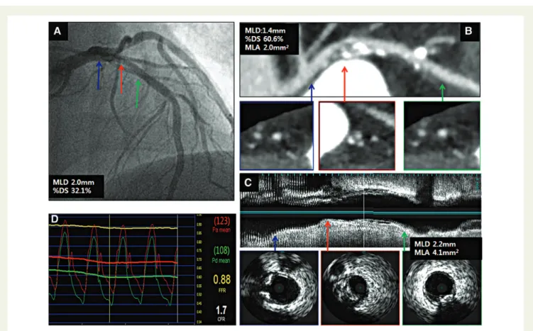
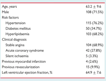
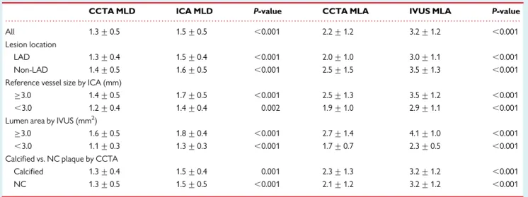
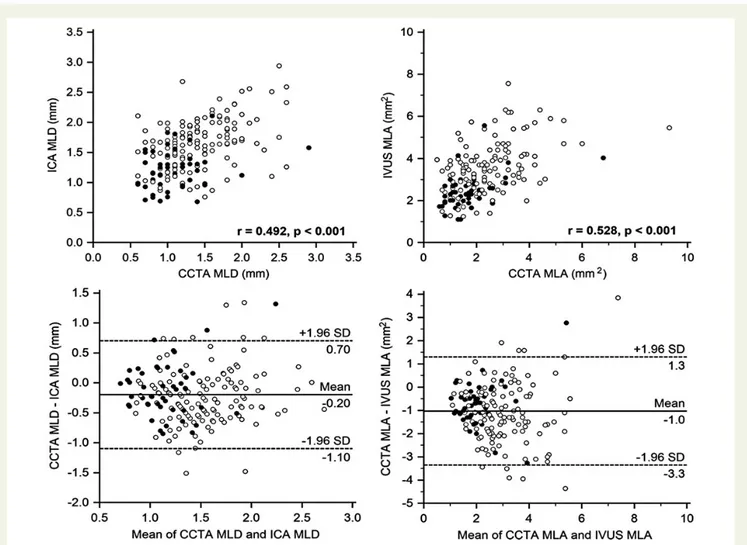
Diagnostic value of coronary CT angiography in comparison with invasive coronary angiography and intravascular ultrasound in patients with intermediate coronary artery stenosis: results from the prospective multicentre FIGURE-OUT (Functional Imaging crite
전체 글
수치


관련 문서
Therefore, cTnT is a recommended biomarker for use in the detection of myocardial infarction (MI) and in acute coronary syndromes.[8] Indeed, several authors
The locations of aneurysms were middle cerebral artery in 15 patients, cerebral artery in 15 patients, cerebral artery in 15 patients, cerebral artery in
To assess the clinical usefulness of performing Q-PCR in practice as a diagnostic technique, we compared blindly the Q-PCR results using blood samples of the
CT (Computed Tomography) – TDLAS (Tunable Diode Laser Absorption Spectroscopy) is a non-intrusive diagnostic technique that allows for spatially resolved measurements
Simultaneous Measurements of the Wake Flow of a Circular Cylinder with a Flexible Film and Its Motions using
Incident major cardiovascular events (coronary artery disease, ischemic stroke, hemorrhagic stroke and cardiovascular mortality) were set as primary end points.
The major findings of the present study include the fol- lowing: (1) EM-seq-based methylation profiling produced good quality data, even with limited human cfDNA quan-
images projected on the surface; the peripheral area appears expanded in comparison with the central area as shown in Figure 1 1. If the symmetric and consistent imaging
