Protein Engineering of Flavin-containing Monooxygenase from Corynebacterium glutamicum for Improved Production of Indigo and Indirubin
Hye Sook Jung
1, Hae Bin Jung
1, Hee Sook Kim
1, Chang Gyeom Kim
2and Jin Ho Lee
1*
1
School of Food Biotechnology & Nutrition, Kyungsung University, Busan 48434, Korea
2
Department of Bioinformatics & Biosystems, Korea Polytechnics, Gyeonggi-do 13122, Korea Received February 21, 2018 /Revised May 29, 2018 /Accepted May 30, 2018
Flavin-containing monooxygenases from Corynebacterium (cFMOs) were mutagenized based on homol- ogy modeling to develop variants with an enhanced indigoid production capability. The four mutants, F170Y, A210G, A210S, and T326S, which fused to a maltose-binding protein (MBP), were constructed, and their biochemical properties were characterized. Of these, purified MBP-T326S required a higher concentration of exogenous FAD (100 mM) than the wild-type MBP-cFMO for optimal activity and showed a 3.8-fold increase in the k
cat/K
mvalue at 100 μ M FAD compared to that of MBP-cFMO at 2 μ M FAD. The indole oxygenase activities of MBP-T326S decreased to 63-77% compared to that of the MBP-cFMO In addition, MBP-T326S displayed a very low level of futile NADPH oxidase activities (21-24%) in the absence of a substrate. Mutant proteins except for T326S displayed similar K
mand in- creased k
cat/K
mvalues compared to the wild-type. MBP-F170Y and -A210S mutants showed elevated indole oxygenase activity higher than 3.1- and 2.9-fold, respectively, in comparison with MBP-cFMO.
When indigoid production was carried out in LB broth with 2.5 g/l of tryptophan, Escherichia coli ex- pressing cFMO produced 684 mg/l of indigo and 104 mg/l of indirubin, while cells harboring T326S produced 1,040 mg/l of indigo and 112 mg/l of indirubin. The results indicate that the production of indigo was 13% higher when compared to a previous report in which an E. coli expressing FMO from Methylophaga produced 920 mg/l of indigo. The protein engineering of cFMO based on homol- ogy modeling provided a more rational strategy for developing indigoid-producing strains.
Key words : Corynebacterium glutamicum, flavin-containing monooxygenase, indigoid, protein engineering, T326S mutant
*Corresponding author
*Tel : +82-51-633-4716, Fax : +82-51-622-4986
*E-mail : [email protected]
This is an Open-Access article distributed under the terms of the Creative Commons Attribution Non-Commercial License (http://creativecommons.org/licenses/by-nc/3.0) which permits unrestricted non-commercial use, distribution, and reproduction in any medium, provided the original work is properly cited.
Journal of Life Science 2018 Vol. 28. No. 6. 656~662 DOI : https://doi.org/10.5352/JLS.2018.28.6.656
Introduction
Flavin-containing monooxygenases (FMOs, EC 1.14.13.8) belong to the single-component flavoprotein monooxygenase subfamilies and can oxidize the nucleophilic nitrogen, sulfur, phosphorus, and selenium atoms in a range of structurally diverse compounds [2, 17]. FMOs from mammals are im- portant biocatalysts involved in detoxification of drugs and other xenobiotics such as trimethylamine (TMA), benzyd- amine, clozapine, itopride, ranitidine, and cimetidine [14].
Meanwhile, the bacterial FMO from Methylophaga aminisulfi- divorans (mFMO) was known to have the oxygenation activ- ity for N-containing amines and C-atom in indole [4].
Indigo is one of the oldest blue dyes for the textile in- dustry and currently produced by chemical synthesis [7].
Indirubin and its derivatives have significant therapeutic ef-
fects on several human cancers and Alzheimer’s disease [5,
6, 11]. To date, several eco-friendly biological approaches
have been attempted for production of these indigoids, and
high titers of indigo and indirubin were achieved by re-
combinant cells expressing bacterial FMOs from M. amini-
sulfidivorans and optimization of culture media [9, 10]. We
previously identified an FMO from Corynebacterium gluta-
micum (cFMO) with indole oxygenase activity and demon-
strated E. coli harboring cFMO produced higher concen-
tration of both indigo and indirubin than recombinant cells
with other oxygenases [1]. Although bacterial FMOs have
a high potential usefulness in biological production of indigo
and indirubin, the production performance by them is still
much lower than that by chemical synthesis. Therefore, it
is necessary to develop mutant FMOs with enhanced cata-
lytic activity and/or indigoid production capability through
rational design and mutagenesis.
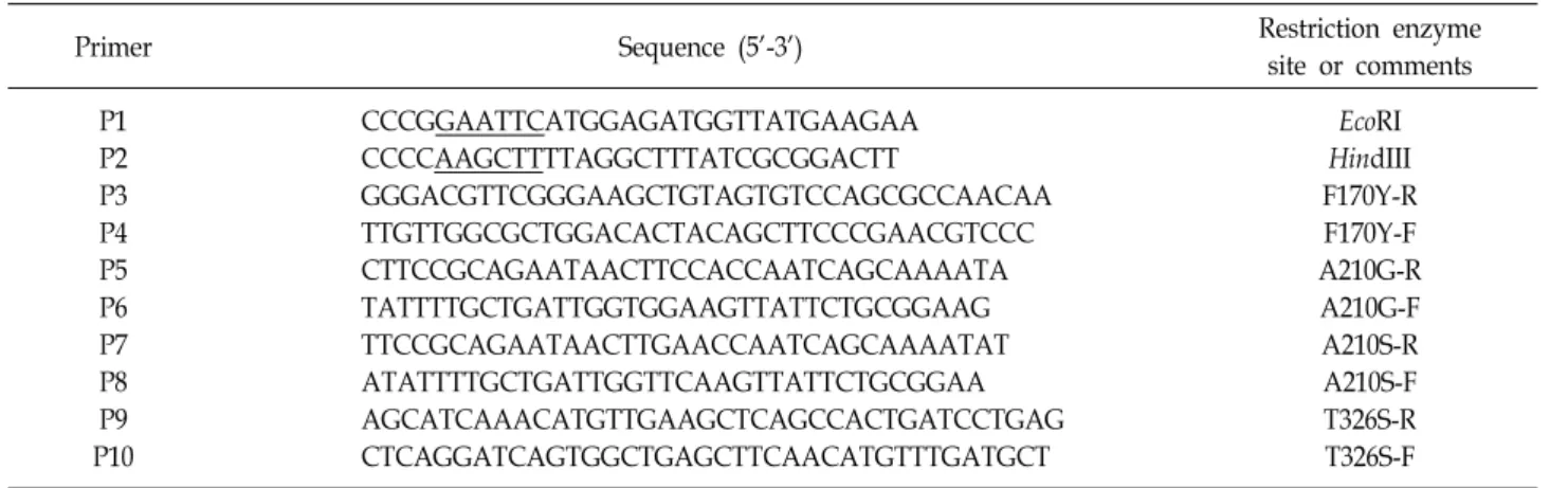
Table 1. The bacterial strains and plasmids used in this study
Strain or plasmid Characteristics
*Source or reference
Escherichia coli Top10
W3110 WCO21 WTS326
F
-mcrA Δ(mrr-hsaRMS-mcrBC) φ80lacZΔM15 ΔlacX74 recA1 araD139 Δ(ara-leu)7679 galU galK rps (Str
R) endA1 nupG
F
-IN(rrnD-rrnE) W3110 with pPIO1 W3110 with pK-T326S
Invitrogen, USA
This lab.
This work This work Plasmids
pKK223-3 pPIO1 pMAL-c2x pMCF14 pM-F170Y pM-A210G pM-A210S pM-T326S pK-F170Y pK-A210G pK-A210S pK-T326S
Expression vector with tac promoter, Amp
RpKK223-3 derivative; 1.4 kb cfmo ORF
Expression vector with tac promoter and an ORF of maltose binding protein, Amp
RpMAL-c2x derivative; 1.4 kb cfmo ORF
pMAL-c2x derivative; 1.4 kb cfmo ORF with mutation at T509A (F170Y) pMAL-c2x derivative; 1.4 kb cfmo ORF with mutation at C629G (A210G) pMAL-c2x derivative; 1.4 kb cfmo ORF with mutation at G628T (A210S) pMAL-c2x derivative; 1.4 kb cfmo ORF with mutation at CG977-978GC (T326S) pKK223-3 with cfmo with F170Y
pKK223-3 with cfmo with A210G pKK223-3 with cfmo with A210S pKK223-3 with cfmo with T326S
This lab.
Ameria et al. (2015) New England Biolabs Ameria et al. (2015) This work
This work This work This work This work This work This work This work
*
Str
R, streptomycin resistance, Amp
R, ampicillin resistance.
FMOs require an FAD prosthetic group and an NADPH cofactor [17]. According to crystal structures of yeast and bacterial FMOs, they consist of a large FAD binding domain and a small domain, and the NADPH binding site lies in the crevice between the two domains [3, 8]. In addition, NADPH binding motif sequence that is also involved in di- verse substrate binding showed some variations, i.e. GASYA in cFMO, GSSYS in mFMO, and GASSA in Shizosaccharomy- ces pombe FMO [3, 8, 13].
Herein, we engineered three interesting amino residues near FAD and NADPH binding sites based on homology modeling of Corynebacterium FMO. Moreover, the indigoid- producing ability of recombinant E. coli cells with mutant enzymes was evaluated in terms of production level.
Materials and Methods
Homology modeling
Homology modeling was performed by MODELER ver- sion 9.14 [16]. The crystal structure of Methylophaga FMO (PDB ID: 2XVH) was used as template for building the ho- mology models [3]. Although the structure of mFMO is con- sisted of homomultimer, we took only one monomer struc- ture for using as a template since the catalytic active site of that seems not to be affected by intermolecular interaction.
Amino acid residues including F170, A210, and T326 inter-
acting with prosthetic groups, FAD and NADPH, by hydro- gen bonds and/or hydrophobic interaction were chosen for mutagenesis.
Site-directed mutagenesis
The Corynebacterium fmo gene in pMCF14 was used as a template DNA for mutagenesis (Table 1). F170, A210, and T326 residues in cFMO based on homology modeling were mutated by site-directed mutagenesis. To introduce muta- tion at F170Y, A210G, A201S, and T326S, 5’-and 3’-regions of fmo gene were amplified by using primer sets P1-P3 and P4-P2, P1-P5 and P6-P2, P1-P7 and P8-P2, and P1-P9 and P10-P2, respectively (Table 2). The resulting fragments of both regions were combined each other and used for the 2
ndround of PCR with P1 and P2, respectively. The final 1.4 kb PCR products with 4 types of mutations were digested with EcoRI and HindIII, ligated with pMAL-c2x/EcoRI/
HindIII, and transformed into competent E. coli Top10 cells.
Constructed plasmids were named as pM-F170Y, pM-A210G, pM-A210S, and pM-T326S, respectively. The introduced mu- tations were analyzed by DNA sequencing.
Subcloning of mutant cFMOs
To construct plasmids harboring mutant cFMOs without
MBP (maltose-binding protein)-tag, the 1.4 kb fmo-ORFs of
MBP-F170Y, -A210G, -A210S, and -T326S mutants were di-
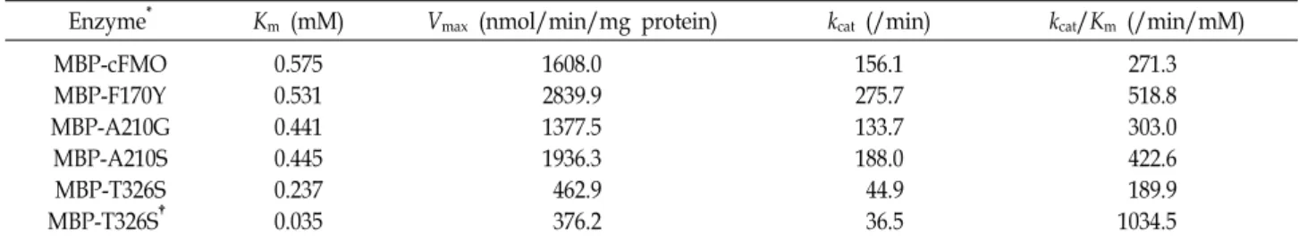
Table 2. Primer lists used in this study
Primer Sequence (5'-3') Restriction enzyme
site or comments P1
P2 P3 P4 P5 P6 P7 P8 P9 P10
CCCGGAATTCATGGAGATGGTTATGAAGAA CCCCAAGCTTTTAGGCTTTATCGCGGACTT
GGGACGTTCGGGAAGCTGTAGTGTCCAGCGCCAACAA TTGTTGGCGCTGGACACTACAGCTTCCCGAACGTCCC CTTCCGCAGAATAACTTCCACCAATCAGCAAAATA TATTTTGCTGATTGGTGGAAGTTATTCTGCGGAAG TTCCGCAGAATAACTTGAACCAATCAGCAAAATAT ATATTTTGCTGATTGGTTCAAGTTATTCTGCGGAA AGCATCAAACATGTTGAAGCTCAGCCACTGATCCTGAG CTCAGGATCAGTGGCTGAGCTTCAACATGTTTGATGCT
EcoRI HindIII F170Y-R F170Y-F A210G-R A210G-F A210S-R A210S-F T326S-R T326S-F
gested with EcoRI and HindIII, ligated with EcoRI/HindIII- cleaved pKK223-3, respectively, and yielded plasmids pK- F170Y, pK-A210G, pK-A210S, and pK-T326S (Table 1).
Expression and purification of mutant MBP-cFMOs E. coli Top10 cells expressing mutant MBP-cFMOs were cultured at 28℃ in LB (10 g/l tryptone, 5 g/l yeast extract, and 10 g/l NaCl) broth with shaking at 180 rpm. Expression of mutant MBP-cFMOs was induced by adding 0.1 mM IPTG into culture broth when cell OD
600nmreached to 0.5-0.6. The purification of mutant MBP-cFMOs was performed accord- ing to previously described method [1]. The purified en- zymes were analyzed by SDS-PAGE and protein concen- trations were measured by the Bradford method (Bio-Rad) with bovine serum albumin as the standard.
Enzyme assay
To investigate biochemical properties of mutant cFMOs, NADPH oxidase activity was measured according to pre- viously reported method [1] with TMA as a substrate. Futile NADPH oxidase activity was determined in the absence of substrate at the same assay condition. Indole oxygenase ac- tivity was determined at 5 mM indole [1]. All enzyme assays described above were performed in triplicate experiments.
Production of indigoid and analyses of indigo, in- dirubin, and tryptophan
Production of indigo and indirubin was performed in LB medium containing 2.5 g/l of tryptophan with IPTG in- duction in a shaking incubator at 32℃ for 48 hr using re- combinant E. coli W3110 expressing mutant cFMOs. The amounts of indigo, indirubin, and tryptophan were analyzed by HPLC [12]. All cell cultures were performed in triplicate
experiments.
Results and Discussion
Construction of mutant MBP-cFMOs based on rational design
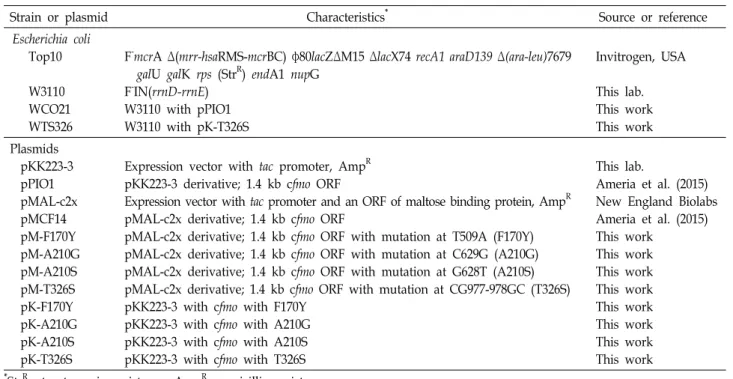
Although the structure of cFMO has not been resolved so far, we could build a reliable 3D structure model by ho- mology modeling because its sequence has a quite high iden- tity with mFMO (56%), of which structure data were pub- lished (PDB ID: 2XVE) [3]. The model of cFMO is super- imposed very well with crystal structure of mFMO, and the RMS (root mean square) of the overlapped structures was only 0.063 Å. Under the visual inspection of the cFMO mod- el, three residues which are located closely enough to co- factors and substrates at the catalytic site were chosen (Fig.
1). First, the benzyl side chain of F170 was found to be lo- cated at a distance of 3.4 Å and 3.6 Å from the hydroxyl group of C-2 in the ribitol of FAD and that of C-3 in the nicotinamide ribose of NADPH, respectively, which seems to affect affinity between cFMO and cofactors. Second, in order to accommodate the cofactor NADPH, A210 in GASYA loop region (209-213 amino acid residues) which is thought to an important role for NADPH binding was picked and replaced to glycine or serine. Third, T326 is lo- cated closer than 3.5 Å from isoalloxazine ring of FAD and may be had an impact on the interaction with FAD. Based on these ideas, four MBP-cFMO mutants F170Y, A210G, A210S, and T326S were constructed in E. coli by site-directed mutagenesis.
Biochemical properties of mutant MBP-cFMOs
In order to characterize biochemical properties of mutant
Table 3. Kinetic parameters for mutant MBP-cFMOs from C. glutamicum using TMA as the substrate
Enzyme
*K
m(mM) V
max(nmol/min/mg protein) k
cat(/min) k
cat/K
m(/min/mM) MBP-cFMO
MBP-F170Y MBP-A210G MBP-A210S MBP-T326S MBP-T326S
†0.575 0.531 0.441 0.445 0.237 0.035
1608.0 2839.9 1377.5 1936.3 462.9 376.2
156.1 275.7 133.7 188.0 44.9 36.5
271.3 518.8 303.0 422.6 189.9 1034.5
*
Kinetic parameters for wild-type and mutant MBP-cFMOs were determined at 2 μM FAD.
†
Kinetic parameters for mutant MBP-T326S were determined at 100 μM FAD.
Fig. 1. The detailed 3D structure model around catalytic active site in cFMO. FAD and NADP are represented by red and white colors, respectively, and three mutation points are shown by balls and sticks, which are F170, A210, and T326.
Fig. 2. Indole oxygenase activity of mutant MBP-cFMOs. Indole oxygenase activity of mutant MBP-cFMOs was meas- ured by the fluorescence of indoxyl in the presence of 5 mM indole and 2 μM FAD except MBP-T326S shown in the rightmost bar in which the activity was de- termined at 100 μM FAD.
enzymes, kinetic studies of the purified four mutants as well as wild-type MBP-cFMO were performed using TMA as a substrate (Table 3). Mutant proteins except for T326S dis- played similar K
mand increased k
cat/K
mvalues compared to the wild-type. In particular, MBP-F170Y had 1.9-fold high- er k
cat/K
mthan control protein. When MBP-cFMOs were purified by using amylose resin, most purified mutants showed yellow color, but not MBP-T326S protein in which the binding affinity for FAD seemed to be greatly decreased due to the structural change. Wild-type and other mutants required 2 μ M of FAD for optimal activity [1], while T326S mutant required 100 μ M of exogenous FAD (data not shown). T326S had 16.4- and 3.8-folds increased substrate affinity and k
cat/K
mfor TMA, respectively, at 100 μ M FAD compared to those of MBP-cFMO at 2 μ M FAD (Table 3).
The indole oxygenase activity was also determined using wild-type and mutant MBP-cFMO proteins (Fig. 2). F170Y and A210S mutants showed elevated indoxyl production ac- tivity more than 3.1- and 2.9-folds, respectively, in compar-
ison to MBP- cFMO, whereas T326S displayed a decreased activity regardless of FAD concentrations. According to the proposed catalytic scheme for mFMO [3], NADPH reduces oxidized FAD bound to mFMO, which interacts with an oxy- gen molecule and is converted to peroxyFAD and NADP
+in the reductive half of the reaction. When substrate indole is present, peroxyFAD returns to the oxidized form with re- leasing indoxy and water molecules. However, in the ab- sence of substrate, peroxyFAD might be slowly oxidized and released H
2O
2, which is called as futile NADPH oxidase activity. In this study, wild MBP-cFMO and most mutants showed similar futile activities in the absence of substrate, whereas T326S at both 2 μM and 100 μM FAD had sig- nificantly decreased futile activity compared to the wild-type (Fig. 3).
Effect of mutant cFMOs on indigo and indirubin
production
Fig. 3. Futile NADHP oxidase activity of mutant MBP-cFMOs.
Futile NADHP oxidase activity of mutant MBP-cFMOs was determined at 2 μM FAD in the absence of substrate except for MBP-T326S shown in the rightmost bar in which the activity was determined at 100 μM FAD.
A
B
C
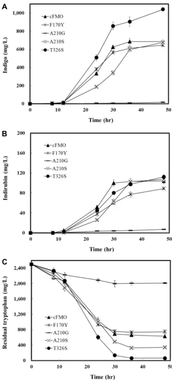
Fig. 4. Production of indigo and indirubin by recombinant E.
coli cells expressing mutant FMOs. (A) and (B) show the production level of indigo and indirubin, respectively, by E. coli cells with mutant FMOs. (C) shows the trypto- phan concentration during cultivation time. Recombinant strains were cultured in LB medium with 2.5 g/l of tryp- tophan for 48 hr at 32℃. Indigo, indirubin, and trypto- phan concentrations were determined by HPLC analyses.
Because the production of indigo and indirubin in E. coli cells harboring wild-type or mutant cFMOs fused to MBP was poor, the production performance of indigoid was eval- uated by using E. coli with wild-type or mutant cFMOs with- out MBP-tag in flask culture. Although the purified MBP- F170Y and MBP-A210S mutants showed higher indole oxy- genase activity, indigoid production was not increased by introduction of the mutant F170Y or A210S into E. coli com- pared with WCO21 (Fig. 4A, Fig. 4B). When E. coli express- ing A210G was cultured for 48 h, less than 20 mg/l of indigo and indirubin were obtained with little consumption of tryp- tophan (Fig. 4). Recombinant E. coli WTS326 harboring pK- T326S produced 1,040 mg/l of indigo and 112 mg/l of in- dirubin with consuming 2.44 g/l of tryptophan, which were 52% and 8%, respectively, higher than those obtained with the control strain expressing cFMO (Fig. 4). This result in- dicated that indigo titer was increased about 13% compared to the previous report in which the highest level, up to date, was 920 mg/l in E. coli expressing mFMO [10]. Since FMOs requires NADPH for a reducing power, an efficient supply or strong regeneration of NADPH pool is crucial for oxygen- ation of indole [15]. Even though MBP-T326S showed 63-77%
indole oxygenase activities compared to MBP-cFMO, low levels of the futile NADPH oxidase activities (21-24%) in MBP-T326S would lead to save the NADPH pool in cells, resulting in higher production of indigoid. Thus, our result suggests that a decreased futile NADPH oxidase activity plays an important role in WTS326 strain for production of indigoid. A previous result regarding indigo production sys-
tem also demonstrated that the expression of glucose de-
hydrogenase in indigo-producing E. coli led to the efficient
regeneration of NADPH and the improved production of
Q323
S326
FAD
T326
2.8 Å 2.9 Å