Factors Influencing Prognosis of Traumatized Tooth in Primary Tooth Intrusion
Yongkwon Chae1, Yoonkyung Han2, Okhyung Nam1, Misun Kim3, Hyoseol Lee1, Kwangchul Kim3, Sungchul Choi1
1Department of Pediatric Dentistry, School of Dentistry, Kyung Hee University
2Chamjoeun Dental Hospital
3Department of Pediatric Dentistry, Kyung Hee University Dental Hospital at Gangdong
The purpose of this study was to investigate the characteristics of intrusion in primary dentition and to evaluate factors influencing complications of primary and permanent dentition during long-term follow-up period.
61 patients (84 teeth) were selected in this study. Medical records of 61 patients were reviewed and age, gender, cause of injury, site of injury, severity of traumatic injury, other injuries associated with trauma, treatment method, and complications of primary and permanent dentition were examined. Collected data were statistically evaluated using Chi- square test and Fisher’s exact test.
Intrusion in primary anterior teeth was predominant in boys over girls and fall was the most common cause of trauma.
It was most common at home and occurred most in the primary maxillary central incisors. Severity had an effect on the incidence of sequelae in permanent successors (p = 0.014). The incidence of complications was significantly lower in patients with soft tissue injuries than in patients with other periodontal injuries (p = 0.000).
Key words : Tooth Injuries, Primary Teeth, Tooth Abnormalities Abstract
Corresponding author : Sungchul Choi
Department of Pediatric Dentistry, School of Dentistry, Kyung Hee University, Kyungheedaero 23, Dongdaemun-gu, Seoul, 02447, Republic of Korea Tel: +82-2-958-9339 / Fax: +82-2-966-4572 / E-mail: [email protected]
Received June 15, 2018 / Revised August 7, 2018 / Accepted August 1, 2018
I. Introduction
Intrusion refers the condition that the traumatized tooth is driven into its socket and this can lead pulp and/or peri- odontal tissue damage and alveolar bone fracture, resulting in pulp necrosis, crown discoloration, pathologic root resorption, or ankylosis[1-3]. Primary tooth intrusion commonly occurs between the ages of 1 and 3 years which corresponds to the time of calcification of the tooth germ of permanent succes- sor[4-6]. Therefore, primary tooth intrusion can affect a wide range of pathologic changes to permanent successors, includ-
ing hypoplasia, crown/root dilacerations, cessation of root de- velopment, and eruption disturbance[1,2,7,8].
In primary dentition, intrusion usually does not occur solely but in the complicated form. Thus when anticipating the prog- nosis of intruded primary teeth, it is reasonable to examine whether the other adjacent tissue such as soft tissue is in- volved together.
Due to spatially and anatomically close relationship between apex of the primary tooth and its permanent successor, intru- sive injuries in primary dentition can lead to sequelae in per- manent dentition. Therefore, the treatment of primary tooth
intrusion should aim to prevent the possible damage to the tooth germ of permanent successors[9]. Therefore, it is neces- sary to identify complications in the intruded primary tooth as well as effect on permanent successors.
The International Association for Dental Traumatology (IADT) recommends different treatment strategies according to the root orientation of affected primary tooth. If apex is labially displaced, the tooth is left for anticipating spontaneous repo- sitioning. Otherwise, the tooth needs removed when apex is displaced palatally[9].
The purpose of this study was to investigate characteristics of primary tooth intrusion and to evaluate factors influenc- ing prognosis of primary and permanent dentition during the long-time follow-up period.
II. Materials and Methods
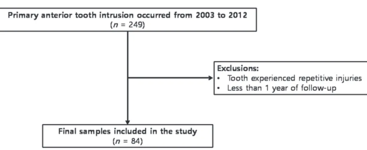
All patients who visited the Department of Pediatric Dentist- ry, School of Dentistry, Kyung Hee University, Seoul, Republic of Korea with chief complaints of traumatic dental injuries (TDIs) in the primary dentition between 2003 and 2012 were ana- lyzed. Only patients who were diagnosed as primary anterior tooth intrusion were included. Patients with special medical history or repetitive TDIs during follow-up periods were ex- cluded (Fig. 1). The study proposal was reviewed and approved by the ethics committee of Kyung Hee University Dental Hos- pital, Kyung Hee University, Seoul, Republic of Korea (KHD- IRB-1408-1).
Based on trauma medical records, we collected data includ- ing clinical history, intraoral photographs and radiographic documentation of all patients. These data were organized ac- cording to age, gender, cause and site of trauma, severity of intrusion, other type of injuries accompanied at the time of in- juries and complications during the follow-up periods in both primary and permanent dentition.
Severity of intrusion was classified into mild, moderate, or severe type by evaluation of clinical photographs and peri- apical radiographs at the time of injury. An adjacent primary incisor with no evidence of displacement was established as a baseline. Comparing with the baseline, it was defined as mild type when greater than half of clinical crown exposure was presented upon the gingival tissue, and moderate type as less than half of clinical crown exposure. Severe type was defined as a total of clinical crown was intruded.
Other types of injury accompanied at the time of TDI oc- curred were divided by classification of Andreasen et al.[2];
soft tissue injuries, hard dental tissue injuries, and periodontal tissue injuries.
1. Injuries to the soft tissue: abrasion, laceration and contusion;
2. Injuries to the hard tissue and the pulp: enamel crack, enamel fracture, enamel-dentin fracture without pulp exposure, enamel-dentin fracture with pulp exposure, enamel-dentin-cementum fracture without pulp exposure, enamel-dentin-cementum fracture with pulp exposure and root fracture;
3. Injuries to the periodontal tissue: concussion, subluxation,
Fig. 1. Flowchart of sample selection for evaluating the clinical outcomes.
displacement, intrusion, extrusion and avulsion.
To evaluate clinical outcomes after primary tooth intrusion, only patients who were followed-up more than 1 year were included. Patients were scheduled for clinical and radiographic examination every 3 - 6 months until the physiologic exfolia- tion of intruded primary tooth and eruption of permanent successor. The follow-up period varied from 0.1 to 8.10 years (mean [SD], 1.8 [1.10] years).
Complication was grouped into 2 categories; (1) complica- tion in primary tooth, and (2) complication in permanent suc- cessor. Crown discoloration, pulp canal obliteration, periapical abscess formation, inflammatory root resorption, and ankylosis were included as complication in primary tooth. To assess clini- cal outcome, clinical success was defined as discoloration only, pulp canal obliteration, and no complication and failure was defined when the traumatized tooth needed further treatment;
periapical abscess formation, inflammatory root resorption, an- kyloses. In permanent successor, enamel hypoplasia and crown shape malformation were recorded.
Data were analyzed using SPSS 22.0 software (SPSS Inc., Chicago, IL, USA). In order to determine the factors that af- fect prognosis, age, severity and type of accompanied injury were examined statistically. As the number of teeth which were followed-up until their successors erupt was small, influence of age and severity on prognosis of permanent tooth was statis- tically analyzed using Fisher’s exact test. For the same reason, difference in incidence of primary tooth complication accord- ing to a type of accompanied injury was analyzed using Fisher’
s exact test. On the other hand, effect of age and severity on primary tooth complication was statistically analyzed with Chi- square test.
III. Results
Of 176 children (249 teeth) who were diagnosed as primary tooth intrusion, 61 children (84 teeth) who met the inclusion criteria were evaluated. The mean age of these samples was 3.1 ± 1.3 years old and most prevalent age was 2 - 3 years in both boy and girl group. The final samples consisted of 40 (65.6%) boys and 21 (34.4%) girls. Intrusion occurred predomi- nantly in males than in females, with a ratio of 1.90:1 (Table 1).
Complication in primary and permanent tooth according to age is presented in Table 2. However, there was no statistical difference in incidence of complication in primary (Table 3, p = 0.197) and permanent tooth (Table 4, p = 0.237) according to age.
Causes and sites of intrusion were as follows (Table 5): Falls (51.2%) were the major sources of intrusion and most of intru- sion occurred at home (56%) and the area around the home (31%).
Intrusion was commonly observed in primary maxillary cen- tral incisors (81%) with no difference between right and left teeth followed by primary maxillary lateral incisors (19%). 40 patients (65.6%) presented with a single affected tooth; 19 patients (31.1%) with 2 affected teeth; 2 patients (3.3%) with multiple affected teeth.
Table 1. Age and gender distribution of patients
Age (year) Male Female Total Number of Patients n (%)
0-1 1 (2.5) 1 (4.8) 2 (3.3)
1-2 8 (20.0) 4 (19.0) 12 (19.7) 2-3 13 (32.5) 5 (23.8) 18 (29.5) 3-4 6 (15.0) 4 (19.0) 10 (16.4) 4-5 10 (25.0) 4 (19.0) 14 (23.0) More than 5 3(7.5) 2 (9.5) 5 (8.2)
Total 40 21 61
Table 2. Age and complication in primary and permanent dentition
Age (year) Total Complication in Primary Tooth Followed-up Cases1 Sequelae in Permanent Successor n (%)
0-2 25 11 (44.0) 11 4 (36.4)
2-5 54 34 (63.0) 12 1 (7.1)
5- 5 1 (20.0) 2 0 (0.0)
1Cases which were followed-up until permanent successors erupt.
According to severity of intrusion, approximately half of tooth intrusion was mild type, followed by moderate type (32.1%) and severe type (14.3%). The severity of intrusion did not affect significantly the incidence of complication (Table 6, p = 0.082). However, it had an effect on the incidence of se-
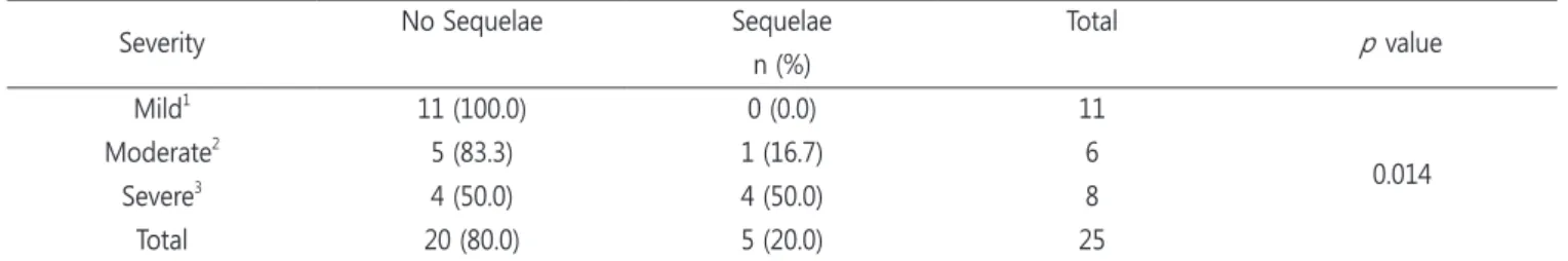
quelae in permanent successors (Table 7, p = 0.014).
Of 84 teeth, 45 teeth (53.6%) exhibited clinical complications in affected primary teeth during the follow-up periods, and 39 teeth (46.4%) did not. Table 8 shows the clinical complications in primary tooth intrusion according to presence of other type of TDIs accompanied on the adjacent primary teeth. When
primary tooth intrusion occurred with other periodontal tissue injuries simultaneously, the affected tooth was more likely to develop clinical complications. However, when intrusion was accompanied with soft tissue injuries, complications were sig- nificantly fewer (Fig. 2).
During the follow-up periods, eruption of 25 permanent successors had completed and complications of permanent successors were observed in 5 teeth (Table 9). In patient 2, though intruded teeth were extracted immediately, consider- able deformation of permanent successor crown was observed (Fig. 3).
Table 5. Cause and site of intrusion according to the age group
Age (year) 0-1 1-2 2-3 3-4 4-5 5- Total
n (%) Cause of Intrusion
Fall 0 (0.0) 11 (57.9) 13 (46.4) 6 (50.0) 11 (64.6) 2 (33.3) 43 (51.2) Collision 2 (100.0) 2 (10.5) 5 (17.9) 3 (25.0) 2 (11.8) 4 (66.7) 18 (21.4)
Drop 0 (0.0) 4 (21.1) 8 (28.6) 3 (25.0) 2 (11.8) 0 (0.0) 17 (20.2)
Traffic accident 0 (0.0) 0 (0.0) 0 (0.0) 0 (0.0) 2 (11.8) 0 (0.0) 2 (2.4)
Unknown 0 (0.0) 2 (10.5) 2 (7.1) 0 (0.0) 0 (0.0) 0 (0.0) 4 (4.8)
Total 2 19 28 12 17 6 84
Site of Intrusion
Inside home 2 (100) 10 (52.6) 21 (75.0) 7 (58.3) 5 (29.4) 2 (33.3) 47 (56.0) Outside home 0 (0.0) 6 (31.6) 4 (14.3) 4 (33.3) 10 (58.8) 2 (33.3) 26 (31.0) Kindergarten 0 (0.0) 1 (5.3) 0 (0.0) 0 (0.0) 1 (5.9) 2 (33.3) 4 (4.8)
Others 0 (0.0) 0 (0.0) 0 (0.0) 1 (8.3) 1 (5.9) 0 (0.0) 2 (2.4)
Unknown 0 (0.0) 2 (10.5) 3 (10.7) 0 (0.0) 0 (0.0) 0 (0.0) 5 (6.0)
Total 2 19 28 12 17 6 84
Table 3. Age and complication in primary tooth Age
(year)
No Complication Complication Total
p value n
0-2 14 11 25
0.197
2- 24 35 59
Total 38 46 84
p value from Chi-square test
Table 4. Age and complication in permanent tooth Age
(year)
No Sequelae Sequelae Total
p value n
0-2 7 4 11
0.237
2-5 11 1 12
5- 2 0 2
Total 20 5 25
p value from Fisher’s exact test
Table 6. Severity of intrusion and complication in primary tooth
Severity No Complication Complication Total
p value n (%)
Mild1 18 (40.0) 27 (60.0) 45
0.082
Moderate2 11 (40.7) 16 (59.3) 27
Severe3 9 (75.0) 3 (25.0) 12
Total 38 (45.2) 46 (54.8) 84
p value from Chi-square test
1When greater than half of clinical crown exposure was presented upon the gingival tissue, 2When less than half of clinical crown exposure, 3When a total of clinical crown was intruded.
Table 7. Severity of intrusion and sequelae in permanent successor
Severity No Sequelae Sequelae Total
p value n (%)
Mild1 11 (100.0) 0 (0.0) 11
0.014
Moderate2 5 (83.3) 1 (16.7) 6
Severe3 4 (50.0) 4 (50.0) 8
Total 20 (80.0) 5 (20.0) 25
p value from Fisher’s exact test
1When greater than half of clinical crown exposure was presented upon the gingival tissue, 2When less than half of clinical crown exposure, 3When a total of clinical crown was intruded.
Fig. 2. Clinical outcomes of primary tooth intrusion accord- ing to presence of accompanying other TDIs to the adja- cent primary teeth.
IN = Intrusion only, INHT = Intrusion accompanied with hard tissue injuries, INPT = Intrusion accompanied with periodontal tissue injuries, INST = Intrusion accompanied with soft tissue injuries, INCT = Intrusion accompanied with complicated tissue injuries.
Table 8. Clinical outcomes of primary teeth intrusions
Favorable Unfavorable
p value None Crown
Discoloration Pulp Canal
Obliteration Total Periapical Abscess
Inflammatory Root
Resorption Ankylosis Total
IN1 11 3 2 14 2 9 3 11
0.000
INHT2 0 1 0 1 3 2 0 3
INPT3 1 0 0 1 5 5 2 10
INST4 15 4 0 19 1 7 0 7
INCT5 4 0 0 4 5 9 2 14
p value from Fisher’s exact test
1Intrusion only, 2Intrusion accompanied with hard tissue injuries, 3Intrusion accompanied with periodontal tissue injuries, 4Intrusion accompanied with soft tissue injuries, 5Intrusion accompanied with complicated tissue injuries.
The same tooth can represent 1 or more complications.
Some of crown discoloration were associated with unfavorable outcome in both groups and were classified into unfavorable outcome.
IV. Discussion
TDIs in the primary dentition can negatively affect the qual- ity of life of children[10,11]. Especially in the children with de- veloping permanent teeth, all kinds of TDIs can have a harmful effect on their quality of life for the whole their lives by caus- Table 9. Sequelae of permanent successors after primary tooth intrusion
Patient No. Age (Month) Gender Tooth No. Intrusion severity Treatment Sequelae of Permanent Successors
1 23 M #61, 62 Severe Reposition Enamel hypoplasia
2 20 M #61, 62 Severe Extraction Crown deformation
3 37 M #62 Moderate Extraction Enamel hypoplasia
ing developmental disorder on permanent teeth. Particularly the intrusive TDI to the primary teeth is likely to harm the successor of theirs due to spatial and anatomical position of primary and permanent teeth.
Most children suffer from primary tooth intrusion in the early stage of their lives. In the present study, higher incidence of intrusion was observed in 2 - 3 years. In this age group, children are likely to experience TDIs due to insufficient motor skills[4,12]. However, even in patients of the same age group, each patient may have all the different degrees of severity.
And this can demonstrate why age was not a factor affecting the prognosis of primary and permanent tooth.
From this study, fall was predominant cause of intrusive dental injury accounting for 51.2% followed by collision and this result is in agreement with previous studies[13-15].
Moreover, approximately 84% of children in this age expe- rienced intrusive luxation at home and around home in the present study. This is because children in this age spend most of their life time at home[16]. Based on these findings, educa- tional program for caregivers regarding prevention and home care of TDIs can be beneficial.
Because of their anterior position in the dental arch, most intrusive luxation occurs in maxillary central incisors[5,7]. In this study, intrusive luxation usually involved a single primary tooth, which corresponded with previous studies[5,12].
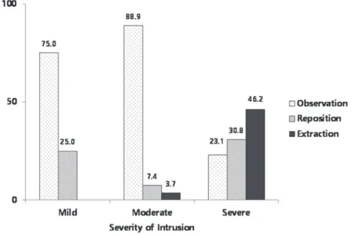
In terms of severity, mild type was most common followed by moderate type in this study. In severe intrusion cases, per- manent successor presented unfavorable outcomes more fre- quently. Therefore, it is reasonable to rely not only on the path or direction of intrusion but also on the remaining portion of intruded tooth when selecting the treatment method and anticipating the prognosis of involved tooth. In the present study, 75% of mild type teeth, 88.9% of moderate type teeth and 23.1% of severe type teeth were observed without any in- tervention. And reposition was performed to 25% of mild type teeth, 7.4% of moderate type teeth and 30.8% of severe type tooth (Fig. 4).
Fig. 3. Long-time follow up of patient 2. (A) Initial periapi- cal radiograph showing intrusion of left maxillary primary incisors. Displacement of permanent tooth germ was revealed. (B) Progressing periapical radiograph at the 3 months follow-up. (C) Progressing periapical radiograph at the 3 years follow-up showing crown deformation in both permanent incisors. (D) Progressing periapical radiograph at the 6 years follow-up. Large extent of crown deformation was observed in both permanent incisors.
A B
C D
In this study, 45 teeth (53.6%) of 84 teeth presented unfa- vorable outcomes in the primary teeth during the periodical check-up. Borum and Andreasen[17] reported inflammatory root resorption accounted for 14% of complications in the primary teeth following intrusion. Whereas in this study the inflammatory root resorption (71.1%) was the most common complication and followed by periapical abscess (35.6%). In the previous studies, the percentage of developmental disturbanc- es of the permanent successors after intrusive dental injury ranges from 18% to 69%[6,18,19]. Likewise, 5 teeth (20.0%) of 25 successors presented complication in the permanent teeth in the study.
As most of intrusive dental injuries were consequences of fall accident, intrusive dental injuries usually occur in multiple teeth and are accompanied by multiple site injuries such as hard and soft tissues. In the present study, the intruded teeth accompanied with periodontal tissue injury showed unfavor- able outcomes more frequently. While the intruded teeth ac- companied with soft tissue injury were more likely to present favorable outcomes. This can be explained by the fact soft tissues like gingiva and buccal mucosa are softer than hard tissue and can absorb and favorably distribute impact from trauma[20-22].
However, there are several limitations in this study. First, since this study was performed retrospectively based on medi-
Fig. 4. Distribution of treatment modalities according to severity of intrusion.
cal records, there was a possibility that majority of complicated type of injuries were included in ‘intrusion only’ group. For example concussion or subluxation of adjacent primary inci- sors could be ignored during the examination due to young patient’s age. Second, as the number of intruded primary teeth which were followed-up until their permanent successors erupt was small, we could not statistically determine the effect of in- trusive injuries on permanent successors development. None- theless, this study is significant in that a research on sequelae of permanent successors was carried out through long-term follow-up and in that this study determined factors influencing the outcome of intruded primary teeth and their successors.
This retrospective study suggested that the severity of primary tooth intrusion can influence prognosis of permanent succes- sor and when intrusion occurs with soft tissue injuries, trauma- tized primary tooth is likely to show favorable prognosis.
V. Conclusion
In this retrospective study, the intrusion occurred most fre- quently at home and fall accident was major cause of intru- sion. Therefore caregivers such as parents should be trained about prevention of TDIs and should be encouraged to visit dental hospital in order to confirm whether the permanent successor is involved. Furthermore the clinician should provide
periodic recall plan to the parents and exact periodic check with radiographs.
Within the limits of this study, age was not a factor which influence incidence of complication. However, severity of intru- sion had an effect on incidence of sequelae in permanent suc- cessor and intrusion accompanied with soft tissue injury was likely to present better prognosis and show fewer complica- tion.
On the basis of present study, when choosing a treatment method and predicting prognosis of intruded teeth, the clini- cian needs to depend not only on the intrusion path or direc- tion, but also on the depth of intrusion or portion of crown above the gingiva and type of accompanied injuries.
Since the number of samples used in this study is limited, further studies with larger number of samples and longer follow-up period are needed.
References
1. Diab M, elBadrawy HE : Intrusion injuries of primary inci- sors. Part II: Sequelae affecting the intruded primary inci- sors. Quintessence Int, 31:335-341, 2000.
2. Andreasen JO, Andreasen FM, Andersson L : Textbook and color atlas of traumatic injuries to the teeth, 4th ed. Black- well Munksgaard, Cophenhagen, 337-371, 516-541, 542- 576, 2007.
3. Tarján J, Balaton P, Kéri I : Consequence and therapy of pri- mary tooth intrusion. J Int Assoc Dent Child, 19:25-28, 1988.
4. Altun C, Cehreli ZC, Güven G, Acikel C : Traumatic intrusion of primary teeth and its effects on the permanent succes- sors: a clinical follow-up study. Oral Surg Oral Med Oral Pathol Oral Radiol Endod, 107:493-498, 2009.
5. Colak I, Markovic D, Milenkovic A, et al. : A retrospective study of intrusive injuries in primary dentition. Dent Trau- matol, 25:605-610, 2009.
6. Zilberman Y, Fuks A, Lustmann J, et al. : Effect of trauma to primary incisors on root development of their permanent successors. Pediatr Dent, 8:289-293, 1986.
7. Gondim JO, Moreira Neto JJ : Evaluation of intruded pri- mary incisors. Dent Traumatol, 21:131-133, 2005.
8. Onetto JE, Flores MT, Garbarino ML : Dental trauma in children and adolescents in Valparaiso, Chile. Endod Dent Traumatol, 10:223-227, 1994.
9. Malmgren B, Andreasen JO, Tsukiboshi M, et al. : Interna- tional Association of Dental Traumatology guidelines for
the management of traumatic dental injuries: 3. Injuries in the primary dentition. Dent Traumatol, 28:174-182, 2012.
10. Cortes MI, Marcenes W, Sheiham A : Impact of traumatic injuries to the permanent teeth on the oral health-related quality of life in 12-14-year-old children. Community Dent Oral Epidemiol, 30:193-198, 2002.
11. Ramos-Jorge ML, Bosco VL, Peres MA, Nunes AC : The im- pact of treatment of dental trauma on the quality of life of adolescents - a case-control study in southern Brazil. Dent Traumatol, 23:114-119, 2007.
12. Kramer PF, Zembruski C, Ferreira SH, Feldens CA : Trau- matic dental injuries in Brazilian preschool children. Dent Traumatol, 19:299-303, 2003.
13. Fleming P, Gregg TA, Saunders ID : Analysis of an emer- gency dental service provided at a children’s hospital. Int J Paediatr Dent, 1:25-30, 1991.
14. Sandalli N, Cildir S, Guler N : Clinical investigation of trau- matic injuries in Yeditepe University, Turkey during the last 3 years. Dent Traumatol, 21:188-194, 2005.
15. Skaare AB, Jacobsen I : Primary tooth injuries in Norwegian children (1-8 years). Dent Traumatol, 21:315-319, 2005.
16. Carvalho V, Jacomo DR, Campos V : Frequency of intrusive luxation in deciduous teeth and its effects. Dent Traumatol, 26:304-307, 2010.
17. Borum MK, Andreasen JO : Therapeutic and economic implications of traumatic dental injuries in Denmark: an estimate based on 7549 patients treated at a major trauma centre. Int J Paediatr Dent, 11:249-258, 2001.
18. von Arx T : Developmental disturbances of permanent teeth following trauma to the primary dentition. Aust Dent J, 38:1-10, 1993.
19. Tewari N, Mathur VP, Pandey RK, et al. : Long-term effects of traumatic dental injuries of primary dentition on perma- nent successors: A retrospective study of 596 teeth. Dent Traumatol, 34:129-134, 2018.
20. Choi WJ, Russell CM, Robinovitch SN, et al. : Age-related changes in dynamic compressive properties of trochanteric soft tissues over the hip. J Biomech, 48:695-700, 2015.
21. Choi WJ, Robinovitch SN : Pressure distribution over the palm region during forward falls on the outstretched hands. J Biomech, 44:532-539, 2011.
22. Robinovitch SN, McMahon TA, Hayes WC : Force attenua- tion in trochanteric soft tissues during impact from a fall. J Orthop Res, 13:956-962, 1995.
국문초록
유치 함입 시 외상 치아의 예후에 영향을 미치는 요인
채용권1ㆍ한윤경2ㆍ남옥형1ㆍ김미선3ㆍ이효설1ㆍ김광철3ㆍ최성철1
1경희대학교 치의학전문대학원 소아치과학교실
2참조은치과병원
3강동경희대학교병원 치과병원 소아치과
본 연구는 유치열에서 발생하는 함입성 손상의 특징을 조사하고 장기간의 추적 조사 기간 동안의 유치열 및 영구치열에 발생하는 합병증에 영향을 미칠 수 있는 요인을 평가하고자 하였다.
이를 위해 총 61명의 84개의 치아가 연구대상으로 선정되어 환자의 연령, 성별, 외상의 원인과 발생장소, 외상 치아의 손상 정도, 외 상 발생 시 동반된 다른 손상, 치료방법, 추적조사 기간 중 유치열과 영구치열에 발생한 합병증이 조사되었다. 연령, 외상 치아의 손상 정도, 외상 발생 시 동반된 다른 손상의 종류가 유치 및 영구치의 예후에 미치는 영향을 확인하기 위해 조사된 자료는 카이제곱 검정 과 Fisher의 정확검정으로 평가하였다.
유전치의 함입은 남아에게 여아보다 우세하게 나타났으며, 함입의 원인으로는 낙상의 빈도가 가장 높았다. 유전치의 함입은 집에서 가장 많이 발생했으며, 상악 유중절치에서 가장 많이 발생했다. 유치열에 발생한 함입의 중증도는 영구치열에 발생하는 후유증의 발생 에 유의하게 영향을 미쳤다(p = 0.014). 유치의 함입성 손상에 연조직 손상이 동반된 경우, 다른 치주조직 손상이 동반된 경우 보다 합 병증의 발생이 유의하게 적었다(p = 0.000).