J Nuclear Medicine Technology Vol. 12, No. 1, May 2008
∙ Received: October 15, 2007. Accepted: October 30, 2007.
∙ Corresponding author: Jae Sam Kim
Department of Nuclear Medicine, Severance Hospital, Yonsei University Health System, Seoul, 120-749, Korea
Tel: +82-2-2228-6077, Fax: +82-2-2227-7062 E-mail: nucmed@yuhs.ac
Original Article
두경부 혈관종 진단시99m
Tc-RBC Scanand SPECT 검사의 유용성
연세의료원 세브란스병원 핵의학과
오신현⋅노동욱⋅안샤론⋅박훈희⋅이승재⋅강천구⋅김재삼⋅이창호
Usefulness of
99mTc-labeled RBC Scan and SPECT in the Diagnosis of Head and Neck Hemangiomas
Shin Hyun Oh, Dong Wook Roh, Sha Ron Ahn, Hoon-Hee Park, Seung Jae Lee, Chun Goo Kang, Jae Sam Kim, Chang Ho Lee
Department of Nuclear Medicine, Severance Hospital, Yonsei University Health System
Purpose: There are various methods to diagnose hemangioma, such as ultrasonography (US), computed tomography (CT), magnetic resonance imaging (MRI) and nuclear medicine. However, by development of SPECT imaging, the blood-pool scan using 99mTc-labeled red blood cell has been used, because it was non-invasive and the most economical method. Therefore, in this study, we proposed that the usefulness of 99mTc-RBC scan and SPECT of the head and neck to diagnose unlocated hemangiomas. Materials and Methods: 99mTc-RBC scan and SPECT was performed on 6 patients with doubtful hemangioma (4 person, head; 1 person, neck; 1 person, another). We labeled radiopharmaceutical using modified in vivo method and then, centrifuged it to remove plasma.
After a bolus injection of tracer, dynamic perfusion flow images were acquired. Then, anterior, posterior, both lateral static blood-pool images were obtained as early and 4 hours delayed. SPECT was progressed 64 projections per 30 seconds. Each image was interpreted by physicians, Nuclear medicine specialist, and technologist blinded to patient’s data. Results: In 5 patients of all the radioactivity of doubtful site didn’t change in flow images, but, in blood-pool, delayed and SPECT images, it was increased. So, it was a typical hemangioma finding. The size of lesion was over 2 cm, and it could discriminate as comparing to the delayed and SPECT imaging. On the other hand, in 1 patient, the radioactivity was increased in blood-pool images, but, not in delayed and SPECT images, so, it was proved no hemangioma. Conclusion: Using 99mTc-RBC Scan and SPECT, we could diagnose the hemangiomas in head and neck, as well as, liver, more non-invasive, economical, and easy. Therefore, it considered that 99mTc-RBC scan and SPECT would offer more useful information for diagnosis of hemangioma, rather than otherimaging such as US, CT, MRI.
(Korean J Nucl Med Technol 2008;12(1):39-43)
Key Words : Hemangioma, Head and Neck, 99mTc-labeled RBC, SPECT
서 론 혈관종의 진단에 초음파(US), 전산화단층촬영영상(CT), 자기공명영상(MRI), 핵의학적 검사 등 다양한 검사방법이 사 용될 수 있지만 99mTc 표지 적혈구를 이용한 혈액풀 영상검사 는 1970년대에 처음으로 소개된 이래 오늘날까지도 혈관종을 확진하고 배제할 수 있는 가장 경제적이고 확실한 방법으로 사용되고 있다.1) 더욱이 감마카메라가 발달됨으로, 특히 단일 광자단층촬영(SPECT)의 발달로 삼차원적 영상정보를 얻을
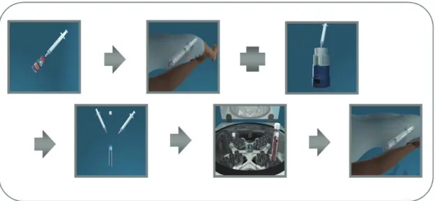
Fig. 1. RBCs were labeled radiopharmaceutical using modified in vivo method.
Fig. 2. Dynamic perfusion flow images, anterior, posterior, both lateral static blood-pool images were obtained as early and 4 hours delayed, and then SPECT images.
수 있는 장점을 가지고 있기 때문에 혈관종을 발견하는데 자 기공명영상의 몇 가지 예외적인 경우를 제외하면 다른 영상 검사가 능가할 수 없을 정도의 예민도와 특이도가 높은 검사 로 발전되어 왔다.2-6) 또한 표지 적혈구 혈액풀 영상검사는 가 격면에서 비교적 저렴하여 경제적일 뿐 아니라 비침습적이고 시행하기도 쉽고 비교적 정확하게 혈관종을 진단할 수 있다.7,8)
본 연구에서는 간 혈관종 외 두경부(head and neck) 혈관 종 진단시 99mTc-RBC scan and SPECT 검사의 유용성에 대 하여 알아보고자 한다.
대상 및 방법
1. 환자 정보
혈관종의 진단을 위해 2006년 11월부터 2007년 8월까지 본원 핵의학과에 내원한 6명의 환자를 (연령범위 32-62세, 평 균연령 42.8세, 남자 4명, 여자 2명) 대상으로 하였다. 6명의 환자 중 head 부위 4명, neck 부위 1명은 혈관종이라 추정 하였고 전산화단층촬영영상, 자기공명영상 검사로 혈관종 으로 확진하기 어렵다고 생각된 환자는 1명이었다.
2. 방사성 의약품
표지방법은 modified in vivo method를 사용하여 20 mCi
오신현 외 7인. 두경부 혈관종 진단시 99mTc-RBC Scan and SPECT 검사의 유용성
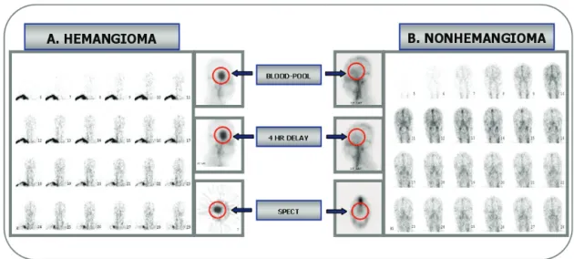
Fig. 3. (A) The radioactivity didn’t change in flow images. But, in blood-pool, delayed and SPECT images, it was increased. (B) The radioactivity was increased in blood-pool images, but, not in delayed and SPECT images.
Table 1. Comparison of Patients Analysis
Patients A B C D E F
Size (cm) 2 3 4.3 5.6 5.4 4
Blood-pool (cnts) 35144 41323 46985 50661 47894 49769
4 HR delayed (cnts) 36449 45625 51058 60332 72787 44322
Rate of increase & decrease (%) +3.71 +10.41 +8.67 +19.1 +51.2 -10.94
99mTc를 적혈구에 표지하고 원심침전시켜 혈장을 제거한 다음 99mTc-RBC를 순간주사 하였다(Fig. 1).
3. 영상획득과정
검사장비로는 PHILIPS FORTE (JET stream-R Forte version 1.2) 감마카메라를 사용하였으며, LEHR (Low Energy High Resolution) collimator를 장착하여 검사하였다.
평면영상은 2초당 24 frame의 혈류기 영상을 얻은 후 즉시 80만 계수의 전면상, 후면상, 양측면상의 혈액풀 영상을 얻었 고 4시간 후 80만 계수의 전면상, 후면상, 양측면상의 지연 영 상을 얻었다.9)
SPECT 영상은 4시간 지연 영상 획득 후 Dual head 검출 기를 사용하여 64 projections (180°)을 시행하였고 각 pro- jection당 30초간 집적하였으며 Butterworth Filter를 사용 하여 pixel당 0.58 mm 두께로 재구성하였다(Fig. 2).
모든 영상을 재구성한 후에 3 명의 핵의학과 전문의, 핵의 학과 전공의 및 핵의학과 방사선사가 특별한 정보 없이 영상 을 검토하였다.
결 과
혈관종으로 추정된 5명의 환자는 혈류기 영상에서 방사 능은 증가하지 않았고 혈액풀 영상에서 증가하였으며 지연 영상과 SPECT 영상에서 증가된 소견을 보여 전형적인 혈 관종 소견을 보였다.10-16) 혈관종이 아닌 1명의 환자는 혈류기 영상과 혈액풀 영상에서 방사능 증가를 보였고, 지연 영상과 SPECT 영상에서 감소된 소견을 보여 혈관종이 아닌 것으로 판정되었다(Fig. 3).
5명의 혈관종 환자의 병소 크기는 모두 2 cm 이상이었고 혈액풀 영상과 4시간 지연영상에 ROI를 그려 카운트를 측정 해본 결과 초기 혈액풀 영상과 비교하여 최고 51.1%의 증가 율을 보여 혈관종 소견을 보였고, 1명의 환자는 10.94%의 감 소율을 보여 비혈관종이라 판정되었다(Table 1).
CT, MRI, SPECT 영상을 비교하여 혈관종과 수막종을 감별을 할 수 있었다(Fig. 4).
연구의 제한점
Fig. 4. Comparison of CT, MRI, SPECT images between hemangioma and meningioma.
이 연구는 단 6명의 환자에게서 얻은 영상을 분석하였기 때문에 통계학적인 타당성에 있어 연구의 제한점이 나타날 가능성이 있다. 초음파 검사는 혈관종 진단에 특이한 소견을 보이나, 뼈로 구성된 두경부 검사에는 적용이 어려워 타 검사 영상과 비교하지 못했다.
결 론
본 연구에서는 간혈관종뿐만 아니라 두경부 혈관종이 의 심되는 경우에는 가격면에서 비교적 경제적이고 비침습적 이며 쉽게 시행할 수 있는 99mTc-RBC scan and SPECT 검사 를 시행함으로써 초음파, 전산화단층촬영영상, 자기공명영상 검사와 같은 다른 영상진단법과 함께 혈관종을 확진하고 배제하는데 의미있는 영상의학적 정보를 제공할 수 있다고 사료된다.
요 약
혈관종의 진단에 초음파(US), 전산화단층촬영영상(CT), 자기공명영상(MRI), 핵의학 검사 등 다양한 검사방법이 사용 될 수 있지만 99mTc 표지 적혈구를 이용한 혈액풀 영상검사는 단일광자단층촬영(SPECT)의 발달로 혈관종을 확진하고 배 제할 수 있는 가장 경제적일 뿐 아니라 비침습적이고 시행 하기도 쉬운 확실한 방법으로 사용되고 있다. 본 연구에서는 간 혈관종 외 두경부 (head and neck) 혈관종 진단시 99mTc- RBC scan and SPECT 검사의 유용성에 대하여 알아보고자
한다.
혈관종의 진단을 위해 본원 핵의학과에 내원한 6명의 환자 를 대상으로 하였다. 6명의 환자 중 두부 부위 4명, 경부 부위 1명은 혈관종이라 추정하였고, 1명은 혈관종으로 의심되었 다. 변형 체내 표지법(modified in vivo method)을 사용하여 표지하고 원심침전시켜 혈장을 제거한 다음 99mTc-RBC를 순 간주사하였다. 혈류기 영상을 얻은 후 즉시 전면상, 후면상, 양측면상의 혈액풀 영상을 얻었고 4시간 후 동일한 지연 영 상을 얻었다.
SPECT 영상은 이중 검출기(dual head detector)를 이용하 여 64 projections (180°)을 시행하였고 각 projection당 30초간 집적하였다. 모든 영상을 재구성한 후에 3명의 핵의학과 전 문의, 핵의학과 전공의 및 핵의학과 방사선사가 특별한 정보 없이 영상을 검토하였다.
혈관종으로 추정된 5명의 환자는 혈류기 영상에서 방사능 은 증가하지 않았고 혈액풀 영상에서 증가하였으며 지연 영 상과 SPECT 영상에서 증가된 소견을 보여 전형적인 혈관종 소견을 보였다. 혈관종이 아닌 1명의 환자는 혈류기 영상과 혈액풀 영상에서 방사능 증가를 보였고, 지연 영상과 SPECT 영상에서 감소된 소견을 보여 혈관종이 아닌 것으로 판정되 었다. 5명의 혈관종 환자의 병소 크기는 모두 2 cm 이상이었 고 SPECT 영상이 지연영상과 비교하여 뚜렷하게 병소 감별 을 할 수 있었다.
간혈관종뿐만 아니라 두경부 혈관종이 의심되는 경우에는 비교적 경제적이고 비침습적이며 쉽게 시행할 수 있는 99mTc- RBC scan and SPECT 검사를 시행함으로써 초음파, 전산화
오신현 외 7인. 두경부 혈관종 진단시 99mTc-RBC Scan and SPECT 검사의 유용성
단층촬영영상, 자기공명영상 검사와 같은 다른 영상진단법과 함께 혈관종을 확진하고 배제하는데 의미있는 영상의학적 정 보를 제공할 수 있다고 사료된다.
REFERENCES
1. Front D, Hardoff R, Israel O, Schneck SO. Perfusion vascularity mismatch in liver hemangiomas. Clin Nucl Med 1978;3:212-3.
2. Bree RL, Schwab RE, Glazer GM, Fink-Bennett D. The varied appearances of hepatic cavernous hemangiomas with sonography, computed tomography, magnetic resonance imaging and scinti- graphy. RadioGraphics 1987;7:1153-1175.
3. Birnbaum BA, Weinreb JC, Megibow AJ, et al. Definitive diagnosis of hepatic hemangiomas: MR imaging versus Tc-99m- labeled red blood cell SPECT. Radiology 1990;176:95-101.
4. Choi BI, Han MC, Kim CW. Small hepatocellular carcinoma versus small cavernous hemangioma: Differentiation with MR imaging at 2.0 T. Radiology 1990;176:103-106.
5. Krause T, Hauenstein K, Studier-Fischer B, Schuemichen C, Moser E. Improved evaluation of technetium-99m-red blood cell SPECT in hemangioma of the liver. J Nucl Med 1993;34:375-80.
6. Ziessman HA, Silverman PM, Patterson J, Harkness B, Fahey FH, Zeman RK, et al. Improved detection of small cavernous hemangiomas of the liver with high-resolution three-headed SPECT. J Nucl Med 1991;32:2086-91.
7. Front D, Israel O, Kieinhaus U. Gdal-On M. Technetium-99m- red blood cells in the evaluation of hemangiomas of the skull and orbit: concise communication. J Nucl Med 1982;23:1080-1084.
8. Front D, Groshar D. Israel O, Robinson E. Hemangioma of tongue demonstrating a perfusion blood-pool mismatch. Clin Nucl Med 1986:11:113-114.
9. Front D, Israel O, Groshar D, Weininger J. Technetium-99m- labeled red blood cell imaging. Semin Nucl Med 1984;14:226-50.
10. Front D, Hardoff R, Israel O, Schneck SO. Perfusion vascularity mismatch in liver hemangiomas. Clin Nucl Med 1978;3:212-3.
11. Rabinowitz SA, McKusick KA, Strauss HW: 99mTc red blood cell scintigraphy in evaluating focal liver lesions. AJR 1984;143:63-8.
12. Front D, Israel O, Joachims H, Brown Y, Eliachar I. Evaluation of hemangiomas with technetium 99m-labeled RBCs. The per- fusion bloodpool mismatch. JAMA 1983;249:1488-90.
13. Murata Y, et al. Perfusion and Blood-Pool Scintigraphy in the Evaluation of Head and Neck Hemangiomas. J Nucl Med 1997;
38:882-885.
14. Ennio P, et al. Technetium Tc 99m–Labeled Red Blood Cells in the Preoperative Diagnosis of Cavernous Hemangioma and Other Vascular Orbital Tumors. Arch Ophthalmol. 2005;123:1678-1683.
15. Burroni L, et al. The Tc 99m-RBC SPET in the diagnosis of orbital cavernous hemangioma. Q J Nucl Med. 2000;44:70.
16. Moon D-H, et al. Diagnosis of Hepatic Hemangioma with 99mTc-Labeled Red Cells and Single Photon Emission Computed Tomography (SPECT). K J Nucl Med. 1991;25:68-75.