Polymerase chain reaction of the vitreous was positive for CMV DNA
3
0
0
전체 글
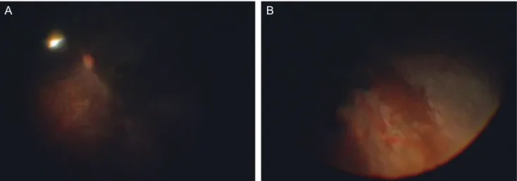
(2) Korean J Ophthalmol Vol.27, No.1, 2013. she had used an anti-glaucoma agent because of increased intraocular pressure of the left eye. At presentation, vision of the left eye was hand motion only. Slit lamp examination demonstrated 4+ cells and hyphema in the anterior chamber and iris neovascularization. Funduscopy revealed dense vitritis and retinal vascular obliteration. Ocular ischemia was suspected through fluorescein angiography which revealed arterial filling delay. No abnormal findings were observed in carotid Doppler sonography which was performed to rule out ocular ischemic syndrome. Examination of the right eye was unremarkable except diabetic retinopathy and scarring from panretinal photocoagulation. Further detailed examination was needed to draw the diagnosis and treatment plan but dense vitritis disturbed further evaluation. As a result, a pars plana vitrectomy was performed. During the vitrectomy, necrotizing retinitis with dense retinal whitening and hemorrhage along the inferotemporal vascular arcade was observed, suggestive of infectious retinitis (Fig. 1). The undiluted vitreous sample acquired by vitrectomy was analyzed by polymerase chain reaction (PCR; Q-CMV real time complete kit, Nanogen Advanced Diagnostics, Turin, Italy) and cultured for herpes simplex virus (HSV), varicellar zoster virus (VZV), and CMV. To rule out other etiologies of infectious retinitis, vitreous was also analyzed by staining and culture for bacteria and fungus. Blood tests did not show any immune dysfunction and complete blood count was normal. CD4 and CD8 cells counts were also within the normal range, 522 and 275 cells/μL. Human immunodeficiency virus (HIV) antigen and antibodies were negative. Her serum CMV IgG level was 244.5 units (range of nonreactive <6.0 AU/mL) and IgM was negative. Although the CMV antigenemia test showed positive results (9/20,000 cells) using the Biotest CMV Brite kit (Biotest Diagnostics, Denville, NJ, USA),. A. there were no other clinical manifestations of CMV infection except retinitis. While the culture for CMV was negative, PCR for CMV DNA was positive in the vitreous and negative in peripheral blood, confirming the diagnosis as CMV retinitis. The others including HSV and VZV were negative in the vitreous and blood. Neither bacteria nor fungus were observed by staining and culture. The patient underwent intravenous administration of ganciclovir (2.5 mg/kg/12 hr), a half dose reduction because of renal insufficiency. Necrotizing retinitis with retinal whitening and iris neovascularization had markedly decreased, but obliterated vessels were sustained without recovery of nonperfusion. After 17 days of administration, intravenous treatment was discontinued. During the follow-up period of 12 months, there has been no recurrence of retinitis, but her left eye could not perceive light because of optic nerve atrophy and retinal ischemia due to obliteration of retinal vessels.. Discussion While CMV retinitis is a well-known opportunistic infection usually affecting immunocompromised patients, there have also been reports of CMV retinitis in immunocompetent patients after intraocular steroid injection [9-11] or fluocinolone acetonide (Retisert) implant [12]. Recent reports revealed CMV retinitis after intravitreal bevacizumab injection combined with subtenon steroid injection [13]. Although the mechanism of CMV retinitis in immunocompetent patients is unclear, these reports suggested the immunosuppressive effect of steroids might provoke the reactivation of latent CMV. However, unlike steroids, bevacizumab, which was used in this case, has no immunosuppressive effects.. B. Fig. 1. Fundus photograph of left eye taken during pars plana vitrectomy. Note the retinal vascular obliteration (A) and inferotemporal confluent necrotizing retinitis associated with retinal whitening (B). Inferior panretinal photocoagulation burns are also can be seen. 62.
(3) SH Bae, et al. CMV Retinitis after Intravitreal Bevacizumab. In this case, fundus examination during vitrectomy revealed a necrotizing retinitis with retinal opacification, but several findings were not compatible with typical CMV retinitis such as severe reactions of the anterior chamber with iris neovascularization and retinal vascular obliteration. The possible explanation for those findings is deterioration of ischemic changes caused by superimposed diabetic retinopathy. To rule out the accompanying active systemic CMV infection, we conducted the following tests; quantitative PCR in the plasma, antigenemia assay in peripheral blood leukocyte and cultures of peripheral blood and urine. The only test showing positive results was the antigenemia assay; all others were negative. However, antigenemia is a semi-quantitative assay and quantitative measurements by quantitative PCR might be more reliable for diagnosis of active CMV disease. PCR using plasma detects only free virion indicative of active viral replication with a higher clinical relevance as compared with antigenemia assay using leukocytes [14,15]. Except for CMV retinitis, the patient did not show clinical manifestations of systemic CMV diseases according to standard criteria [16] at the time of examination and during the follow-up period. Also, there was no evidence of abnormalities in the systemic immune system. After considering these findings, we reached the conclusion that she had no active systemic CMV infection other than CMV retinitis and retinitis was strongly suspected to correlate with the bevacizumab injection. Disruption of blood retinal barrier by diabetic retinopathy might play a role on the development of CMV retinitis [17], but the pathogenesis of CMV retinitis after bevacizumab injection is still unclear. The authors hope this case can promote awareness of the risk of CMV retinitis and the need for close monitoring for a considerable period after intravitreal bevacizumab injection.. Conflict of Interest No potential conflict of interest relevant to this article is reported.. References 1. Markomichelakis NN, Canakis C, Zafirakis P, et al. Cytomegalovirus as a cause of anterior uveitis with sectoral iris atrophy. Ophthalmology 2002;109:879-82. 2. Chee SP, Bacsal K, Jap A, et al. Corneal endotheliitis associated with evidence of cytomegalovirus infection. Oph-. thalmology 2007;114:798-803. 3. Van Boxtel LA, van der Lelij A, van der Meer J, Los LI. Cytomegalovirus as a cause of anterior uveitis in immunocompetent patients. Ophthalmology 2007;114:1358-62. 4. De Schryver I, Rozenberg F, Cassoux N, et al. Diagnosis and treatment of cytomegalovirus iridocyclitis without retinal necrosis. Br J Ophthalmol 2006;90:852-5. 5. Wreghitt TG, Teare EL, Sule O, et al. Cytomegalovirus infection in immunocompetent patients. Clin Infect Dis 2003;37:1603-6. 6. Patra S, Samal SC, Chacko A, et al. Cytomegalovirus infection of the human gastrointestinal tract. J Gastroenterol Hepatol 1999;14:973-6. 7. Holland GN, Tufail A, Jordan C. Cytomegalovirus retinitis. In: Pepose JS, Holland GN, Wilhelmus KR, editors. Ocular infection and immunity. St. Louis: Mosby; 1996. p. 1088129. 8. Freeman WR, Lerner CW, Mines JA, et al. A prospective study of the ophthalmologic findings in the acquired immune deficiency syndrome. Am J Ophthalmol 1984;97:13342. 9. Stewart MW, Bolling JP, Mendez JC. Cytomegalovirus retinitis in an immunocompetent patient. Arch Ophthalmol 2005;123:572-4. 10. Delyfer MN, Rougier MB, Hubschman JP, et al. Cytomegalovirus retinitis following intravitreal injection of triamcinolone: report of two cases. Acta Ophthalmol Scand 2007;85:681-3. 11. Sekiryu T, Iida T, Kaneko H, Saito M. Cytomegalovirus retinitis after intravitreal triamcinolone acetonide in an immunocompetent patient. Jpn J Ophthalmol 2008;52:414-6. 12. Ufret-Vincenty RL, Singh RP, Lowder CY, Kaiser PK. Cytomegalovirus retinitis after fluocinolone acetonide (Retisert) implant. Am J Ophthalmol 2007;143:334-5. 13. Toyokawa N, Kimura H, Kuroda S. Cytomegalovirus retinitis after subtenon triamcinolone acetonide and intravitreal injection of anti-vascular endothelial growth factor in an immunocompetent patient with age-related macular degeneration and diabetes mellitus. Jpn J Ophthalmol 2010;54:166-8. 14. Boeckh M, Leisenring W, Riddell SR, et al. Late cytomegalovirus disease and mortality in recipients of allogeneic hematopoietic stem cell transplants: importance of viral load and T-cell immunity. Blood 2003;101:407-14. 15. Mhiri L, Kaabi B, Houimel M, et al. Comparison of pp65 antigenemia, quantitative PCR and DNA hybrid capture for detection of cytomegalovirus in transplant recipients and AIDS patients. J Virol Methods 2007;143:23-8. 16. Ljungman P, Griffiths P, Paya C. Definitions of cytomegalovirus infection and disease in transplant recipients. Clin Infect Dis 2002;34:1094-7. 17. Zhang M, Xin H, Atherton SS. Murine cytomegalovirus (MCMV) spreads to and replicates in the retina after endotoxin-induced disruption of the blood-retinal barrier of immunosuppressed BALB/c mice. J Neurovirol 2005;11:36575.. 63.
(4)
수치
관련 문서
It is believed that people will reconsider our study -the possibility of connection between zinc and for hair loss treatment methods- and especially people would be
Since it is found that the self-regulation counseling program has an positive effect on the reduction of addiction in psychological factor, behavior․social
Our results also showed that pigment epithelium-derived factor concentration in vitreous was higher than that in serum, suggesting that it is mainly derived from
purpose of introducing the representative lawsuits in such a trend, it is necessary to review the status of representative cases and judicial issues in
Purpose: Calcaneal fracture is a rare fracture, which accounts for about 2% of all fractures, but is one of the most common fractures in the ankle bone.. There is
It was summarized as follows that group participating in after-school basketball activities for 12 weeks showed decrease of the body fat(%) and increase
In this study, it is shows that the stream of CO 2 is more effective in the reduction of unreacted TEGDMA and the increase of surface microhardness than that of N 2
A stress component is positive when it acts in the positive direction of the coordinate axes, and on a plane whose outer normal points in one of the positive coordinate
