INTRODUCTION
The success of an imaging-guided core needle breast biopsy depends on the post-biopsy management as well as the performance of the biopsy procedure (1). Any core biopsy procedure may fail to sample a cancer, resulting in a benign, often nonspecifi c pathologic diagnosis,
Concordant or Discordant? Imaging-Pathology
Correlation in a Sonography-Guided Core Needle Biopsy of a Breast Lesion
Ji Hyun Youk, MD
1, Eun-Kyung Kim, MD
1, Min Jung Kim, MD
1, Kyung Hee Ko, MD
2, Jin Young Kwak, MD
1, Eun Ju Son, MD
1, Junjeong Choi, MD
3, Hae Youn Kang, MD
41Department of Radiology, Research Institute of Radiological Science, Yonsei University College of Medicine, Seoul 120-752, Korea; 2Department of Radiology, Bundang CHA Hospital, CHA University, Gyeonggi-do 463-712, Korea; 3Department of Pathology, Yonsei University College of Medicine, Seoul 120-752, Korea; 4Department of Pathology, Bundang CHA Hospital, CHA University, Gyeonggi-do 463-712, Korea
An imaging-guided core needle biopsy has been proven to be reliable and accurate for the diagnosis of both benign and malignant diseases of the breast, and has replaced surgical biopsy. However, the possibility of a false-negative biopsy still remains. Imaging-pathology correlation is of critical importance in imaging-guided breast biopsies to detect such a possible sampling error and avoid a delay in diagnosis. We will review fi ve possible categories and corresponding management after performing an imaging-pathology correlation in a sonography-guided core needle biopsy of a breast lesion, as well as illustrate the selected images for each category in conjunction with the pathologic fi nding. Radiologists should be familiar with the imaging features of various breast pathologies and be able to appropriately correlate imaging fi ndings with pathologic results after a core needle biopsy.
Index terms: Breast; Ultrasonography; Biopsy, needle; Pathology
Received June 16, 2010; accepted after revision October 5, 2010.
This research was supported by Basic Science Research Program through the National Research Foundation of Korea (NRF) funded by the Ministry of Education, Science and Technology (2009- 0067048).
Corresponding author: Eun-Kyung Kim, MD, Department of Radiology, Yonsei University College of Medicine, 250 Seongsanno, Seodaemun-gu, Seoul 120-752, Korea.
• Tel: (822) 2228-7400 • Fax: (822) 393-3035
• E-mail: ekkim@yuhs.ac
This is an Open Access article distributed under the terms of the Creative Commons Attribution Non-Commercial License (http://creativecommons.org/licenses/by-nc/3.0) which permits unrestricted non-commercial use, distribution, and reproduction in any medium, provided the original work is properly cited.
pISSN 1229-6929 · eISSN 2005-8330 Korean J Radiol 2011;12(2):232-240
despite optimization of the technique (2). Although there are methods such as specimen radiography or post- biopsy mammography to confi rm lesion retrieval after performing a core biopsy, they often provide incomplete information, especially in lesions visualized by sonography only. Correlation of the pathologic result with the imaging fi ndings after biopsy is found to be useful to validate the biopsy result and to offer subsequent management (2-6).
The purpose of this study is to review derived categories and corresponding management for an imaging-pathology correlation after performing a sonography-guided core needle biopsy and to illustrate the selected images for each category, which will provide guidance in the application of this post-biopsy assessment in practice.
Assessment for Concordance
Before beginning the biopsy procedure, all imaging features of a targeted lesion, mammography, sonography, and MRI, should be carefully reviewed. Moreover, the
Categories of Concordance in Imaging- Pathology Correlation
The imaging and pathologic fi ndings are considered to be concordant when the pathologic result provides an acceptable explanation for the imaging feature and discordant when they do not. After the assessment for concordance has been completed, a management plan can be provided. Parikh and Tickman (4) described fi ve possible outcomes of imaging-pathology correlation and suggested corresponding management for each category.
Category 1. Concordant Malignancy
A lesion which showed a suspicious fi nding for
malignancy on images (i.e., Breast Imaging Reporting and Data System [BI-RADS] category 4 or 5) and is diagnosed to be malignant on a subsequent core needle biopsy is a concordant malignancy (4) (Fig. 1). Within this category, diagnosis criteria for the fi nding in question as well as
setting the probability of malignancy based on these set criteria should be predetermined (7). When the pathology result is received after biopsy, the radiologist can compare the pathologic diagnosis with the expected result from the imaging fi nding. Images documented during the biopsy should also be carefully reviewed to verify whether the lesion was accurately targeted with regard to sampling the wrong lesion, suboptimal sampling (i.e., patient movement or insuffi cient penetration), and procedural complications (i.e., bleeding or hematoma formation) (1, 4, 7). A strong working relationship between the radiologist and pathologist is important for imaging-pathology correlation.
The pathologist is critical in assessing and communicating the quantitative and qualitative aspects of the biopsy (1, 4).
A C
B
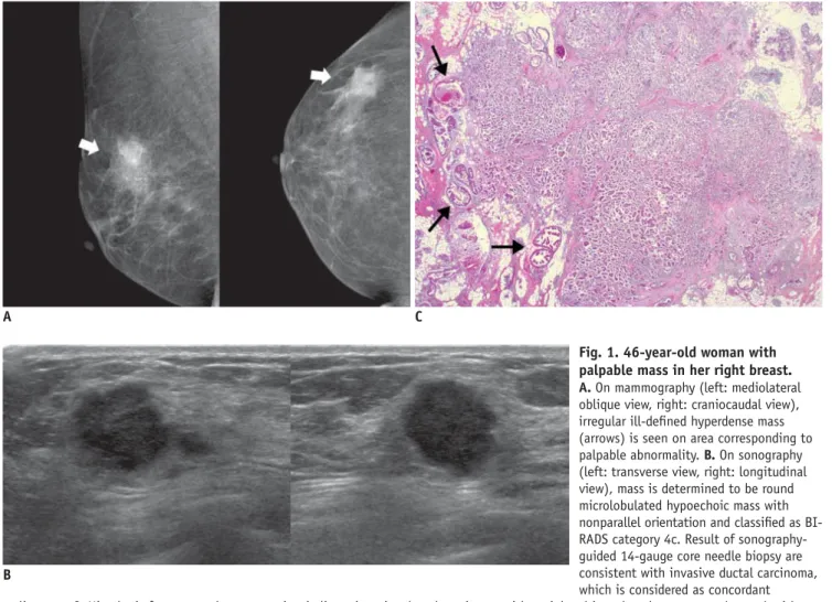
Fig. 1. 46-year-old woman with palpable mass in her right breast.
A. On mammography (left: mediolateral oblique view, right: craniocaudal view), irregular ill-defi ned hyperdense mass (arrows) is seen on area corresponding to palpable abnormality. B. On sonography (left: transverse view, right: longitudinal view), mass is determined to be round microlobulated hypoechoic mass with nonparallel orientation and classifi ed as BI- RADS category 4c. Result of sonography- guided 14-gauge core needle biopsy are consistent with invasive ductal carcinoma, which is considered as concordant malignancy. C. Histologic features on low power view indicate invasive ductal carcinoma with peripheral intraductal component (arrows) without microcalcifi cation. Surrounding breast parenchyma shows fatty changes (Hematoxylin & Eosin staining; original magnifi cation, × 10).
appropriate action should be taken without any delay. The radiologist should communicate the biopsy result to the referring physician, and the patient should be informed of the results and referred to a surgeon or oncologist for proper treatment.
Category 2. Discordant Malignancy
A lesion which typically had benign or benign-favoring imaging features (i.e., BI-RADS category 2 or 3) but proves to be malignant at core needle biopsy falls into this category (Fig. 2). Case management should be identical to that for a concordant malignancy (2, 4). Lesions that usually lie at the circumscribed end of the malignant spectrum can simulate benign nodules; small infi ltrating ductal carcinomas, high-grade invasive ductal carcinomas not otherwise specifi ed, metastatic lesions, lymphoma, and special-type tumors that are well circumscribed such as medullary carcinomas, mucinous carcinomas, and papillary carcinoma (8). The radiologist should notify the discordant result to the pathologist and ask the patient to review and confi rm the diagnosis. Also, the images of
the lesion should be reviewed for image quality, lesion characteristics, and missed associated features which may cause an underestimation of the severity of the lesion. The discrepancy between imaging and pathologic results should be discussed thoroughly.
Category 3. Concordant Benign
A lesion which is initially thought to be benign
radiologically (i.e., BI-RADS category 2, 3, or 4a) and also demonstrates benign pathology at core needle biopsy falls into this category (2, 4) (Fig. 3). This result can offer both the physician and the patient reassurance. However, imaging follow-up should be recommended to patients because of delayed false-negative diagnoses at core biopsy (2). Although there is no standard follow-up guideline, a follow-up sonography at six months after biopsy and then annually for at least two years can be recommended (Fig. 4).
Category 4. Discordant Benign
A lesion in this category is suspicious for malignancy at imaging (i.e., BI-RADS category 4 or 5), but demonstrates
B
A C
Fig. 2. 46-year-old woman with mass in her left breast.
A. On mammography (left: mediolateral oblique view, right: craniocaudal view), there is focal asymmetry (arrows) in upper outer portion of left breast. B. On sonography (left: transverse view, right:
longitudinal view), oval circumscribed hypoechoic mass (arrows) is seen and classifi ed as BI-RADS category 3.
Sonography-guided 14-gauge core needle biopsy was performed at request of patient and pathologic result is consistent with invasive ductal carcinoma, which is considered as discordant malignancy. C.
Photomicrograph of microscopic specimen after surgical excision shows relatively well-differentiated tumor cell nests dispersed in desmoplastic stroma (arrows).
No intraductal component was seen in submitted specimen. Final diagnosis was invasive ductal carcinoma (Hematoxylin &
Eosin staining; original magnifi cation, × 100).
benign pathologic result after performing a core needle biopsy (2, 4) (Figs. 5-8). Benign lesions with spiculated fi ndings can simulate malignant lesions and be considered as differential diagnoses as follows: sclerosing adenosis, fat necrosis, postsurgical scar, mastitis, granular cell tumor, diabetic mastopathy, and sarcoidosis (8). However, the radiologist should give special attention to discordant benign lesions from which a substantial number of missed cancers at core needle biopsy can be detected without any delay in diagnosis (Figs. 6, 7). In published reports, up to 64% of discordant lesions after a percutaneous biopsy were confi rmed as cancer by subsequent surgical excision (5). For a sonography-guided 14-gauge core needle biopsy, discordant lesions had cancer rates of up to 50% (6). If there is concern regarding a discordant benign lesion, it is prudent for the radiologist to immediately contact the interpreting pathologist and thoroughly communicate with each other. According to the discussion, the radiologist should notify the result and discuss the need for a repeat biopsy to the referring physician or the patient. A surgical
biopsy, rather than a core needle biopsy, is recommended for a repeat biopsy because of the inconclusive outcome from the fi rst core biopsy. Recently, the vacuum-assisted core needle biopsy has been reported to be an alternative to surgery to obtain a defi nitive histological diagnosis for discordant benign lesions (9).
Category 5. Borderline or High Risk
A lesion in this category is not malignant but is considered to have an increased lifetime risk for the development of breast cancer (e.g., atypical ductal hyperplasia, lobular neoplasm, radial sclerosing lesion, papillary lesions, possible phyllodes tumors) (2, 4) (Figs. 9, 10). Controversy exists regarding the surgical and oncologic treatment for this lesion. A case-by-case approach is needed to manage the patient in accordance with the active discussion between different subspecialties (4). However, surgical biopsy is usually recommended regardless of concordance, because of the relatively high upgrade rate to malignancy.
A
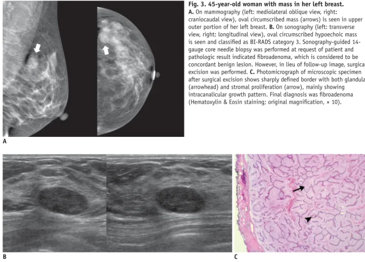
Fig. 3. 45-year-old woman with mass in her left breast.
A. On mammography (left: mediolateral oblique view, right:
craniocaudal view), oval circumscribed mass (arrows) is seen in upper outer portion of her left breast. B. On sonography (left: transverse view, right: longitudinal view), oval circumscribed hypoechoic mass is seen and classifi ed as BI-RADS category 3. Sonography-guided 14- gauge core needle biopsy was performed at request of patient and pathologic result indicated fi broadenoma, which is considered to be concordant benign lesion. However, in lieu of follow-up image, surgical excision was performed. C. Photomicrograph of microscopic specimen after surgical excision shows sharply defi ned border with both glandular (arrowhead) and stromal proliferation (arrow), mainly showing intracanalicular growth pattern. Final diagnosis was fi broadenoma (Hematoxylin & Eosin staining; original magnifi cation, × 10).
C B
A B
C
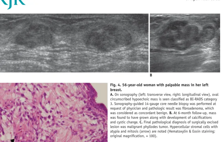
Fig. 4. 56-year-old woman with palpable mass in her left breast.
A. On sonography (left: transverse view, right: longitudinal view), oval circumscribed hypoechoic mass is seen classifi ed as BI-RADS category 3. Sonography-guided 14-gauge core needle biopsy was performed at request of physician and pathologic result was fi broadenoma, which was considered as concordant benign. B. At 6-month follow-up, mass was found to have grown along with development of calcifi cations and cystic change. C. Final pathological diagnosis of surgically excised lesion was malignant phyllodes tumor. Hypercellular stromal cells with atypia and mitosis (arrow) are noted (Hematoxylin & Eosin staining;
original magnifi cation, × 100).
A
B
C Fig. 5. 52-year-old woman with palpable mass in her right breast.
A. On mediolateral oblique view of mammography, there is architectural distortion (arrow) in right breast. B. On sonography (left: transverse view, right: longitudinal view), irregular spiculated hypoechoic mass is seen and classifi ed as BI-RADS category 4c. Result of sonography- guided 14-gauge core needle biopsy indicates stromal fi brosis, which was considered as discordant benign. C. Surgically excised specimen reveals widespread dense stromal fi brosis without ductal cell hyperplasia (arrow). Final diagnosis was stromal fi brosis (Hematoxylin
& Eosin staining; original magnifi cation, × 100).
BI-RADS Final Assessment Category in Imaging- Pathology Correlation
Since the establishment of BI-RADS for sonography in 2003, the reliability of the sonographic BI-RADS lexicon or classifi cation in evaluating sonographic masses for the likelihood of malignancy have been assessed and reported to have a good performance (10-13). Based on the BI-RADS category, the indication for biopsy of particular lesions can be clarifi ed.
A category 3 lesion is judged to have a 2% or lower probability of malignancy and suggested to be followed up with short-term imaging surveillance. While performing a biopsy is contradictory to a category 3 lesion, it is performed in a specifi c circumstance such as physician or
patient preference. For a category 3 lesion, a benign core biopsy result can be regarded as concordant benign and a malignant core biopsy result as discordant malignant.
However, subtle suspicious sonographic features are sometimes overlooked because the fi nal assessment category is determined based on the individual radiologist’s experience and training as well as published criteria. In imaging-pathology correlation, therefore, the sonographic features, even for category 3 lesions, should be reviewed based on the strict criteria to avoid missing cancer.
While BI-RADS category 4 lesions have been
recommended for biopsy, a wide range of positive predictive value (3% to 94%) is problematic. A new recommendation in the fourth edition of BI-RADS is for category 4 to be subdivided internally into three subgroups (4a, 4b, and
A C
B
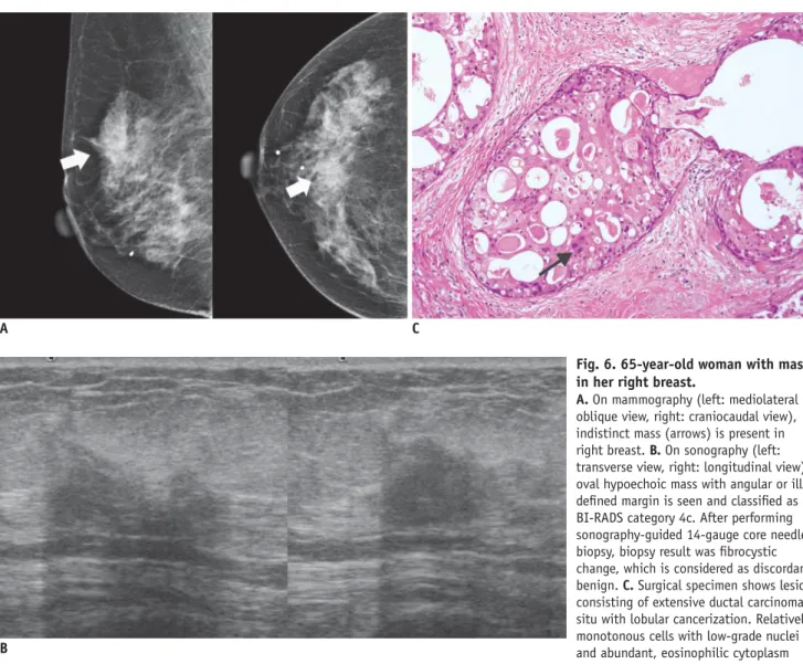
Fig. 6. 65-year-old woman with mass in her right breast.
A. On mammography (left: mediolateral oblique view, right: craniocaudal view), indistinct mass (arrows) is present in right breast. B. On sonography (left:
transverse view, right: longitudinal view), oval hypoechoic mass with angular or ill- defi ned margin is seen and classifi ed as BI-RADS category 4c. After performing sonography-guided 14-gauge core needle biopsy, biopsy result was fi brocystic change, which is considered as discordant benign. C. Surgical specimen shows lesion consisting of extensive ductal carcinoma in situ with lobular cancerization. Relatively monotonous cells with low-grade nuclei and abundant, eosinophilic cytoplasm in form of cribriform pattern (arrow) are noted (Hematoxylin & Eosin staining;
original magnifi cation, × 200).
4c) on the basis of the likelihood of malignancy, although optional (14). The BI-RADS did not set out specifi c guidelines regarding what was the risk of malignancy for each of the subcategories should represent. However, Bent et al. (15) suggested that the guidance range of malignancy likelihood should be 2-10% for category 4a, 11-50% for category 4b, and 51-95% for category 4c. In a study of categorizing lesions by mammography or sonography (11), PPV was 6%, 15%, and 53% for categories 4a, 4b, and 4c, respectively. Whereas, Lee et al. (13) reported that PPV for sonography was 26%, 83%, and 91% for category 4a, 4b, and 4c, respectively. The interobserver variability and poor stratifi cation for the risk of malignancy in the subcategories could be explained by the lack of known factors clearly and objectively defi ning each subdivision. Because the use of subcategories is optional and clinical data of those
subcategories are limited, management is not standardized.
Based on BI-RADS (14), the benign core biopsy result may be regarded as concordant benign and the malignant core biopsy result as discordant malignant for a category 4a lesion. The benign core biopsy result may be regarded as discordant benign and the malignant core biopsy result as concordant malignant for a category 4c lesion. For category 4b lesions, close imaging and pathologic correlations are needed. Further studies are needed to evaluate the role of these subdivisions in stratifying the level of suspicion and the use of this information in management decisions.
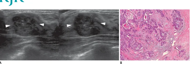
In conclusions, careful imaging-pathology correlation and appropriated post-biopsy management should be the cornerstone of a successful core biopsy program. It will allow the detection of a substantial number of false- negative results immediately after core needle biopsy Fig. 7. 36-year-old woman with palpable mass in her right breast.
A. On sonography (left: transverse view, right: longitudinal view), round microlobulated hypoechoic mass with microcalcifi cations and nonparallel orientation (arrowheads) is seen and classifi ed as BI-RADS category 4c. Results of sonography-guided 14-gauge core needle biopsy indicate presence of stromal sclerosis, which is considered as discordant benign. B. Final diagnosis after surgical excision was invasive ductal carcinoma.
Photomicrograph of microscopic specimen after surgical excision shows carcinoma within sclerotic stroma (Hematoxylin & Eosin staining; original magnifi cation, × 40).
B A
Fig. 8. 40-year-old woman with palpable mass in her left breast.
A. On sonography (left: transverse view, right: longitudinal view), oval microlobulated hypoechoic mass with echogenic halo and nonparallel orientation is seen and classifi ed as BI-RADS category 4b. Result of sonography-guided 14-gauge core needle biopsy indicates fi broadenoma, which is considered as discordant benign. B. Surgically excised specimen reveals well circumscribed, lobulated mass with tumoral fl orid adenosis, consistent with adenosis tumor (Hematoxylin & Eosin staining; original magnifi cation, × 100).
B A
B A
Fig. 9. 49-year-old woman with mass in her left breast.
A. On sonography (left: transverse view, right: longitudinal view), oval ill-defi ned hypoechoic mass (arrowheads) is seen and classifi ed as BI- RADS category 4a. Results of sonography-guided 14-gauge core needle biopsy indicate presence of atypical intraductal papilloma and atypical ductal hyperplasia, which is considered as borderline or high-risk. B, C. At photomicrograph of microscopic specimen after surgical excision (B: Hematoxylin & Eosin staining; original magnifi cation, × 10), ductal carcinoma in situ (upper and lower left; arrowheads) and atypical intraductal papilloma (upper right; arrow) are shown. At high-power fi eld (C: Hematoxylin & Eosin staining; original magnifi cation, × 100), ductal carcinoma in situ with low grade, cribriform pattern (left), as well as an atypical papilloma with central intraductal papilloma pattern and peripheral atypical ductal hyperplasia pattern in largest duct (right) are shown.
C
A
B
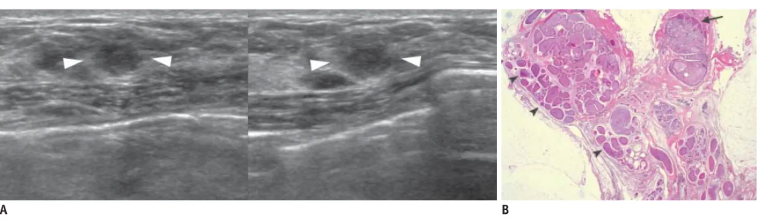
C Fig. 10. 49-year-old woman with palpable mass in her left breast.
A. On mediolateral oblique view of mammography, oval obscured mass (arrow) is shown in left breast. B. On sonography (left: transverse view, right: longitudinal view), oval circumscribed hypoechoic mass is seen and classifi ed as BI-RADS category 3. Results of sonography-guided 14- gauge core needle biopsy performed by request of physician indicate presence of benign phyllodes tumor, which is considered as borderline or high-risk. C. Photomicrograph of microscopic specimen after surgical excision shows relatively ill-defi ned border, elongated epithelial-lined clefts, and mild increase in stromal cellularity with periductal stromal accentuation (arrowheads), consistent with benign phyllodes tumor (Hematoxylin & Eosin staining; original magnifi cation, × 100).
by identifying discordant lesions prospectively, thereby avoiding delays in the diagnosis of cancer. Although the communication between the radiologist and pathologist is the basis of imaging-pathology correlation, establishing concordance is subject to the experience of the radiologist.
The radiologist performing the biopsy must be familiar with the imaging features of a vast array of pathologic breast lesions and they must be able to correlate with each other.
REFERENCES
1. Bassett LW, Mahoney MC, Apple SK. Interventional breast imaging: current procedures and assessing for concordance with pathology. Radiol Clin North Am 2007;45:881-894 2. Youk JH, Kim EK, Kim MJ, Lee JY, Oh KK. Missed breast
cancers at US-guided core needle biopsy: how to reduce them.
Radiographics 2007;27:79-94
3. Liberman L, Drotman M, Morris EA, LaTrenta LR, Abramson AF, Zakowski MF, et al. Imaging-histologic discordance at percutaneous breast biopsy. Cancer 2000;89:2538-2546 4. Parikh J, Tickman R. Image-guided tissue sampling: where
radiology meets pathology. Breast J 2005;11:403-409 5. Liberman L. Percutaneous image-guided core breast biopsy.
Radiol Clin North Am 2002;40:483-500
6. Comstock CE. US-guided interventional procedures. In: Feig SA, ed. 2005 Syllabus: categorical course in diagnostic radiology- breast imaging. Oak Brook, IL: Radiological Society of North America, 2005:155-168
7. Whitman GJ, Erguvan-Dogan B, Yang WT, Wilson J, Patel P, Krishnamurthy S. Ultrasound-guided breast biopsies.
Ultrasound Clin 2006;1:603-615
8. Starvros AT. False-negative and false-positive examinations. In:
McAllister L, Donnellan K, Martin SP, Rothschild R, eds. Breast ultrasound. Philadelphia, PA: Lippincott Williams & Wilkins, 2004:947-978
9. Kim MJ, Kim EK, Lee JY, Youk JH, Park BW, Kim SI, et al.
Breast lesions with imaging-histologic discordance during US-guided 14G automated core biopsy: can the directional vacuum-assisted removal replace the surgical excision? Initial fi ndings. Eur Radiol 2007;17:2376-2383
10. Kim EK, Ko KH, Oh KK, Kwak JY, You JK, Kim MJ, et al.
Clinical application of the BI-RADS fi nal assessment to breast sonography in conjunction with mammography. AJR Am J Roentgenol 2008;190:1209-1215
11. Lazarus E, Mainiero MB, Schepps B, Koelliker SL, Livingston LS. BI-RADS lexicon for US and mammography: interobserver variability and positive predictive value. Radiology
2006;239:385-391
12. Raza S, Chikarmane SA, Neilsen SS, Zorn LM, Birdwell RL. BI- RADS 3, 4, and 5 lesions: value of US in management--follow- up and outcome. Radiology 2008;248:773-781
13. Lee HJ, Kim EK, Kim MJ, Youk JH, Lee JY, Kang DR, et al. Observer variability of Breast Imaging Reporting and Data System (BI-RADS) for breast ultrasound. Eur J Radiol 2008;65:293-298
14. American College of Radiology. Breast imaging reporting and data system-mammography. In: American College of Radiology, ed. Breast imaging reporting and data system, 4th ed. Reston, VA: American College of Radiology, 2003
15. Bent CK, Bassett LW, D’Orsi CJ, Sayre JW. The positive predictive value of BI-RADS microcalcifi cation descriptors and fi nal assessment categories. AJR Am J Roentgenol 2010;194:1378-1383