Copeptin in Hemodialysis Patients with Left Ventricular Dysfunction
Jae Seok Kim, Jae Won Yang, Moon Hee Chai, Jun Young Lee, Hyeoncheol Park, Youngsub Kim, Seung Ok Choi, and Byoung Geun Han
Division of Nephrology, Department of Internal Medicine, Yonsei University Wonju College of Medicine, Wonju, Korea.
Received: July 17, 2014 Revised: August 20, 2014 Accepted: August 26, 2014
Corresponding author: Dr. Byoung Geun Han, Division of Nephrology,
Department of Internal Medicine,
Yonsei University Wonju College of Medicine, Wonju Severance Christian Hospital, 20 Ilsan-ro, Wonju 220-701, Korea.
Tel: 82-33-741-0509, Fax: 82-33-741-5884 E-mail: neptune@yonsei.ac.kr
∙ The authors have no financial conflicts of interest.
© Copyright:
Yonsei University College of Medicine 2015 This is an Open Access article distributed under the terms of the Creative Commons Attribution Non- Commercial License (http://creativecommons.org/
licenses/by-nc/3.0) which permits unrestricted non- commercial use, distribution, and reproduction in any medium, provided the original work is properly cited.
Purpose: Copeptin has been considered as a useful marker for diagnosis and pre- diction of prognosis in heart diseases. However, copeptin has not been investigated sufficiently in hemodialysis patients. This study aimed to investigate the general features of copeptin in hemodialysis and to examine the usefulness of copeptin in hemodialysis patients with left ventricular dysfunction (LV dysfunction). Materials and Methods: This study included 41 patients on regular hemodialysis. Routine laboratory data and peptides such as the N-terminal of the prohormone brain natri- uretic peptide and copeptin were measured on the day of hemodialysis. Body fluid volume was estimated by bioimpedance spectroscopy, and the E/Ea ratio was esti- mated by echocardiography. Results: Copeptin increased to 171.4 pg/mL before hemodialysis. The copeptin had a positive correlation with pre-dialysis body fluid volume (r=0.314; p=0.04). The copeptin level decreased along with body fluid vol- ume and plasma osmolality during hemodialysis. The copeptin increased in the pa- tients with LV dysfunction more than in those with normal LV function (218.7 pg/
mL vs. 77.6 pg/mL; p=0.01). Receiver operating characteristic curve analysis showed that copeptin had a diagnostic value in the hemodialysis patients with LV dysfunc- tion (area under curve 0.737; p=0.02) and that the cut-off value was 125.48 pg/mL (sensitivity 0.7, specificity 0.8, positive predictive value 0.9, negative predictive val- ue 0.6). Conclusion: Copeptin increases in hemodialysis patients and is higher in patients with LV dysfunction. We believe that copeptin can be a useful marker for the diagnosis of LV dysfunction in hemodialysis patients.
Key Words: Copeptin, hemodialysis, ventricular dysfunction
INTRODUCTION
Copeptin is the peptide at the C-terminal of preprovasopressin. Recent studies have demonstrated that copeptin, a surrogate marker for vasopressin, could predict the prognosis of heart failure1-5 and myocardial infarction.6,7 In addition, it could also be useful in the diagnosis of left ventricular dysfunction (LV dysfunction) and myocar- dial infarction.6,8
Dialysis patients are usually classified as high-risk for heart diseases. Therefore, the clinical use of copeptin should be considered for dialysis patients. For this, our
the index of overhydration (OH, liter). For example, OH, 1 means that body fluid excess is one liter. The OH value was also measured pre-dialysis (pre-OH) and post-dialysis (post- OH).
Statistical analysis
The statistical program PASW 18.0 (SPSS Inc., Chicago, IL, USA) was used for all statistical analyses. The Shapiro- Wilk test was conducted to check for normality. The result showed that the variables had non-normal distributions;
therefore, we performed non-parametric tests. Spearman’s correlation analysis was used to examine correlations be- tween copeptin and other variables. The Mann-Whitney U test was conducted to investigate the differences between the patients with normal LV function and those with LV dysfunction. Statistical significance was defined as p<0.05.
RESULTS
Baseline characteristics of subjects
The average (±standard deviation) age of the patients was 57±10 years. In total, 17 male and 24 female patients were included in the study. Hemodialysis was performed three times per week for 4 hours per session. The average dura- tion of hemodialysis was 2721±2367 days. Diabetes was found in 20 (48.8%) patients, and high blood pressure was found in 36 (87.8%) patients. Three (7.3%) patients had a past history of cardiovascular disease, and four (9.8%) pa- tients had been diagnosed with heart failure. Six (14.6%) pa- tients had a history of stroke. The average Kt/V was 1.44±
0.23. Laboratory data [median (interquartile range)] yielded the following values: whole blood hemoglobin 9.7 (9.3, 10.7) g/dL, serum albumin 3.9 (3.8, 4.1) g/dL, calcium 9.0 (8.7, 9.3) mg/dL, phosphorus 6.5 (5.0, 6.8) mg/dL, high-sen- sitivity C-reactive protein 0.13 (0.06, 0.34) mg/dL, and intact parathyroid hormone 191.2 (111.8, 272.8) pg/mL. NT-proB- NP was elevated to 13071 (3984, 35000) pg/mL, and the E/
Ea ratio was 5.5 (4.4, 7.2).
study aimed to investigate the general features of copeptin in hemodialysis. In addition, we hypothesized that copeptin could be useful in the diagnosis of LV dysfunction in he- modialysis patients. To determine the usefulness of co- peptin, our study investigated the difference in copeptin lev- el between patients with normal LV function and those with LV dysfunction.
MATERIALS AND METHODS
Patients and data collection
This study targeted 41 patients who had received hemodial- ysis regularly for three times per week. When the patients visited the hospital for dialysis, we collected clinical and lab- oratory data. In particular, serum sodium concentration, plas- ma osmolality, body fluid volume, and copeptin were mea- sured both before and after dialysis in order to evaluate ch- anges during hemodialysis. Body fluid volume was measur- ed by bioimpedance spectroscopy (Body Composition MonitoringTM, Fresenius Medical Care, Bad Homburg, Ger- many). Copeptin was quantified using an ELISA kit (co- peptin: USCNK Life Science Inc., sensitivity 5.7 pg/mL, CV intra-assay<10%, inter-assay<12%).
We also measured the N-terminal of the prohormone brain natriuretic peptide (NT-proBNP) and performed echocar- diography to evaluate heart dysfunction. These tests were performed before dialysis. In echocardiography, we mea- sured the E/Ea ratio, which was used to estimate LV end di- astolic pressure. We used an NT-proBNP level as a standard to determine LV dysfunction in our study, as the accuracy of an echocardiogram depends on the skill of the performer.
We applied an NT-proBNP level of 5300 pg/mL as a thresh- old for the determination of LV dysfunction according to a study by David, et al.9 in which this value was considered to indicate LV dysfunction in hemodialysis patients.
Parameters
Copeptin was measured pre-dialysis (pre-copeptin) and post-dialysis (post-copeptin). The value of body fluid excess measured by bioimpedance spectroscopy was presented as
Table 1. Changes of Overhydration (OH) Index, Plasma Osmolality, and Copeptin during Hemodialysis
Pre-dialysis Post-dialysis Intradialytic change
OH (liter) 2.5 (1.7, 3.9) 0.7 (-0.6, 2.0) -1.8 (-2.2, -1.3)
Osmolality (mmol/kg) 303.1 (300.1, 308.6) 288.2 (284.4, 291.6) -14.9 (-20.2, -11.2)
Copeptin (pg/mL) 171.4 (75.6, 289.8) 127.2 (66.6, 259.6) -9.9 (-61.5, 38.2)
Data are presented as median (interquartile range).
in Table 2. A Mann-Whitney U test was then conducted to investigate the differences between the two groups. The re- sults showed that the Kt/V, E/Ea ratio, pre-OH, and pre-co- peptin values were all significantly different. Pre-copeptin increased in the patients with LV dysfunction more than in those with normal LV function (Fig. 2).
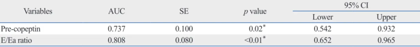
Receiver operating characteristic (ROC) curve analysis of copeptin and the E/Ea ratio for the diagnosis of LV dysfunction
ROC curve analysis for the diagnosis of LV dysfunction showed that pre-copeptin and the E/Ea ratio had significant area under the curve values (Table 3). In addition, the cut-off value of pre-copeptin for the diagnosis of LV dysfunction was 125.48 pg/mL (sensitivity 0.7, specificity 0.8, positive pre- dictive value 0.9, negative predictive value 0.6).
DISCUSSION
Previous literature reported that copeptin increased in dialy- sis patients with cardiovascular disease and predicted cardio- Changes of overhydration (OH) index, plasma
osmolality, and copeptin during hemodialysis
The changes of OH, plasma osmolality, and copeptin during hemodialysis are shown in Table 1. The copeptin level was higher than the reference value of 4.0‒48.0 pg/mL10 and de- creased along with body fluid volume and plasma osmolali- ty during hemodialysis.
Correlations between variables
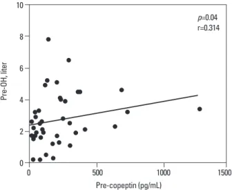
Spearman’s correlation analysis indicated that pre-copeptin had a significant positive correlation with pre-OH (p=0.04) (Fig. 1). When adjusted for age, sex, plasma osmolality, NT- proBNP, and E/Ea ratio, pre-copeptin still showed a signifi- cant positive correlation with pre-OH (p=0.04).
Comparison of characteristics between patients with normal LV function and those with LV dysfunction We divided subjects into normal LV function (n=12) and LV dysfunction (n=29) groups, based on a study by David, et al.9 We used the NT-proBNP level of 5300 pg/mL as a standard for the determination of LV dysfunction.9 The com- parisons of characteristics between both groups are shown
Fig. 1. Correlation between pre-dialysis copeptin (pre-copeptin) and pre-di- alysis overhydration (pre-OH). OH, overhydration.
10 8 6 4 2 0
Pre-OH, liter
0 500 1000 1500 p=0.04 r=0.314
Pre-copeptin (pg/mL)
Table 2. Comparisons of Characteristics between Patients with Normal LV Function and Those with LV Dysfunction
Normal LV function (n=12) LV dysfunction (n=29) p value
Kt/V 1.3 (1.2–1.4) 1.5 (1.3–1.7) 0.04*
Serum albumin (g/dL) 3.9 (3.9–4.1) 3.9 (3.8–4.1) 0.53
Sodium (mmol/L) 136.0 (133.8–137.8) 135.0 (134.0–138.0) 0.85
E/Ea ratio 3.9 (3.8–4.7) 6.1 (5.2–7.9) <0.01*
Pre-OH (liter) 1.8 (1.3–2.5) 2.9 (1.9–4.5) 0.02*
Pre-copeptin (pg/mL) 77.6 (37.9–101.1) 218.7 (113.4–299.3) 0.01*
LV, left ventricular-; OH, overhydration.
Data are presented as median (interquartile range).
*p<0.05.
Fig. 2. Comparison of pre-dialysis copeptin (pre-copeptin) levels between patients with normal LV function and those with LV dysfunction. LV, left ventricular-.
400
300
200
100
0
Copeptin (pg/mL)
p=0.01
Normal LV dysfunction
plasma osmolality. According to previous research, vaso- pressin and plasma osmolality did not decrease during iso- lated ultrafiltration, though both decreased during conven- tional hemodialysis, which indicated that a decrease in vasopressin during hemodialysis was not due to the direct re- moval of vasopressin by hemodialysis.13,15,16 We believe that copeptin also changes in the same way as the vasopressin described previously in these studies.
Previous studies also indicated that copeptin was related to LV dysfunction in the patients who did not receive dialy- sis.8,19 Our study investigated whether copeptin could be re- lated to LV dysfunction in hemodialysis patients. Based on a previous study, an NT-proBNP of 5300 pg/mL was set as a standard for the determination of LV dysfunction.9 When compared to the patients with normal LV function, the pa- tients with LV dysfunction had higher pre-copeptin levels.
Such a result is similar to the findings of copeptin from pre- vious studies conducted on heart failure patients who did not receive dialysis. As a result, our study indicates that co- peptin can be a useful marker for the diagnosis of LV dys- function in hemodialysis patients. ROC curve analysis showed that pre-copeptin had a diagnostic value in the pa- tients with LV dysfunction. The cut-off value was 125.48 pg/mL.
Collectively, our study indicates that copeptin increases in hemodialysis patients and is higher in hemodialysis patients with LV dysfunction. We believe that copeptin can be a use- ful marker for hemodialysis patients with LV dysfunction.
REFERENCES
1. Tentzeris I, Jarai R, Farhan S, Perkmann T, Schwarz MA, Jakl G, et al. Complementary role of copeptin and high-sensitivity troponin in predicting outcome in patients with stable chronic heart failure.
Eur J Heart Fail 2011;13:726-33.
2. Maisel A, Xue Y, Shah K, Mueller C, Nowak R, Peacock WF, et al.
Increased 90-day mortality in patients with acute heart failure with elevated copeptin: secondary results from the Biomarkers in Acute Heart Failure (BACH) study. Circ Heart Fail 2011;4:613-20.
3. Neuhold S, Huelsmann M, Strunk G, Stoiser B, Struck J, Morgen-
vascular disease mortality.11 However, the general features of copeptin in hemodialysis patients were not investigated sufficiently, although copeptin was considered as a useful biomarker for dialysis patients with heart disease. In the case of vasopressin, several studies described a change in blood level during hemodialysis.12-15 They went on to report that vasopressin level was high before hemodialysis due to increased plasma osmolality and that vasopressin decreased if there was a fall in plasma osmolality, although body flu- ids were removed during hemodialysis. Furthermore, the studies indicated that the decreased vasopressin during he- modialysis was found to be associated with intradialytic hy- potension and that hypertonic saline injection stimulated the release of vasopressin, which prevented intradialytic hypotension.16,17
Our results showed that the copeptin level in hemodialy- sis patients was higher than in healthy people. This result is similar to the case of vasopressin described by previous studies. However, our study also showed that copeptin had no significant correlation with plasma osmolality. Rather, copeptin had a significant positive correlation with pre-dial- ysis body fluid volumes. This result is confusing, as it is dif- ferent from that of previous studies and body fluid amounts usually have a negative association with vasopressin levels.
Our hypothesis is that hemodialysis patients may have vari- ous stimulating factors for vasopressin release irrelevant to plasma osmolality and body fluid volume, for example, a decrease in effective circulating volume, LV dysfunction, or nonspecific stress such as uremia. Schrier, et al.18 also indi- cated that vasopressin could be controlled by non-osmotic factors other than osmotic stimuli. We believe that hemodi- alysis patients with uncontrolled volume status usually have more of these non-osmotic stimulating factors for vasopres- sin release than patients with controlled volume status. Th- erefore, it is believed that copeptin demonstrates a positive association with body fluid volume and that the higher level of copeptin in hemodialysis patients is attributed to various non-osmotic factors as well as plasma osmolality. Addition- ally, our results showed that copeptin decreased together with
Table 3. Receiver Operation Characteristics Curve Analysis of Pre-Copeptin and the E/Ea Ratio for the Diagnosis of LV Dys- function
Variables AUC SE p value 95% CI
Lower Upper
Pre-copeptin 0.737 0.100 0.02* 0.542 0.932
E/Ea ratio 0.808 0.080 <0.01* 0.652 0.965
LV, left ventricular-; AUC, area under the curve; SE, standard error; CI, confidence interval.
*p<0.05.
11. Fenske W, Wanner C, Allolio B, Drechsler C, Blouin K, Lilienthal J, et al. Copeptin levels associate with cardiovascular events in pa- tients with ESRD and type 2 diabetes mellitus. J Am Soc Nephrol 2011;22:782-90.
12. Horký K, Srámková J, Lachmanová J, Tomásek R, Dvoráková J.
Plasma concentration of antidiuretic hormone in patients with chronic renal insufficiency on maintenance dialysis. Horm Metab Res 1979;11:241-6.
13. Fasanella d’Amore T, Wauters JP, Waeber B, Nussberger J, Brun- ner HR. Response of plasma vasopressin to changes in extracellu- lar volume and/or plasma osmolality in patients on maintenance hemodialysis. Clin Nephrol 1985;23:299-302.
14. Hegbrant J, Thysell H, Mårtensson L, Ekman R, Boberg U. Chang- es in plasma levels of vasoactive peptides during standard bicar- bonate hemodialysis. Nephron 1993;63:303-8.
15. Os I, Nordby G, Lyngdal PT, Eide I. Plasma vasopressin, catechol- amines and atrial natriuretic factor during hemodialysis and se- quential ultrafiltration. Scand J Urol Nephrol 1993;27:93-9.
16. Thompson AM, Oliver JA. Endogenous and exogenous vasopres- sin during hemodialysis. Semin Dial 2009;22:472-5.
17. Ettema EM, Kuipers J, Groen H, Kema IP, Westerhuis R, de Jong PE, et al. Vasopressin release is enhanced by the Hemocontrol bio- feedback system and could contribute to better haemodynamic stability during haemodialysis. Nephrol Dial Transplant 2012;27:
3263-70.
18. Schrier RW, Berl T, Anderson RJ. Osmotic and nonosmotic con- trol of vasopressin release. Am J Physiol 1979;236:F321-32.
19. Pitkin SL, Maguire JJ, Bonner TI, Davenport AP. International Union of Basic and Clinical Pharmacology. LXXIV. Apelin recep- tor nomenclature, distribution, pharmacology, and function. Phar- macol Rev 2010;62:331-42.
thaler NG, et al. Comparison of copeptin, B-type natriuretic pep- tide, and amino-terminal pro-B-type natriuretic peptide in patients with chronic heart failure: prediction of death at different stages of the disease. J Am Coll Cardiol 2008;52:266-72.
4. Voors AA, von Haehling S, Anker SD, Hillege HL, Struck J, Hart- mann O, et al. C-terminal provasopressin (copeptin) is a strong prognostic marker in patients with heart failure after an acute myocardial infarction: results from the OPTIMAAL study. Eur Heart J 2009;30:1187-94.
5. Alehagen U, Dahlström U, Rehfeld JF, Goetze JP. Association of copeptin and N-terminal proBNP concentrations with risk of car- diovascular death in older patients with symptoms of heart failure.
JAMA 2011;305:2088-95.
6. Potocki M, Reichlin T, Thalmann S, Zellweger C, Twerenbold R, Reiter M, et al. Diagnostic and prognostic impact of copeptin and high-sensitivity cardiac troponin T in patients with pre-existing cor- onary artery disease and suspected acute myocardial infarction.
Heart 2012;98:558-65.
7. Morawiec B, Kawecki D. Copeptin: a new marker in cardiology. J Cardiovasc Med (Hagerstown) 2013;14:19-25.
8. Kelly D, Squire IB, Khan SQ, Quinn P, Struck J, Morgenthaler NG, et al. C-terminal provasopressin (copeptin) is associated with left ventricular dysfunction, remodeling, and clinical heart failure in survivors of myocardial infarction. J Card Fail 2008;14:739-45.
9. David S, Kümpers P, Seidler V, Biertz F, Haller H, Fliser D. Diag- nostic value of N-terminal pro-B-type natriuretic peptide (NT- proBNP) for left ventricular dysfunction in patients with chronic kidney disease stage 5 on haemodialysis. Nephrol Dial Transplant 2008;23:1370-7.
10. Morgenthaler NG, Struck J, Alonso C, Bergmann A. Assay for the measurement of copeptin, a stable peptide derived from the precur- sor of vasopressin. Clin Chem 2006;52:112-9.