Plasma Total Homocysteine Level Is Associated with the Pulsatility Index of Cerebral Arteries in Lacunar Infarction
Se-A An, Han-Bin Lee, Yoon Kim, Jinkwon Kim, Hyun-Sook Kim, Won-Chan Kim, Ok-Joon Kim, and Seung-Hun Oh
Department of Neurology, CHA Bundang Medical Center, CHA University, Seongnam, Korea.
Received: August 16, 2012 Revised: October 3, 2012 Accepted: October 16, 2012
Corresponding author: Dr. Seung-Hun Oh, Department of Neurology, CHA Bundang Medical Center, CHA University, 59 Yatap-ro, Bundang-gu, Seongnam 463-712, Korea.
Tel: 82-31-780-1904, Fax: 82-31-780-5269 E-mail: ohsh72@chamc.co.kr
∙ The authors have no financial conflicts of interest.
© Copyright:
Yonsei University College of Medicine 2013 This is an Open Access article distributed under the terms of the Creative Commons Attribution Non- Commercial License (http://creativecommons.org/
licenses/by-nc/3.0) which permits unrestricted non- commercial use, distribution, and reproduction in any medium, provided the original work is properly cited.
Purpose: The pulsatility index (PI), measured by transcranial Doppler (TCD), is a surrogate marker for distal vascular resistance in cerebral arteries, and elevated plasma total homocysteine (tHcyt) is regarded as a cause of ischemic stroke, in- cluding lacunar infarction. We investigated the relationship between the PI of cere- bral arteries and plasma tHcyt in patients with lacunar infarction. Materials and Methods: Plasma tHcyt level and TCD examination were performed in 94 pa- tients with lacunar infarction. Mean flow velocity (MFV) and PI were assessed at the ipsilateral middle cerebral artery (MCA) and contralateral MCA, relative to the infarction, and the basilar artery (BA). Multivariate regression analysis was con- ducted between log-transformed tHcyt levels (logHcyt) and the PI of individual ar- teries. Results: There was a significant correlation between logHcyt and the PI in all tested arteries (ipsilateral MCA: r=0.21, p=0.03; contralateral MCA: r=0.21, p=0.04; BA: r=0.35, p=0.01). In multivariate regression analysis, this significance remained unchanged after adjusting for vascular risk factors, creatinine, hematocrit and platelet count (ipsilateral MCA: β=0.26, p=0.01; contralateral MCA: β=0.21, p=0.04; BA: β=0.39, p=0.001). There was no significant association between logHcyt and MFV of individual arteries. Conclusion: A significant association be- tween plasma tHcyt and the PI of cerebral arteries indicates that homocysteine plays a role in the increase of distal arterial resistance in lacunar infarction.
Key Words: Homocysteine, pulsatility index, lacunar infarction
INTRODUCTION
Lacunar infarction is a subtype of ischemic stroke, resulting from occlusion of a perforating artery originating from a major cerebral artery. Distinct from other types of ischemic stroke, lacunar infarctions have different pathogeneses, including lipo- hyalinosis, microatheroma of the cerebral microvessels, and segmental demyelin- ation of white matter in the brain.1 Recent studies have demonstrated that endotheli- al dysfunction has a pivotal role in the development of lacunar infarction.2
Hyperhomocysteinemia is an established risk factor for major vascular events, including stroke.3 Elevated plasma total homocysteine (tHcyt) levels increase oxy-
amination.
Vascular risk factors were assessed using patients’ inter- view and laboratory data from hospitalization. The demo- graphic data included age, gender, and previous history of hypertension, diabetes, hyperlipidemia, or cardiac disease.
Laboratory data were obtained from the morning following admission, using a standard protocol, and included hemato- crit, platelet count, serum creatinine, total cholesterol, low- density lipoprotein (LDL) cholesterol, fasting blood glucose, and plasma tHcyt. Based on the result of patients’ interviews and blood tests, individual vascular risk factors were deter- mined. Hypertension was diagnosed when a patient had a high baseline blood pressure, consisting of a systolic blood pressure (SBP) ≥140 mm Hg or a diastolic blood pressure
≥90 mm Hg on more than 1 occasion; treatment with an an- tihypertensive medication also qualified. Diabetes mellitus was diagnosed when a patient had a high fasting plasma glu- cose level (≥126 mg/dL) or had been treated with either oral hypoglycemic agents or insulin. Hyperlipidemia was de- fined as fasting serum total cholesterol of ≥240 mg/dL or a history of antihyperlipidemic medication. Ischemic heart disease was defined to include a history of myocardial in- farction, unstable angina, coronary angioplasty by balloon or stent, or coronary bypass graft surgery. The measurement of plasma tHcyt levels were evaluated by fluorescence po- larization immunoassay using a Hitachi D-2400 analyzer (Hitachi High-Technologies Corporation, Tokyo, Japan).
Transcranial Doppler study
TCD examination was performed within 3 days of stroke onset in all patients. Blood flow velocities at the BA and the ipsilateral and contralateral MCA were measured using the Power M mode transcranial Doppler technology (PMD- 150, 2 channel, Spencer Technologies Inc., Seattle, WA, USA) with a 2-MHz probe. The parameters obtained from TCD examination were systolic flow velocity (SFV), dia- stolic flow velocity (DFV), and mean flow velocity (MFV);
these were automatically calculated by TCD instrument.
The PI was automatically calculated with the equation: PI=
(SFV-DFV)/MFV.11 All SFV, DFV, and MFV values were measured along full segment of each MCA (45-60 mm) and BA (80-115 mm).12 In the present study, we selected the highest PI value detected from several measurements.
Statistical analysis
Demographic and laboratory data were represented as mean values for continuous variables, and percentages for cate- gen free radical generation, inducing the acceleration of ath-
erosclerosis and endothelial dysfunction through the reduc- tion of bioavailability of endothelial nitric oxide synthetase (eNOS).4,5 The majority of previous studies have shown that elevated plasma tHcyt level is associated with the small vessel disease (SVD) type of ischemic stroke.6-8
The pulsatility index (PI), measured by transcranial Dop- pler (TCD), represents peripheral resistance downstream from tested arteries. Several studies have demonstrated that the PI is an independent predictor of SVD, including lacunar infarction.9,10 One previous study reported that elevated plas- ma tHcyt level is associated with the increased PI of cerebral arteries in patients with lacunar infarction, indicating close relationship between plasma tHcyt and PI in this disease.9 Because of the paucity of data, however, this relationship re- quires validation in another cohort. Previous studies have non-selectively included cases with anterior and posterior cir- culation infarcts, without considering the effect of plasma tH- cyt on arteries relevant or irrelevant to the infarct lesion.
We, therefore, investigated the impact of plasma tHcyt on the PI of the middle cerebral artery (MCA), ipsilateral and contralateral to the infarct lesion, and on the basilar artery (BA) in patients with anterior circulation lacunar infarction.
MATERIALS AND METHODS
Patients and vascular risk factor assessment
This study included 94 patients with acute lacunar infarction who admitted to the Department of Neurology between Jan- uary 2009 and December 2010. The Institutional Review Board at CHA Medical Center approved this study. All pa- tients showed clinical symptoms of lacunar stroke within 24 hours of stroke onset. Patients also demonstrated a lesion,
<1.5 cm in diameter, in a perforating artery of the MCA on diffusion-weighted imaging. There could be no significant arterial stenosis in territories relevant to the infarction on computed tomography or magnetic resonance (MR) angi- ography. All patients satisfied the criteria of SVD subtype of infarction according to the Trial of ORG 10172 in Acute Stroke Treatment classification. Study exclusion criteria were: 1) age below 40 years; 2) significant vascular stenosis on CT- or MR-angiography; 3) presence of cardioembolic sources; 4) unobtainable demographic information; 5) unob- tainable TCD data due to a poor temporal window; 6) vita- min supplementation; and 7) severe systemic disease (severe anemia, acute infection, hyperthyroidism) at the time of ex-
tion was also observed when partial correlation analysis ex- cluded the effect of other variables (age, gender, SBP, glu- cose, hematocrit, platelet count, creatinine, and LDL- cholesterol). There was no significant association between gorical variables. For linear correlation analysis, we first
conducted the Kolmogorov-Smirnov test to find out wheth- er plasma tHcyt, PI, and MFV values in each examined ar- tery have a normal standard distribution. The plasma tHcyt level was log-transformed (logHcyt), as this variable showed a skewed distribution. We evaluated the zero-order correla- tion coefficient (Pearson correlation coefficient) and the partial correlation coefficient (excluding the effects of age, gender, SBP, creatinine, hematocrit, glucose, LDL choles- terol, and platelet count) on the relationship between logH- cyt and PI or MFV in each examined artery. Additionally we conducted multivariate linear regression analysis after adjusting for possible confounding factors (age, gender, SBP, creatinine, hematocrit, glucose, LDL cholesterol, and platelet count) to examine linear association between logH- cyt and PI or MFV in each examined artery. To examine the presence of auto-correlation and multi-collinearity among variables, we included the Durbin-Watson test and the vari- ance inflation factor (VIF) in the analysis. Statistical signifi- cance was given to p values of <0.05, and analyses were conducted using Statistical Package for the Social Science software (SPSS ver. 18.0, SPSS Inc., Chicago, IL, USA).
RESULTS
In 94 patients with lacunar infarction, the mean age was 59 years and 31% were female. Left-sided lacunar infarctions were found in 57 (61%) patients. The demographic charac- teristics and laboratory findings of study subjects are sum- marized in Table 1. Detailed data on MFV and PI values in the ipsilateral MCA and contralateral MCA, and the BAs are summarized in Table 2. In pair-wise comparison be- tween the ipsilateral MCA and contralateral MCA, no dif- ferences were found in SFV (p=0.48), DFV (p=0.86), MFV (p=0.93), and PI (p=0.52) between the two arteries. In pair- wise comparison between the ipsilateral or contralateral MCA and the BA, the SFV, DFV, and MFV values in either MCA were higher than those in the BA (all p<0.05). There were no differences in the PI values of the ipsilateral MCA (p=0.60) or the contralateral MCA (p=0.98) and those of the BA.
We calculated a Pearson’s correlation coefficient between logHcyt, the PI and MFV of individual cerebral arteries.
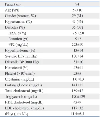
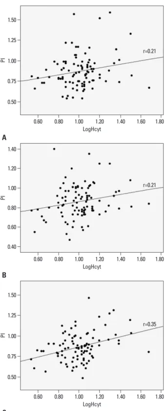
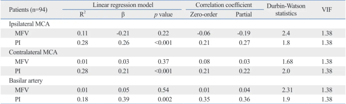
The logHcyt value was correlated with the PI of the ipsilat- eral MCA (r=0.21, p=0.03), the contralateral MCA (r=0.21, p=0.04), and the BA (r=0.35, p=0.01) (Fig. 1). This correla-
Table 1. Demographic Characteristics of Patients with Lacu- nar Infarction
Patient (n) 94
Age (yrs) 59±10
Gender (women, %) 29 (31)
Hypertension (%) 43 (46)
Diabetes (%) 35 (37)
HbA1c (%) 7.9±2.0
Duration (yr) 9±2
PP2 (mg/dL) 223±19
Hyperlipidemia (%) 13±14
Systolic BP (mm Hg) 130±14
Diastolic BP (mm Hg) 81±10
Hematocrit (%) 43±11
Platelet (×104/mm3) 23±5
Creatinine (mg/dL) 1.0±0.3
Fasting glucose (mg/dL) 141±72
Total cholesterol (mg/dL) 189±42
Triglyceride (mg/dL) 170±129
HDL cholesterol (mg/dL) 43±9
LDL cholesterol (mg/dL) 117±32
tHcyt (μmol/L) 11.4±6.5
HbA1c, glycosylated hemoglobin; PP2, 2 hours post-prandial plasma glucose level; BP, blood pressure; LDL, low-density lipoprotein; tHcyt, total homocysteine; HDL, high-density lipoprotein.
Table 2. Results of Transcranial Doppler (TCD) in Patients with Lacunar Infarction
Total (n=94) Ipsilateral MCA
SFV 97±38*
DFV 42±15*
MFV 63±25*
PI 0.87±0.20
Contralateral MCA
SFV 95±30*
DFV 42±16*
MFV 63±22*
PI 0.86±0.17
Basilar artery
SFV 63±21
DFV 26±8
MFV 40±13
PI 0.86±0.17
MCA, middle cerebral artery; SFV, systolic flow velocity; DFV, diastolic flow velocity; MFV, mean flow velocity; PI, pulsatility index.
*p<0.05 in comparison with individual TCD indices of the basilar artery.
0.04), and the BA (β=0.39, p=0.001), after adjusting for cardiovascular risk factors (age, SBP, glucose, and LDL-cho- lesterol), gender, hematocrit, platelet count, and creatinine (Table 3). There was no linear association between logHcyt and the MFVs of tested arteries. Neither auto-correlation (Durbin-Watson statistics: 1.8-2.2) nor multi-collinearity (VIF <3) was found on individual regression analysis.
DISCUSSION
The present study demonstrated that plasma tHcyt levels show a significant association with the PI of major cerebral arteries in patients with lacunar infarction. This association remains significant after adjusting for possible confounders, including age, gender, SBP, creatinine, hematocrit, glucose, LDL cholesterol, and platelet count. This finding indicates that plasma tHcyt plays a role in the increase of distal arteri- al resistance of major cerebral arteries in lacunar infarction.
Hyperhomocysteinemia induces endothelial dysfunction due to the reduced bioavailability of eNOS, brought by in- creased oxygen free radical species.4,5 One experimental study reported that hyperhomocysteinemia produces endo- thelial dysfunction in the cerebral arterioles, at the concen- tration that is significantly lower than that is necessary to produce the same effect in the aorta.13 This result provides evidence that cerebral arterioles are more sensitive to the vascular effects of hyperhomocysteinemia than are larger vessels. Clinical studies also have shown that the associa- tion between plasma tHcyt and the SVD subtype of isch- emic stroke is stronger than that between tHcyt and any other subtype of stroke.6-8 In a healthy population, an in- creased tHcyt level is closely linked to the development of silent lacunar lesion14,15 and cerebral white matter hyperin- tensity lesions,15,16 both of which are radiological indices of SVD in the brain.
PI reflects distal vascular resistance of cerebral vessels, and is useful in predicting the risk of ischemic cerebrovas- cular disease.10,17-19 In lacunar infarction, it is expected that distal arterial resistance increases due to occlusion or insuf- ficient blood flow in a perforating artery, originated from a major cerebral artery. Therefore, the PI of a major cerebral artery is a good candidate for a marker to evaluate local he- modynamic change in the branch artery or microvessel.
Several studies support this finding, having demonstrated that the PI is increased in pathologic conditions of cerebral microangiopathy,20,21 including lacunar infarction and logHcyt and the MFV values of tested arteries (p>0.05).
In multivariate linear regression analysis, logHcyt showed a significant linear correlation with the PI of the ipsilateral MCA (β=0.26, p=0.01), the contralateral MCA (β=0.21, p=
Fig. 1. Correlation analysis between log-transformed homocysteine (logH- cyt) and pulsatility index (PI) of ipsilateral MCA (A), contralateral MCA (B), and basilar artery (C). r, Pearson’s correlation coefficient; MCA, middle ce- rebral artery.
0.50
0.40
0.50 0.75
0.60
0.75 1.00
0.80
1.00 1.25
1.00
1.25 1.50
1.20 1.40
1.50
PIPIPI
0.60
0.60
0.60 0.80
0.80
0.80 1.00
1.00
1.00 1.20
1.20
1.20 1.40
1.40
1.40 1.60
1.60
1.60 1.80
1.80
1.80 LogHcyt
LogHcyt
LogHcyt
r=0.21
r=0.21
r=0.35
A
B
C
point of blood sampling and TCD examination was not ex- actly the same. There might have been some effect occur- ring in the intervening time span. Plasma tHcyt has been re- ported to fluctuate after vascular events,23 and TCD results are also variable after 24 hours of vascular accidents.24 Third, we did not include control subjects. It is difficult to include sufficient number of healthy subjects who agreed to perform blood test and TCD. In previous study, Jeong, et al.9 showed linear trend between cerebral PIs and tHcyt ter- tiles in lacunar infarction, whereas this trend was not ob- served in control subjects. In patients with lacunar infarc- tion, our results are similar to those of previous study.9 Based on the biological role of plasma Hcyt on endothelial dysfunction, reduced vascular elastic compliance and vas- cular blood flows, we believe that plasma tHcyt level is sur- rogate marker for increased resistance of small cerebral ar- teries or arterioles, which can be a pathological substrate for lacunar infarction, such as lipohyalonosis and/or arterio- sclerosis. Fourth, a causal relationship between plasma tH- cyt and increased PI could not be defined due to the lack of a serial measurement. Therefore, our findings require future validation.
In conclusion, we found a positive correlation between plasma tHcyt levels and PI, and suggests that plasma tHcyt induces a circulatory disturbance in the perforating arteries or microvessels originating from major cerebral arteries, leading to the development of cerebral SVD.
ACKNOWLEDGEMENTS
This study was supported by a grant of the Korea Health- SVD.10,22 There is, however, a paucity of data regarding the
relationship between plasma tHcyt and PI in lacunar infarc- tion. Jeong, et al.9 reported that, in patients with lacunar in- farction and hyperhomocysteinemia, the PI values are in- creased: they examined MFV and PI values of the right and left MCA and the BA in patients with lacunar stroke, and found that the PI in all tested arteries increased as homocys- teine levels rose. Our study was designed to test the PI of the ipsilateral and contralateral MCA relative to the infarc- tion, in order to examine whether acute hemodynamic change occurs differently between the relevant MCA and the opposite MCA. However, no differences were found in MFV and PI values of the ipsilateral MCA and contralateral MCA, and correlation with plasma tHcyt was found in all tested arteries, regardless of lesion locality. Consistent with Jeong’s study,9 we found a strong correlation between plas- ma tHcyt and the PI of the BA, a part of the posterior circu- lation. Based on the previous TCD study and our own re- sults, an increased PI represents general pathological status of SVD rather than local hemodynamic changes following acute lacunar stroke.
One strength of the present study is the inclusion of cases with lacunar infarction only in the distribution of the anterior cerebral circulation, enabling us to examine the effect of tH- cyt on the PI of arteries which are both relevant and irrele- vant to the infarction site. In addition, we conducted TCD ex- aminations and collected the plasma tHcyt levels in the narrow time window (within 72 hours after stroke) compared with previous studies, which may reduce the impact of tem- poral variations in cerebral hemodynamics following stroke.
The limitations of the present study should be addressed.
First, our sample size was relatively small. Second, the time
Table 3. Results of Linear Regression Model of Log-Transformed tHcyt Levels (LogHcyt) and TCD Indices in the Ipsilateral and Contralateral Middle Cerebral Arteries and the Basilar Artery
Patients (n=94) Linear regression model Correlation coefficient Durbin-Watson
statistics VIF
R2 β p value Zero-order Partial
Ipsilateral MCA
MFV 0.11 -0.21 0.22 -0.06 -0.19 2.4 1.38
PI 0.28 0.26 <0.001 0.21 0.27 1.8 1.38
Contralateral MCA
MFV 0.01 0.03 0.37 0.08 0.03 1.68 1.38
PI 0.28 0.21 <0.001 0.21 0.22 2.0 1.38
Basilar artery
MFV 0.01 0.05 0.54 0.01 0.04 2.31 1.38
PI 0.18 0.39 0.002 0.35 0.36 1.9 1.38
VIF, variance inflation factor; MCA, middle cerebral artery; MFV, mean flow velocity; PI, pulsatility index; TCD, transcranial Doppler; BP, blood pressure.
R2, adjusted R square; β, standardized coefficients.
Adjusted for age, gender (dummy variable), systolic BP, hematocrit, platelet count, fasting glucose, creatinine, and low-density lipoprotein cholesterol.
LW, et al. Utility of transcranial Doppler ultrasound for the inte- grative assessment of cerebrovascular function. J Neurosci Meth- ods 2011;196:221-37.
13. Dayal S, Arning E, Bottiglieri T, Böger RH, Sigmund CD, Faraci FM, et al. Cerebral vascular dysfunction mediated by superoxide in hyperhomocysteinemic mice. Stroke 2004;35:1957-62.
14. Kohara K, Fujisawa M, Ando F, Tabara Y, Niino N, Miki T, et al.
MTHFR gene polymorphism as a risk factor for silent brain in- farcts and white matter lesions in the Japanese general population:
the NILS-LSA Study. Stroke 2003;34:1130-5.
15. Vermeer SE, van Dijk EJ, Koudstaal PJ, Oudkerk M, Hofman A, Clarke R, et al. Homocysteine, silent brain infarcts, and white matter lesions: the Rotterdam Scan Study. Ann Neurol 2002;51:
285-9.
16. Wright CB, Paik MC, Brown TR, Stabler SP, Allen RH, Sacco RL, et al. Total homocysteine is associated with white matter hy- perintensity volume: the Northern Manhattan Study. Stroke 2005;
36:1207-11.
17. Hassler W, Steinmetz H, Gawlowski J. Transcranial Doppler ul- trasonography in raised intracranial pressure and in intracranial circulatory arrest. J Neurosurg 1988;68:745-51.
18. Lee KY, Sohn YH, Baik JS, Kim GW, Kim JS. Arterial pulsatility as an index of cerebral microangiopathy in diabetes. Stroke 2000;
31:1111-5.
19. Bellner J, Romner B, Reinstrup P, Kristiansson KA, Ryding E, Brandt L. Transcranial Doppler sonography pulsatility index (PI) reflects intracranial pressure (ICP). Surg Neurol 2004;62:45-51.
20. Park JS, Cho MH, Lee KY, Kim CS, Kim HJ, Nam JS, et al. Ce- rebral arterial pulsatility and insulin resistance in type 2 diabetic patients. Diabetes Res Clin Pract 2008;79:237-42.
21. Wijnhoud AD, Koudstaal PJ, Dippel DW. Relationships of tran- scranial blood flow Doppler parameters with major vascular risk factors: TCD study in patients with a recent TIA or nondisabling ischemic stroke. J Clin Ultrasound 2006;34:70-6.
22. Lee KO, Lee KY, Lee SY, Ahn CW, Park JS. Lacunar infarction in type 2 diabetes is associated with an elevated intracranial arterial pulsatility index. Yonsei Med J 2007;48:802-6.
23. Howard VJ, Sides EG, Newman GC, Cohen SN, Howard G, Ma- linow MR, et al. Changes in plasma homocyst(e)ine in the acute phase after stroke. Stroke 2002;33:473-8.
24. Kaps M, Teschendorf U, Dorndorf W. Haemodynamic studies in early stroke. J Neurol 1992;239:138-42.
care Technology R&D Project, Ministry for Health, Wel- fare & Family Affairs, Republic of Korea (A090057).
REFERENCES
1. Fisher CM. The arterial lesions underlying lacunes. Acta Neuro- pathol 1968;12:1-15.
2. Wardlaw JM, Farrall A, Armitage PA, Carpenter T, Chappell F, Doubal F, et al. Changes in background blood-brain barrier integ- rity between lacunar and cortical ischemic stroke subtypes. Stroke 2008;39:1327-32.
3. Homocysteine Studies Collaboration. Homocysteine and risk of ischemic heart disease and stroke: a meta-analysis. JAMA 2002;288:2015-22.
4. Devlin AM, Arning E, Bottiglieri T, Faraci FM, Rozen R, Lentz SR. Effect of Mthfr genotype on diet-induced hyperhomocystein- emia and vascular function in mice. Blood 2004;103:2624-9.
5. Ungvari Z, Pacher P, Rischák K, Szollár L, Koller A. Dysfunction of nitric oxide mediation in isolated rat arterioles with methionine diet-induced hyperhomocysteinemia. Arterioscler Thromb Vasc Biol 1999;19:1899-904.
6. Perini F, Galloni E, Bolgan I, Bader G, Ruffini R, Arzenton E, et al. Elevated plasma homocysteine in acute stroke was not associ- ated with severity and outcome: stronger association with small artery disease. Neurol Sci 2005;26:310-8.
7. Iso H, Moriyama Y, Sato S, Kitamura A, Tanigawa T, Yamagishi K, et al. Serum total homocysteine concentrations and risk of stroke and its subtypes in Japanese. Circulation 2004;109:2766-72.
8. Khan U, Crossley C, Kalra L, Rudd A, Wolfe CD, Collinson P, et al. Homocysteine and its relationship to stroke subtypes in a UK black population: the south London ethnicity and stroke study.
Stroke 2008;39:2943-9.
9. Jeong SK, Kim DH, Cho YI. Homocysteine and pulsatility index in lacunar infarction. Clin Neurol Neurosurg 2011;113:459-63.
10. Kidwell CS, el-Saden S, Livshits Z, Martin NA, Glenn TC, Saver JL. Transcranial Doppler pulsatility indices as a measure of diffuse small-vessel disease. J Neuroimaging 2001;11:229-35.
11. Gosling RG, King DH. Arterial assessment by Doppler-shift ultra- sound. Proc R Soc Med 1974;67(6 Pt 1):447-9.
12. Willie CK, Colino FL, Bailey DM, Tzeng YC, Binsted G, Jones