40(1) : 65 69 (2009)
65
까마귀쪽나무(Litsea japonica)의 HL-60/ADR 세포 Apoptosis 유도효과
김엘비라·부혜진·현재희·김상철·강정일·김민경·유은숙·강희경
*
제주대학교의학전문대학원약리학교실
,
의과학연구소The Effects of Litsea japonica on the Induction of Apoptosis in HL-60/ADR Elvira Kim, Hye-Jin Boo, Jae-Hee Hyun, Sang-Cheol Kim, Jung-Il Kang, Min-Kyoung Kim,
Eun-Sook Yoo and Hee-Kyoung Kang
*Department of Pharmacology, School of Medicine, Institute of Medical Sciences, Jeju National University, 66 Jejudaehakno, Jeju 690-756, South Korea.
Abstract −
The present study investigated the antiproliferative effect of Litsea japonica in HL-60/ADR, adriamycin resistant human promyelocytic leukemia cells. The 80 % ethanol extract of L. japonica markedly inhibited the growth of HL-60/ADR cells. When HL-60/ADR cells were treated with the extract, several apoptosis events like as DNA fragmentation, chromatin condensation and the increase of the population of sub-G1 hypodiploid cells were observed. In the mechanism of apoptosis induction by L. japonica , we examined the changes of Bcl-2 and Bax protein expression levels, and activation of caspases. After the HL-60/ADR cells were treated with the extract, the Bcl-2 expression was decreased, whereas the expression of Bax was increased in a time-dependent manner compared to the control. In addition, the active forms of caspase-9 and -3 were increased and the cleavage of poly (ADP-ribose) polymerase, a vital substrate of effector caspase, was observed. The results suggest that the inhibitory effect of L. japonica on the growth of the HL-60/ADR appears to arise from the induction of apoptosis via the down-regulation of Bcl-2 and the activation of caspases.
Key words −
Litsea japonica , HL-60/ADR, Apoptosis, Bcl-2, Caspases
항암물질에대한개발및그기전에대한연구가활발해 지면서
,
효과적인암치료가이루어지고있지만새롭게대두되고있는문제점은암자체의다양성및발병기전의다양 화로인해생기는부작용과항암제에대한암세포의내성
(resistance)
이다.
항암제에대한내성은장기적인항암제사용으로약물에노출된세포들의세포내축적을감소시키거 나
,
1,2)해독작용또는배출을활성화하거나,
3,4)표적이되는단백질을변형5)시키는등의여러가지기작을통하여일 어나게된다
.
까마귀쪽나무
( Litsea japonica (Thunb.) Jussieu)
는상록엽소교목으로서수직적으로표고
700 m
이내의바닷가및산기슭에분포한다
.
이식물의화학적구성에대한기존의연구에서
,
여러 종류의essential oils, fatty acids, lactones, alkaloids
및terpenoids
가발견되었다고보고된바가있다.
6,7)Lactones
의 종류로 알려진hamabiwalactone A, hamabiwa-
latone B, akolactone B, litsealactone A
및litsealactone B
가분리되었다
.
8)또한,
잎으로부터분리된flavonoids
성분인epicatechin, afzelin, quercitrin
및tiliroside
의anti-complement activity
를조사한결과, tiliroside
가complement system
에대해가장강력한억제작용을나타냄이보고되었다
.
9)한국에서까마귀쪽나무는식용으로사용하였다는보고가있으나
,
어떤용도로사용하였는지에대한연구보고는미미한실 정이므로이식물에대한항암효능을비롯한생리활성을탐 색하는연구가필요하다
.
본연구에서는까마귀쪽나무
80%
에탄올추출물을사용하여 항암제인
adriamycin
에내성을 가지고있는HL-60/
ADR (adriamycin resistant human promyelocytic leukemia)
세포의 증식억제효과를조사하였다
.
또한까마귀쪽나무80%
에탄올추출물의HL-60/ADR
세포에대한세포증식억제효과가
apoptosis
유도에의한것인지알아보기위하여,
핵의형태학적변화
, DNA
절편화및DNA content
변화를알아보았으며
, apoptosis
유도신호전달기전에중요한Bcl- 2
와Bax
의증가와이에따른caspase-9, caspase-3 protease
*교신저자(E-mail):[email protected] (Tel):064-754-3846
활성화를조사하였다
.
이와같은연구로HL-60/ADR
세포와같이항암제에대해내성을가지고있는내성암세포주에 대한까마귀쪽나무추출물의항암효과를밝혀
,
효과적인항암물질개발의재료로사용가능한근거를제시하고자하였다
. 실험 방법
까마귀쪽나무, 환삼덩굴, 호랑가시나무 및 유칼립투스의 추출물 제조 − 제주도의해안가에자생하는까마귀쪽나무
( L. japonica )
의잎을2005
년11
월경에채집하여시료185g
을음지에서건조시킨후
,
마쇄기로분쇄하여각각의미세말시료를
80%
에탄올에침적하고초음파를이용하여1
시간씩
3
회추출하였으며,
그후상층액을회수하여감압농축하여얻은
(43 g)
에탄올추출물을2007
년제주생물종다양성연구소로부터분양받았다
.
본실험에서는이추출물을
PBS (phosphate-buffered saline)/
에탄올(1:1)
에용해시킨후원하는농도에따라실험용배지로희석하여사용하였 다
.
그리고환삼덩굴( Humulus japonicas Siebold & Zucc.) 80%
메탄올추출물,
호랑가시나무( Ilex cornuta Lundi. &
Paxton) 80%
메탄올 추출물 및 유칼립투스( Eucalyptus globulus Labill.) 100%
메탄올추출물또한제주생물종다양성연구소로부터분양받아사용하였으며
,
분양받은각각의 추출물들을
PBS (phosphate-buffered saline)/
메탄올(1:1)
에용해시킨후원하는농도에따라실험용배지로희석하여사용하였다
.
세포 배양 −
HL-60/ADR (adriamycin resistant human promyelocytic leukemia)
세포는내성세포연구센터(Research Center for Resistant Cell)
로부터분양받아서사용하였다. 100 units/ml
의penicillin-streptomycin (GIBCO Inc, Grand Island, NY, USA)
과10% heat-inactivated fetal bovine serum (FBS; GIBCO Inc, Grand Island, NY, USA)
이첨가된
RPMI 1640
배지를사용하여37
oC, 5% CO
2, humidified incubator
에서 배양하였으며,
계대배양은 약70~80%
의confluence
가되는3~4
일마다한번씩시행하였다.
세포 독성 측정 − 여러식물추출물에의한
HL-60/ADR
세포의성장증식에대한효과를
MTT [3-(4,5-dimethylthiazol- 2-yl)-2,5-diphenyl tetrazolium bromide] assay
를이용하여검색하였다
.
10)살아있는암세포mitochondria
의탈수소효소작용에의하여수용성의노란색
MTT
가환원되어형성되는자주색을띠는비수용성의
formazan
을microplate (ELISA) reader
로540 nm
에서흡광도를측정하여,
대사적으로왕성하게생존하는세포를조사하였다
. HL-60/ADR (2.5×10
5cells/
ml)
세포를96 well plate
에 넣고 여러 식물 추출물을100
µg/ml
의농도로처리하였다.
이를4
일간배양한후, MTT (Sigma chemical Co., St. Louis, USA) 50
µl (2 mg/ml)
를첨가하고
4
시간동안반응시킨후, 1000 rpm
에서10
분간원심분리 한 후 상층액을 제거하였다
.
그 후dimethyl- sulfoxide (DMSO: Sigma chemical Co., St. Louis, USA) 150
µl
를가하여침전물을용해시킨다음, microplate reader (Amersham Pharmacia Biotech, NY, USA)
를 사용하여540 nm
에서흡광도를측정하였다.
각시료군에대한평균흡광도값을구하여
,
대조군의흡광도값과비교하여세포증식억제정도를조사하였다
.
세포주기변화 측정 − 까마귀쪽나무
80%
에탄올추출물을HL-60/ADR (2.5×10
5cells/ml)
세포에 농도별(25, 50
및100
µg/ml)
로처리한다음, 48
시간동안배양한후,
세포를수확하여
PBS
로세척하였다.
그후70%
에탄올로-20
oC
에서
30
분동안 고정시키고, PBS
로 세척한다음RNase A (1 mg/ml)
를처리한후propidium iodide(PI: Sigma chemical Co., St. Louis, USA)
로 염색하고, COULTER
®EPICS
®XL
TMFlow Cytometer(Coulter, Miami, FL, USA)
를사용하여세포주기를분석하였다
.
핵의 형태학적 변화 관찰 − 까마귀쪽나무
80%
에탄올추출물을
HL-60/ADR (2.5×10
5cells/ml)
세포에농도별(25, 50
및100
µg/ml)
로처리한다음, 48
시간동안배양하였다.
그후
DNA
에특이적으로결합하는형광색소인Hoechst 33342 (H33342: Sigma chemical Co., St. Louis, USA)
용액
(1
µg/ml)
을가하여37
oC
에서10
분간염색한후형광현미경
(IX-71, Olympus, Japan)
하에서관찰하였다.
11)Western Blot Analysis − 까마귀쪽나무
80%
에탄올추출물을
100
µg/ml
의농도로처리한다음3, 6, 9
및12
시간동안배양하고
,
세포를수확한후PBS (phosphate-buffered saline)
로2~3
회세척하였다.
그후500
µl
의lysis buffer
를첨가한다음
, 1
시간동안lysis
시킨후12,000 rpm
에서15
분간원심분리하여상층액을얻었다
.
단백질의농도는BSA (bovine serum albumin)
를 표준물질로 사용하여Protein Assay Kit (BIO-RAD, HC, USA)
로 정량하였다.
12) 그 후20~30
µg
의lysate
를12% mini gel SDS-PAGE (Sodium dodecyl sulfate-polyacrylamide gel electrophoresis)
로 변성분리하여
polyvinylidene difluoride membrane (BIO-RAD, HC, USA)
에200 mA
로2
시간동안transfer
하였다.
그리고5% skim milk
가함유된TTBS (TBS+0.1% Tween 20)
용액에서
membrane
의blocking
을overnight
실시하였다. Bcl- 2
의 발현량을 검토하기 위하여mouse monoclonal anti- human Bcl-2 Ab (1:1000) (Santa Cruz Biotech, MA, USA)
항체를, Bax
의 발현량을 검토하기 위하여rabbit polyclonal anti-human Bax Ab (1:1000) (Santa Cruz Biotech, MA, USA)
항체를, PARP
의발현량을검토하기위하여
rabbit polyclonal anti-human PARP Ab (1:1000)
(Santa Cruz Biotech, MA, USA)
항체를사용하였다.
또한caspase-3
의발현량을검토하기위하여rabbit polyclonal anti-
human caspase-3 Ab (1:1000) (Cell Signaling Technology,
Beverly, MA, USA)
항체를, caspase-9
의발현량을검토하기위하여
rabbit polyclonal anti-human caspase-9 Ab (1:1000) (Cell Signaling Technology, Beverly, MA, USA)
항체를사용하였다
.
각각의항체들은모두TTBS
용액에희석하여사용하였으며
,
상온에서2
시간동안반응을진행하였다
. 2
차항체로는HPR (Horse Radish Peroxidase)
이결합된
anti-rabbit IgG (American Prarmacia Biotech, NY, USA)
와anti-mouse IgG (American Pharmacia Biotech, NY, USA)
를TTBS
용액에희석(1:5000)
하여사용하였으며,
상온에서
30
분동안 반응을진행하였다.
그후TTBS
로membrane
을3
회세척한후, ECL
기질(American Pharmacia Biotech, NY, USA)
과1~3
분간반응시킨다음X-ray
필름에감광하였다
.
통계처리 − 표시된결과들은
3
번이상의독립적인실험결과들이며
,
각각의데이터를mean±standard error
값으로나타내었다
. Student’s t-test
를사용하여실험군사이의통계적유의성검증을평가하였다
. 결과 및 고찰
세포 증식 저해 효과항암제인
adriamycin
에내성을가지고있는암세포인HL-
60/ADR
세포의증식에대한여러식물추출물의효과를측정하기위하여
MTT assay
를이용하였다. HL-60/ADR
세포주에까마귀쪽나무
80%
에탄올추출물외3
종의추출물을처리하여세포증식저해효과를조사하였다
(Fig. 1).
각각의추출물을
100
㎍/
㎖ 의 농도로처리하였을때, HL-60/
ADR
의세포증식저해효과는까마귀쪽나무( L. japonica )
80%
에탄올 추출물의 경우는64.5%,
환삼덩굴( H.
japonicus ) 80%
메탄올추출물의경우는17.3%,
호랑가시나무
( I. cornuta ) 80%
메탄올추출물의경우는37.7%,
유칼립투스
( E. globulus ) 100%
메탄올추출물의경우는6.6%
의저해효과를보였다
.
이중저해효과가가장높은까마귀Fig. 1.
Inhibitory effect of several extracts of plants on the growth of HL-60/ADR cells.
HL-60/ADR cells (2.5×10
5/ml) were treated for 4 day with 100
µg/ml of the extracts in 96-microwell plates. After incubation, MTT [3-(4,5-dimethylthiazol-2-yl)-2,5-diphenyl tetrazolium bromide] was added and incubation was continued for further 4 hours. The formazan salt formed was dissolved in dimethyl- sulfoxide, and quantified using a microplate reader at 540 nm.
Data were presented as a mean±SD from three separate experiments. *p<0.05, **p<0.01 compared with the control.
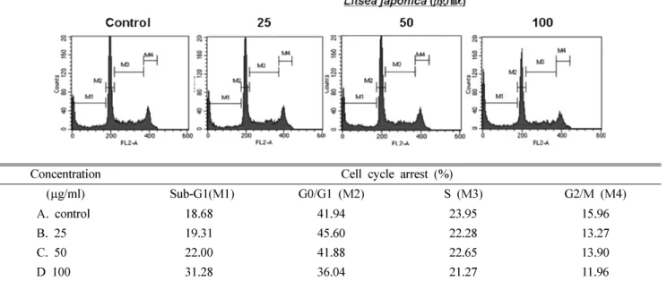
Concentration Cell cycle arrest (%)
(
µg/ml) Sub-G1(M1) G0/G1 (M2) S (M3) G2/M (M4)
A. control 18.68 41.94 23.95 15.96
B. 25 19.31 45.60 22.28 13.27
C. 50 22.00 41.88 22.65 13.90
D 100 31.28 36.04 21.27 11.96
Fig. 2.
The degree of apoptosis represented as the DNA content measured by flow cytometric analysis.
The HL-60/ADR cells (2.5×10
5/ml) were treated with various concentrations (25, 50 and 100
µg/ml) of 80% EtOH extract from L.
japonica for 48 hours.
쪽나무
80%
에탄올추출물은정상세포인HEL-299
에서현저히낮은세포증식억제효과를나타냄을이미보고하였 다
.
13)이는정상세포에대하여까마귀쪽나무80%
에탄올추출물이독성을갖지않는것으로판단할수있다
.
Adriamycin
에내성을갖는HL-60/ADR
의성장저해효과가탁월한까마귀쪽나무
80%
에탄올 추출물의작용이HL-60/ADR
의apoptosis
유도에의한것인지를조사하였다.
Apoptosis 유도효과
세포주기변화 측정 −
HL-60/ADR
세포에서까마귀쪽나무의
80%
에탄올추출물이apoptosis
를유도하는지를알아보기위하여세포주기를조사하였다
. HL-60/ADR
세포에까마귀쪽나무의
80%
에탄올추출물을25, 50
및100
µg/ml
의농도로
48
시간 처리한후, apoptosis
를 측정하기 위하여DNA helix
에결합하여형광을나타내는물질인PI
를처리하여
,
세포주기에서apoptotic
세포인sub-G1 hypodiploid
세포의증가를유세포분석기로분석하였다
.
그결과,
까마귀쪽나무
80%
에탄올추출물을25, 50
및100
µg/ml
의농도로48
시간처리하였을때,
까마귀쪽나무추출물에의해sub-G1 hypodiploid
세포가19, 22
및31%
로,
농도 의존적으로apoptosis
세포가증가되는것을확인할수있었다(Fig. 2).
핵의 형태학적 변화 −
DNA
에특이적으로결합하는형광염색액인
Hoechst 33342
를사용하여apoptosis
의형태학적특징중의하나인핵의변화를관찰하였다
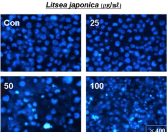
. Hoechst 33342
로
DNA
를염색하고형광현미경으로관찰한결과,
까마귀쪽나무의
80%
에탄올추출물을25, 50
및100
µg/ml
의농도로
48
시간동안처리하였을때,
정상대조군에비하여농도의존적으로
HL-60/ADR
세포의크기가축소되었으며,
핵의모양이불규칙하고부분적인핵의응집현상을관찰할 수있었다
(Fig. 3).
Bcl-2/Bax 발현 양상의 변화
까마귀쪽나무
80%
에탄올추출물에의한HL-60/ADR
의apoptosis
유도의분자적기전을밝히기위하여Bcl-2
와Bax
의발현을조사하였다
. Apoptosis
를조절하는유전자산물중제일먼저알려진암유발유전자산물의하나인
anti-
apoptosis protein Bcl-2
는분자량26 KDa
의단백질로서세포증식에는관여하지않고
chemoresistance
에중요한역할을담당하여여러종류의자극에대해
apoptosis
를억제하는특이적기능을가지고있다고알려져있다
.
반면Bcl-2
family
에속하는pro-apoptosis protein Bax protein
은apoptosis
를촉진시키는
protein
으로밝혀졌다.
11)Bax
는Bcl-2
의anti- apoptosis effects
를방해하는것으로알려져있다.
14)Fig. 4
에서 보는바와같이
, HL-60/ADR
세포에까마귀쪽나무80%
에탄올추출물을3, 6, 9
및12
시간동안100
µg/ml
의농도로처리하였을때
Bcl-2
의발현은9
시간이후부터시간의존적으로감소한반면
Bax
의경우는그발현이큰차이가없었다
(Fig. 4).
Caspase-9 및 caspase-3의 활성화
현재수많은
apoptosis
관련유전자가알려져있음에도불구하고
,
그중공통적인경로는단백질분해효소의활성과관련이깊음이알려졌다
.
특히caspase
가apoptosis
기작의중심적인요소로여겨지고있다
. Tetrapeptide motif
를인식하여기질을절단하는
caspase
는cystein protease
로,
그인식
peptide
의특이성에따라여러종류의isoform
이보고되었다
.
이들중Caspase-3
는다양한apoptosis
자극에의해서Fig. 3.
The degree of apoptosis represented as the fluorescent image of nuclei in cells by fluorescent microscope.
The HL-60/ADR cells (2.5×10
5/ml) were treated with 100
µg/
ml of 80 % EtOH extract of L. japonica for 48 hours. DNA- specific fluorescent dye, H33342 (10
µg/ml medium at final) was directly added to media and apoptosis bodies were observed under inverted fluorescent microscope equipped with a IX-71 Olympus camera. The cells were photographed under microscopy (X 400).
Fig. 4.
Western blot analyses of Bcl-2 and Bax proteins.
The HL-60/ADR cells(3.5×10
5/ml) were treated with 100
µg/
ml of 80% EtOH extract of L. japonica for 3, 6, 9 and 12
hours. Western blots were performed as described in Material
and methods.
공통적으로활성화될수있으며활성화된
Caspase-3
는여러종류의세포내단백질들을절단할수있다
.
핵내에존재하는효소중하나인
PARP
의촉매부위는apoptosis
가진행되는여러세포에서효과적으로
caspase-3
를포함하는여러caspase
에의해DNA
결합부위로부터절단되어분리된다. Caspase-9
과caspase-3
는PARP
분절의상부경로에있는것으로서
, apoptosis
가 일어날 때 활성화되며, caspase-3
가apoptosis
의집행자의역할을하는것으로알려져있다.
본실험에서까마귀쪽나무
80%
에탄올추출물을100
µg/ml
의농도로
3, 6, 9
및12
시간동안처리하여caspase
의활성형인
cleaved caspase
의단백질수준을조사한결과, Fig. 5
에서처럼
9
시간이후부터활성이증가됨을확인할수있었다.
또한
caspase-3
의활성도와 비례하여, caspase-3
기질의하나인
PARP (116 KDa)
는시간의존적으로PARP
분해단백질
(85 KDa)
로나타나는것을확인하였다(Fig. 5).
결 론
본연구에서는까마귀쪽나무의추출물이
adriamycin
에내성을가지고있는
HL-60/ADR
세포에서Bcl-2
의발현감소및
caspase
를활성화함으로서apoptosis
를유도하여증식을저해함을밝혔다
.
이와같은연구결과는까마귀쪽나무추출물이항암제에내성을가지고있는내성암세포주에대한항 암제로이용될수있는근거를시사하며
,
항암치료제또는예방제의유효성분및그작용기전연구에중요한기초자 료가될것이라고사료된다
.
사 사
본논문은
2
단계두뇌한국21(Brain Korea 21; BK21)
의지원에의해서연구되었다
.
인용문헌
1
.Shen, D. W., Goldenberg, S., Pastan, I. and Gottesman M. M.
(2000) Decreased accumulation of [14C] carboplatin in human cisplatin-resistant cells results from reduced energy- dependent uptake. J Cell Physiol .
183: 108-116.
2
.Gottesman, M. M., Fojo, T. and Bates, S. E. (2002) Mul- tidrug resistance in cancer: role of ATP-dependent trans- porters. Nat Rev Cancer .
1: 8-58.
3
.Schuetz, E. G., Beck, W. T. and Schuetz, J. D. (1996) Mod- ulators and substrates of P-glycoprotein and cytochrome P4503A coordinately up-regulate these proteins in human colon carcinoma cells. Mol Pharmacol .
49(2): 311-8.
4
.Goto, S., Kamada, K., Soh, Y., Ihara, Y. and Kondo, T. (2002) Significance of nuclear glutathione S-transferase pi in resis- tance to anti-cancer drugs. Jpn J Cancer Res .
93(9): 1047-56
.5. Urasaki, Y., Laco, G. S., Pourquier, P., Takebayashi, Y., Kohl- hagen, G., Gioffre, C., Zhang, H., Chatterjee, D., Pantazis, P.
and Pommier, Y. (2001) Characterization of a novel topoi- somerase I mutation from a camptothecin-resistant human prostate cancer cell line. Cancer Res .
61(5): 1964-9.
6. Tanaka, H., Nakamura, T. and Ichino, K. (1990) Butanolides from Litsea japonica . Phytochemistry .
29: 857-859.
7. Takeda, K., Sakurawi, K. and Ishii, H. (1972) Components of the Lauraceae family-l. New lactonic compounds from Litsea japonica . Tetrahedron .
28: 3757-3766.
8. Min, B. S., Lee, S. Y., Kim, J. H., Kwon, O. K., Park, B. Y., An, R. B., Lee, J. K., Moon, H. I., Kim, T. J., Kim, Y. H., Joung, H. and Lee, H. K. (2003) Lactones from the leaves of the Litsea japonica and their anti-complement activity. J Nat Prod .
66(10): 1388-1390.
9. Lee, S. Y., Min, B. S., Kim, J. H., Lee, J., Kim, T. J., Kim, C.
S., Kim, Y. H. and Lee, H. K. (2005) Flavonoids from the leaves of Litsea japonica and their anti-complement activity.
Phytotherapy research .
19: 273-276.
10. Carmichael, J., DeGraff, W. G., Gazdar, A. F., Minna, J. D.
and Mitchell, J. B. (1987) Evaluation of a tetrazolium-based semiautomated colorimetric assay: assessment of chemisen- sitivity testing . Cancer Research .
47: 944-946.
11. Luo, Y. and Kessel, D. (1997) Initiation of apoptosis versus necrosis by photodynamic therapy with chloroaluminum phthalocyanine. Photochem Photobiol .
66: 479.
12. Bradford, M. M. (1976) A rapid and sensitive method for the quantitation of microgram quantities of protein utilizing the principle of protein-dye binding. Anal Biochem .
72: 248-54.
13. Kim, E., Boo, H. J., Hyun, J. H., Kim, S. C., Kang, J. I., Kim, M. K., Yoo, E. S. and Kang, H. K. (2009) The effect of Litsea japonica on the Apoptosis Induction of HL-60 leukemia cells. Yakhak Hoeji .
53(1): 6-11.
14. Kroemer, G., Zamzami, N. and Susin, S. A. (1997) Mito- chondrial control of apoptosis. Immunol Today .
18: 44-51.
(2009년 3월 6일) Fig. 5.