• 검색 결과가 없습니다.

Characterization of Yersinia ruckeri isolated from the farm-cultured eel Anguilla japonica in Korea

N/A
N/A
Protected

Academic year: 2021

Share "Characterization of Yersinia ruckeri isolated from the farm-cultured eel Anguilla japonica in Korea"

Copied!
7
0
0

로드 중.... (전체 텍스트 보기)

전체 글

(1)

29

Characterization of Yersinia ruckeri isolated from the farm-cultured eel Anguilla japonica in Korea

Seong Joon Joh*, Chang Hee Kweon, Min Jeong Kim, Min Su Kang, Hwan Jang, Jun Hun Kwon

Laboratory of Aquatic Animal Diseases, Department of Animal Health, National Veterinary Research and Quarantine Service, Anyang 430-757, Korea

(Accepted: January 13, 2010)

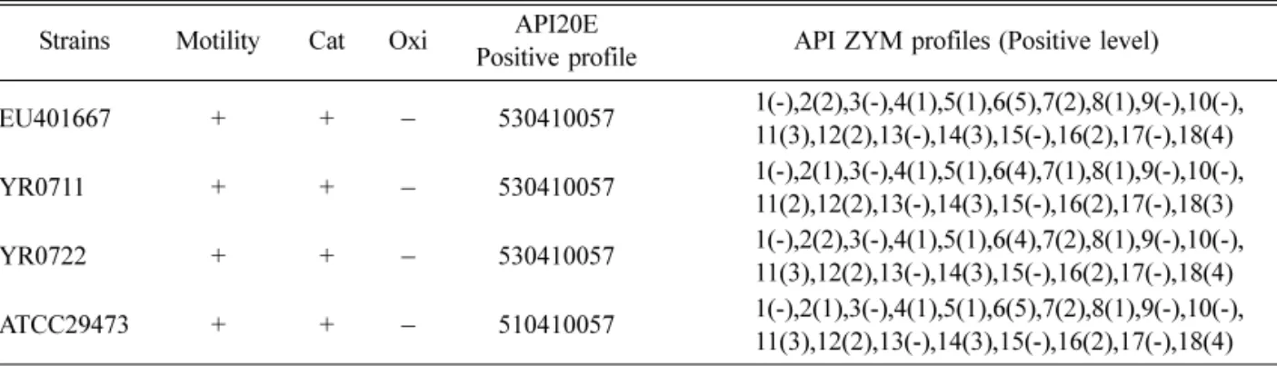
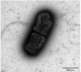
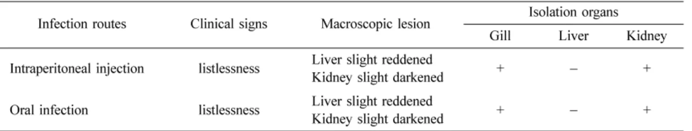
Abstract : Yersinia ( Y. ) ruckeri has been recognized as a serious bacterial pathogen to several kinds of fish, including rainbow trout. However, there are no reports about the characteristics and pathogenicity of Y. ruckeri isolated from farm-cultured eels. In this study, we isolated and characterized Y. ruckeri from the farm-cultured eel Anguilla japonica in Korea. We investigated the phenotypic and genotypic characteristics of Y. ruckeri and tested the virulence of Y. ruckeri isolates on experimentally infected eels. Examination of the flagellar morphology of Y. ruckeri by electron microscopy showed peritrichous flagella in its cell body. Biochemical reaction studies showed overall identical profiles between the isolates and the reference strain of Y. ruckeri in API 20E and API ZYM tests. We sequenced the 16S rRNA of the Y. ruckeri (1,505 bp) for the genotypic characterization (National Center for Biotechnology Information accession number EU401667). Comparison of the 16S rRNA sequences with previously reported Y. ruckeri strains revealed similar phylogenetic relationships. In the virulence assay of the Y. ruckeri on eels, the eels exhibited listlessness, but Y. ruckeri was reisolated from those of the gills and kidneys.

Keywords : characteristics, eel, virulence, Yersinia ruckeri

Introduction

Yersinia ( Y. ) ruckeri was initially isolated from rainbow trout, Oncorhynchus mykiss (Walbaum), in the Hagerman Valley of Idaho, USA, in the 1950s [1, 9, 20] and has been recognized as a causative agent of enteric redmouth disease in rainbow trout [4, 11, 14, 21]. Y. ruckeri is now widely found in fish populations throughout North America, Australia, South Africa, and Europe [2, 3, 7]. Although salmonids are the main fish species susceptible to Y. ruckeri , susceptibility has also been reported in other fish species such as catfish, carp, and the eel Anguilla anguilla [2, 3, 8]. Y. ruckeri belongs to the family Enterobacteriaceae, and the cells are Gram-negative rods with rounded ends [1]. This nonspore-forming bacterium does not possess a capsule, but often has flagella [5, 20]. Since flagella are not always present, Y. ruckeri strains show variable mortality in host animals [9]. Like other members of the Enterobacteriaceae family, Y. ruckeri is glucose-

fermentative, oxidase-negative, and nitrate-reductive [5, 10, 20]. Although biochemical tests can be used to distinguish Y. ruckeri from other species, biochemical reactions in Y. ruckeri strains are homogeneous [23- 25]. Distinguishing phenotypic characteristics of Y.

ruckeri are the presence of

β

-galactosidase, lysine decarboxylase, and ornithine decarboxylase, and the lack of hydrogen sulfide and indole [12, 17]. Molecular characterization of sequences such as 16S rRNA has been helpful in identifying Y. ruckeri and investigating intraspecies differences [15].

The aim of this study was the extensive characterization and comparison of Y. ruckeri isolated from farm-cultured eels using phenotypic and genotypic data.

Materials and Methods

Bacterial isolation

We examined moribund farm-cultured eels that were weak and would not move in response to any

*Corresponding author: Seong Joon Joh

Department of Animal Health, National Veterinary Research and Quarantine Service, Anyang 430-757, Korea

[Tel: +82-31-467-1805, Fax: +82-31-467-1814, E-mail: [email protected]]

(2)

stimulation. There were no mortalities, no increases in body weight or feeding. The eels showed slight discoloration of the skin and a slight redness of the abdomen and gill cover. We examined the gill and internal organs for parasitic and bacterial infections.

For bacterial isolation, samples from moribund fish were aseptically collected from internal organs (liver and kidney) and directly streaked onto blood agar plates (BAP) containing 5% sheep blood (Asan Pharmaceutical, Korea). Plates were incubated at 25

o

C for up to 7 days [9, 12]. All isolates were initially subcultured twice on 5% sheep blood agar at 25

o

C to confirm purity. Gram-stained smears were prepared for microscopic examination after 24 h of incubation.

Motility was determined after overnight incubation of an additional subculture in semisolid sulfide-indole- motility (SIM) medium.

Examination of flagella with transmission elec- tron microscope

The bacteria were streaked onto a BAP and incubated for 48 h at 25

o

C. The single colony was looped, suspended, and washed with phosphate- buffered saline. One drop of the suspension bacteria was placed on parafilm for 5 min, then onto a forma- coated, 300-mesh grid and allowed to dry. The grid was stained negatively with 2% uranyl acetate (pH 6.5) for 30 sec. The grid was dried at room temperature and then examined by transmission electron microscope (TEM; Hitachi, Japan).

Biochemical and enzymatic characterization of the isolates

Biochemical characterization was carried out using API 20E strips (BioMérieux, Spain) supplemented with additional tests. Since studies by other workers showed that acetoin production (Voges-Proskauer (VP) test), citrate use, gelatin hydrolysis, and sorbitol fermentation were likely to be the most variable biochemical traits among Y. ruckeri isolates, we performed the conventional tests three times and confirmed the characteristics.

Enzymatic characterization was carried out using API ZYM (BioMérieux, Spain) strips following the manufacturer’s instructions (API manual ver. 4;

BioMérieux, Spain). Both the API 20E and API ZYM tests were incubated at 25

o

C as the optimum growth temperature for Y. ruckeri , and readings were performed daily from 24 to 72 h depending on growth rates [9,

18]. The API profiles were read and compared with the API database (API Web ver. 1.2.1; bioMérieux, Spain). We also assayed Y. ruckeri for the ability to hydrolyze Tween-80 [9].

Antimicrobial susceptibility test

A disk diffusion susceptibility test was performed to investigate the susceptibility of Y. ruckeri isolates against antimicrobial agents. The disk zone diameter interpretive standard was adapted, in part, in accordance with a principle from the Clinical and Laboratory Standards Institute [6]. All isolates were tested on Mueller-Hinton agar (Difco, USA). The following chemotherapeutic agents (micrograms per disk) were used: sulfonamides (sulfamethoxazole/trimethoprim) (25), quinolones (oxolinic acid) (2), peploxacin (5), norfloxacin (10), ofloxacin (5), penicillins (amoxicillin/clavulanic acid) (30), cephalosporins (cephalothine) (30), cefopera- zone (75), and cefixime (5) from SensiDisk (BBL, USA). Inhibition zone readings were performed after 48 h of incubation at 25

o

C.

16S rRNA sequencing and analysis

The overall procedures for molecular work were conducted as described by Sambrook and Russell [22]

and were modified using kits. Isolates were grown at

25 for 24 h in butylated hydroxyanisole and harvested,

and the bacterial cells were lysed. The DNA was

extracted using a microbial DNA isolation kit (iNtRON

Biotechnology, Korea). The DNA was checked for

purity using NanoDrop (NanoDrop Technologies,

USA). DNA templates were amplified by polymerase

chain reaction (PCR) on an authorized thermal cycler

(Eppendorf, Germany) using universal primers to

amplify a 1,505-bp region of the 16S rRNA gene. The

primers, 27F (5´ AGR GTT TGA TCM TGG CTC AG

3´) and 1492R (5´ GGY TAC CTT GTT ACG ACT

T 3´), were obtained from Bioneer (Bioneer Technologies,

Korea). The amplification mixture (100 µL) comprised

2 µL (50 pmol µ/L) each of the 27F and 1492R

primers, 0.5 µL (2 U µ/L) of Taq DNA polymerase, 10

µL of

×

10 reaction buffer, 10 µL of dNTP mixture,

70 µL of sterile filtered water, and 5.5 µL of DNA

template. The DNA templates were amplified by initial

denaturation at 94

o

C for 10 min, followed by 20 cycles

of denaturation at 94

o

C for 1 min, annealing at 55

o

C

for 1 min, extension at 72

o

C for 1 min, and a final

extension at 72

o

C for 10 min. Controls, without DNA,

(3)

were simultaneously included in the amplification process. The integrity of the PCR products was assayed by the development of single bands following electrophoresis for 1 h at 100 V in 2% (w/v) agarose gels in Tris-borate-EDTA buffer. Each of the resulting products was then cloned into TA cloning vectors (Promega Corporation, USA) according to the manufacturer’s instructions. Nucleotide sequencing was carried out by the dideoxynucleotide chain termination method using T7 DNA and SP6 DNA polymerase with an ABI PRISM 3730 (Applied Biosystems, USA). The nucleotide sequences were analyzed by Vector NTI ver.

9.0 (Hitachi, Japan). The compared genetic sources of Y. ruckeri in this study are listed in Table 1 [16, 19].

Virulence assay of

Y. ruckeri

on eels

Healthy juvenile eels (20-cm average size), obtained

from eel farm, were used for virulence assays. The health of the fish stock was checked upon arrival in the laboratory by collecting samples from the internal organs of 3 out of 15 of the stock for microbiological analysis. Virulence assays were done by intraperitoneal (i.p.) injection, intraoral (i.o.) inoculation, and water bath inoculation [9, 13]. Each group of 4 fish was challenged with doses of 10

6

(i.p.), 10

8

(i.o.), and 10

7

(i.b.) colony-forming units per fish. Fish were maintained at 28

o

C in 20-L tanks containing aerated, static fresh water and equipped with a filtering system.

Fish conditions were recorded daily for 2 weeks. All of the fish were examined microbiologically at 14 days post inoculation. Experimental control groups were challenged with sterile saline solution in a separate aquarium.

Table 1. 16S rRNA sequences of Yersinia ( Y. ) ruckeri compared in this study Accession number (Strain) Nation /

Isolation Year /

deposition Host Sequence

Size (bp) FJ908709 (FF003) China 2009 Channel catfish Ictalurus punctatus 1,503 FJ873802 (LY0903) China 2009 Bighead carp Aristichthys nobilis 1,461

FJ641886 (529-36/85) Russia 2009

1,405

FJ641880 (H527-36/85) Russia 2009

1,405

EU401667 (YR0710) Korea 2008 Eel Anguilla japonica 1,505

YR0711 Korea

Eel Anguilla japonica 1,505

YR0712 Korea

Eel Anguilla japonica 1,505

EF179132 (CCUG14190) France 2008

1,461

AY332849 (WS 10/95) Belgium 2005 Fish 480

AY332850 (WS 20/94) Belgium 2005 Fish 480

AY332851 (WS 25/94) Belgium 2005 Fish 428

AY332852 (WS 34/93) Belgium 2005 Fish 471

AF366385 (ATCC29473) USA 2003 Rainbow trout Onchorhynchus mykiss 1,461

AJ289197 (TS1) Finland 2000 Fish 570

Table 2. Phenotypic characteristics of Y. ruckeri isolates

Strains Motility Cat Oxi API20E

Positive profile API ZYM profiles (Positive level) EU401667 + +

530410057 1(-),2(2),3(-),4(1),5(1),6(5),7(2),8(1),9(-),10(-),

11(3),12(2),13(-),14(3),15(-),16(2),17(-),18(4) YR0711 + +

530410057 1(-),2(1),3(-),4(1),5(1),6(4),7(1),8(1),9(-),10(-),

11(2),12(2),13(-),14(3),15(-),16(2),17(-),18(3) YR0722 + +

530410057 1(-),2(2),3(-),4(1),5(1),6(4),7(2),8(1),9(-),10(-),

11(3),12(2),13(-),14(3),15(-),16(2),17(-),18(4) ATCC29473 + +

510410057 1(-),2(1),3(-),4(1),5(1),6(5),7(2),8(1),9(-),10(-),

11(3),12(2),13(-),14(3),15(-),16(2),17(-),18(4)

(4)

Results

Bacterial isolation and biochemical characteristics Pure bacterial cultures were recovered on BAP from each internal organ sample, collected from the eels.

Fresh isolates exhibited an overall morphologic homogeneity in the colony. They were all Gram- negative rods, oxidase negative, and catalase positive

and all hydrolyzed Tween-80. In addition, all isolates were positive for lysine, ornithine decarboxylase, gelatin, and the VP reaction. The isolates had an API 20E profile of 530410057 (Table 2) and showed a different reaction in citrate use than the Y. ruckeri ATCC 29473 strain. In reading the API ZYM results, we classified a reaction of 2 or more as positive. The positive reaction can be obtained in the number 6, 11, 14, and 18 as API ZYM profiles (Table 2) and showed the same profiles as the Y. ruckeri ATCC 29473 strain.

Fig. 1. The flagellar state of Y. ruckeri as shown by transmission electron micrographs. The cell has 5 flagella in its cell body. ×40,000.

Table 4. Pairwise distances among the 16S rRNA sequence of Y. ruckeri strains 366385 AF AJ

289197 AY 332849 AY

332850 AY 332851 AY

332852 EF 179132 EU

401667 YR 0711 YR

0712 FJ 641880 FJ

641886 FJ 873802 FJ

908709

AF366385 99 100 100 100 100 100 100 100 100 100 100 100 100

AJ289197 1 99 99 99 99 99 98 98 98 99 99 99 99

AY332849 0 1 100 100 100 100 100 100 100 100 100 100 100

AY332850 0 1 0 100 100 100 100 100 100 100 100 100 100

AY332851 0 1 0 0 100 100 100 100 100 100 100 100 100

AY332852 0 1 0 0 0 100 100 100 100 100 100 100 100

EF179132 0 1 0 0 0 0 100 100 100 100 100 100 100

EU401667 0 2 0 0 0 0 0 100 100 100 100 100 100

YR0711 0 2 0 0 0 0 0 0 100 100 100 100 100

YR0712 0 2 0 0 0 0 0 0 0 100 100 100 100

FJ641880 0 1 0 0 0 0 0 0 0 0 100 100 100

FJ641886 0 1 0 0 0 0 0 0 0 0 0 100 100

FJ873802 0 1 0 0 0 0 0 0 0 0 0 0 100

FJ908709 0 1 0 0 0 0 0 0 0 0 0 0 0

Table 3. Antibiotic susceptibility pattern of Y. ruckeri isolates

Group Antibiotic No. sensitive isolates (%) Sulphonamides Sulfamethoxazole/

Trimethoprim 3 (100)

Quinolones

Pefloxacin 3 (100)

Ofloxacin 3 (100)

Oxolinic acid 3 (100) Norfloxacin 3 (100) Penicillins Amoxiciline/

clavulanic acid 3 (100) Cephalosporins Cephalothine 2 (67)

Cefoperazone 3 (100)

Cefixime 3 (100)

(5)

We used Y. ruckeri as biotype 1 to indicate a positive for motility and phospholipase activity.

Flagella morphology by transmission electron microscopy

We used TEM to confirm the ultrastructure of flagella that were attached to the Y. ruckeri . The flagella were usually peritrichous and were anchored firmly at the cell body (Fig. 1).

Antimicrobial sensitivity of the isolates

Antimicrobial susceptibility tests against 3 isolates of Y. ruckeri were performed by the disk diffusion method using 9 antimicrobial agents as shown in Table 1. Each of the 3 isolates was susceptible to most of the antimicrobial agents, except for one isolate tested against cephalothine (Table 3).

Sequence analysis of 16S rRNA

The 1,505-bp region of 16S rRNA from the 3 isolates revealed a 99.9% sequence identity in a BLAST search. One of the sequences has been deposited with accession number EU401667 in a genome database (i.e., GenBank) of the NCBI,

National Institutes of Health, USA (Table 1). The 16S rRNA sequence of the isolate was compared in a BLAST search with previously reported Y. ruckeri

Fig. 2. Cladogram representing the phylogenetic relationships between sequences of Y. ruckeri strains based on nucleotide sequences. The length of each pair of branches represents the distance between the sequence pairs.

Fig. 3 . Macroscopic observation of eels inoculated with Y. ruckeri . The liver slightly reddened following Y. ruckeri inoculation (A) and normal state of the liver in noninoculated eels (B).

Table 5. Pathogenicity test and re-isolation of Y. ruckeri against eels

Infection routes Clinical signs Macroscopic lesion Isolation organs

Gill Liver Kidney

Intraperitoneal injection listlessness Liver slight reddened

Kidney slight darkened +

+

Oral infection listlessness Liver slight reddened

Kidney slight darkened +

+

(6)

strains (data not shown). The comparison of the phylogenetic relationship between the 16S rRNA sequences of Y. ruckeri indicated some pairwise distance between the Korean isolate and other Y.

ruckeri strains (Table 4, Fig. 2).

Experimental infection of

Y. ruckeri

on eels After experimental inoculation of Y. ruckeri on the eels, we observed light clinical symptoms (listlessness in the oral and peritoneal inoculation experimental groups). In the macroscopic examination of the lesions, the liver was found to be slightly reddened and the kidney was slightly darkened in both inoculations. In the microbiological examination, Y. ruckeri was reisolated from those of the gill and kidney. However, it was not possible to reisolate Y. ruckeri from the liver with both of the inoculation methods (Table 5). In addition, there were no clinical symptoms, macroscopic lesions, or reisolation with the water bath inoculation (data not shown).

Discussion

After the first identification of Y. ruckeri as a pathogen of rainbow trout, Y. ruckeri has appeared in various fishes throughout several continents [3].

Outbreaks of yersiniosis are often associated with poor water quality, excessive stocking densities, and the occurrence of environmental stressors. Y. ruckeri can also persist in an asymptomatic carrier state, where infection through carrier fish is especially important under stress conditions [25].

In the present study, we describe the isolation of Y.

ruckeri from farm-cultured eels and the characteristics of the Y. ruckeri isolates. In phenotypes, the Y. ruckeri from the eels showed motility with the SIM test and profile 530410057 with the API 20E test. This result is in agreement with API 20E profiles for Y. ruckeri strains that had been reported previously by other authors [13, 18]. In addition, Fouz et al. [13] reported that 4 Spanish isolates, which are motility negative, show the API 20E profile as 530410057. The report compared the motility strains of Y. ruckeri and showed slightly different biochemical characteristics, especially in VP and gelatinase activity. However, the authors also reported that the API database was not conclusive for Y. ruckeri , presumably due to the lack of information on the biochemical characteristics of Y.

ruckeri in the database. Therefore, PCR for the 16S rRNA partial gene can be used to confirm the isolates as Y. ruckeri [13].

In the antimicrobial susceptibility tests, all of the isolates showed susceptibility to most of the antimicrobial agents, indicating that the isolates might be unexposed or less exposed to many antimicrobial agents. All of the isolates had motility with 3-5 flagella. This result suggests that Y. ruckeri has morphological characteristics similar to those in strains isolated from salmon species and may be potentially pathogenic or virulent to the eels [9].

We also confirmed from the 16S rRNA sequence information that those of the 3 isolates had the same nucleotide sequences. We compared these sequences with previously reported sequences and confirmed that the Y. ruckeri strains isolated from the eels have a few different nucleotide sequences, evident in the phylogenetic relationships. No same-size sequence is available for the 16S rRNA sequence; therefore, we should assess the sequence identity with available short-length sequences. Although the Y. ruckeri isolated from eels in Korea showed clinically very low pathenogenicity or relatively lower virulence compared to the healthy eels in the experimental infection, Y.

ruckeri nevertheless could be isolated from the gill and kidney, suggesting potential cell-associated properties of the isolate. In fact, information about the pathogenicity or exact virulence of Y. ruckeri to the eel Anguilla japonica has been unavailable until now.

Although the Y. ruckeri isolates were not clinically pathogenic or virulent in the eels, the potential pathogenicity in a coinfected situation with other agents or atmospheric stressors cannot be ruled out.

Therefore, it is of future interest to investigate the opportunistic pathogenicity or virulence along with corresponding immune responses in eels.

Acknowledgments

This project was supported by a grant from the National Veterinary Research and Quarantine Service, Ministry of Food, Agriculture, Forestry and Fisheries, Korea.

References

1. Austin DA, Robertson PAW, Austin B. Recovery of

(7)

a new biogroup of Yersinia ruckeri from diseased rainbow trout (Oncorhynchus mykiss, Walbaum). Syst Appl Microbiol 2003, 26 , 127-131.

2. Berc A, Petrinec Z, Matasin Z, Kozari Z. Yersinia ruckeri septicaemia in experimentally infected carp (Cyprinus carpio L.) fingerlings. Acta Vet Hung 1999, 47 , 161-172.

3. Buller NB. Bacteria from Fish and Other Aquatic Animals: A Practical Identification Manual. p. 74, CABI Publishing, Cambridge, 2004.

4. Bullock GL, Stuckey HM, Shotts EB Jr. Enteric redmouth bacterium: comparison of isolates from different geographic areas. J Fish Dis 1978, 1 , 351-356.

5. Cipriano RC, Schill WB, Pyle SW, Horner R. An epizootic in Chinook salmon (Oncorhynchus tshawytscha) caused by a sorbitol-positive serovar 2 strain of Yersinia ruckeri . J Wildl Dis 1986, 22 , 488-492.

6. Clinical and Laboratory Standards Institute.

Performance Standards for Antimicrobial Disk and Dilution Susceptibility Tests for Bacteria Isolated from Animals; Approved Standard - 6th ed. M2-AP. Clinical and Laboratory Standards Institute, Wayne, 2006.

7. Coquet L, Cosette P, Quillet L, Petit F, Junter GA, Jouenne T. Occurrence and phenotypic charac- terization of Yersinia ruckeri strains with biofilm- forming capacity in a rainbow trout farm. Appl Environ Microbiol 2002, 68 , 470-475.

8. Danley ML, Goodwin AE, Killian HS. Epizootics in farm-raised channel catfish, Ictalurus punctatus (Rafinesque), caused by the enteric redmouth bacterium Yersinia ruckeri . J Fish Dis 1999, 22 , 451-456.

9. Davies RL, Frerichs GN. Morphological and biochemical differences among isolates of Yersinia ruckeri obtained from wide geographical areas. J Fish Dis 1989, 12 , 357-365.

10. De Grandis SD, Krell PJ, Flett DE, Stevenson RMW. Deoxyribonucleic acid relatedness of serovars of Yersinia ruckeri , the enteric redmouth bacterium. Int J Syst Bacteriol 1988, 38 , 49-55.

11. Ewing WH, Ross AJ, Brenner DJ, Fanning GR.

Yersinia ruckeri sp . nov., the redmouth (RM) bacterium.

Int J Syst Bacteriol 1978, 28 , 37-44.

12. Frerichs GN. Isolation and identification of fish bacterial pathogens. In: Inglis V, Roberts RJ, Bromage NR (eds.). Bacterial Diseases of Fish. pp. 270-272, Blackwell Scientific, Oxford, 1993.

13. Fouz B, Zarza C, Amaro C. First description of non-

motile Yersinia ruckeri serovar I strains causing disease in rainbow trout, Oncorhynchus mykiss (Walbaum), cultured in Spain. J Fish Dis 2006, 29 , 339-346.

14. Furones MD, Rodgers CJ, Munn CB. Yersinia

ruckeri , the causal agent of enteric redmouth disease (ERM) in fish. Annu Rev Fish Dis 1993, 3 , 105-125.

15. García-López I, Otero A, García-López ML, Santos JA. Molecular and phenotypic characterization of nonmotile Gram-negative bacteria associated with spoilage of freshwater fish. J Appl Microbiol 2004, 96 , 878-886.

16. Kim W, Song MO, Song W, Kim KJ, Chung SI, Choi CS, Park YH. Comparison of 16S rDNA analysis and rep-PCR genomic fingerprinting for molecular identification of Yersinia pseudotuberculosis.

Antonie Van Leeuwenhoek 2003, 83 , 125-133.

17. Llewellyn LC. A bacterium with similarities to the redmouth bacterium and Serratia liquefaciens (Grimes and Hennerty) causing mortalities in hatchery reared salmonids in Australia. J Fish Dis 1980, 3 , 29-39.

18. Kotetishvili M, Kreger A, Wauters G, Morris JG Jr, Sulakvelidze A, Stine OC. Multilocus sequence typing for studying genetic relationships among Yersinia species. J Clin Microbiol 2005, 43 , 2674-2684.

19. Romalde JL, Toranzo AE. Evaluation of the API 20E system for the routine diagnosis of the enteric redmouth disease. Bull Eur Assoc Fish Pathol 1991, 11 , 147-149.

20. Ross AJ, Rucker RR, Ewing WH. Description of a bacterium associated with redmouth disease of rainbow trout (Salmo gairdneri). Can J Microbiol 1966, 12 , 763- 21. 770. Rucker RR. Redmouth disease of rainbow trout (Salmo gairdneri). Bull Off Int Epizoot 1966, 65 , 825- 22. 830. Sambrook J, Russell DW. Molecular Cloning: A Laboratory Manual. 3rd ed. pp. 1.35-1.37, Cold Spring Harbor Laboratory Press, Cold Spring Harbor, 2001.

23. Santos Y, Romalde JL, Bandin I, Magarinos B, Nunez S, Barja JL, Toranzo AE. Usefulness of the API-20E system for the identification of bacterial fish pathogens. Aquaculture 1993, 116 , 111-120.

24. Stevenson RMW, Daly JG. Biochemical and serological characteristics of Ontario isolates of Yersinia ruckeri . Can J Fish Aquat Sci 1982, 39 , 870-876.

25. Tobback E, Decostere A, Hermans K, Haesebrouck F, Chiers K. Yersinia ruckeri infections in salmonid fish. J Fish Dis 2007, 30 , 257-268.

c

í

수치

Table 2.  Phenotypic characteristics of  Y. ruckeri  isolates
Fig. 1.  The flagellar state of  Y. ruckeri  as shown by transmission electron micrographs
Fig. 3 . Macroscopic observation of eels inoculated with  Y. ruckeri . The liver slightly reddened following  Y

참조

관련 문서