http://dx.doi.org/10.14405/kjvr.2015.55.3.199
199
<원례보저>
지질다당류를 투여한 비장세포에서 4,4’-diaminodiphenyl sulfone (dapsone)의 항염증 효과: 염증 관련 사이토카인의 선택적 억제
문선영·주홍구*
제주대학교 수의과대학 수의약리학실
(접수: 2015년 6월 10일, 수정: 2015년 7월 26일, 게재승인: 2015년 7월 28일)
Anti-inflammatory effects of 4,4’-diaminodiphenyl sulfone (dapsone) in lipopolysaccharide- treated spleen cells: selective inhibition of inflammation-related cytokines
Sun-Young Moon, Hong-Gu Joo*
Laboratory of Veterinary Pharmacology, College of Veterinary Medicine, Jeju National University, Jeju 690-756, Korea
(Received: June 10, 2015; Revised: July 26, 2015; Accepted: July 28, 2015)
Abstract : 4,4’-diaminodiphenyl sulfone (dapsone) is a sulfone drug that has antibacterial effects on a variety of bacteria, especially Mycobacterium leprae; thus, it has been used to treat leprosy. Previous studies demonstrated that dapsone inhibits integrin-mediated adherence of neutrophils and production of prostaglandin E2 by polymorphonuclear leukocytes.
Hence, dapsone may act in immune cells and regulate cell-mediated inflammation processes. However, its anti- inflammatory effects remain unclear. The present study demonstrated that dapsone modulates the production of inflammation-related cytokines in immune cells. We employed the spleen cells of mice, which are major immune cells, and lipopolysaccharide (LPS) as a causative agent of inflammation for experiments. Dapsone induced a proportional change in splenocyte subsets and the apoptosis of spleen cells. Interestingly, dapsone decreased the production of tumor necrosis factor-alpha and interleukin (IL)-10, but not IL-6, in LPS-treated spleen cells. In other assays, we measured the dapsone-induced production of nitric oxide (NO) and the expression of activation markers of spleen cells. Dapsone decreased NO production in LPS-treated spleen cells. Taken together, our results demonstrate that dapsone has anti- inflammatory effects in immune cells and provide new insight into the potential uses of this agent.
Keywords : anti-inflammatory activity, dapsone, inflammatory cytokines, spleen cells
서 론
Dapsone은 세균에서 dihydrofolic acid의 생산을 억제하는 항균제로 초기에 결핵균의 치료를 위해 개발되었고, 한센병 을 포함한 피부질환을 치료하는 데 오랜 기간 사용되어왔다.
한센병 치료를 위해서는 일반적으로 clofazimine, rifampicin 을 dapsone과 함께 복합하여 사용한다. Dapsone은 말라리아 또는 특정 염증성 질환에서 사용을 시도한 적도 있다 [12].
또한 dapsone은 integrin이 매개하는 호중구의 부착을 억제하 고 [2], 다형핵백혈구의 프로스타글란딘 E2생산을 억제한다 [1]. 이러한 특징은 dapsone이 세포 매개성 염증 과정을 조 절하고 관련된 면역세포를 억제할 수 있다는 가능성을 보여
주는 것이다. 그러나 현재까지 dapsone의 항균효과와 비교하 면 항염증 효과에 대한 연구는 거의 알려지지 않았다.
본 연구에서는 마우스의 주요 면역세포인 비장세포를 이 용하여 염증 물질의 일종인 lipopolysaccharide(LPS)를 처리 한 후 세포생존율, 면역사이토카인 생산 등을 다양하게 분석 하여 dapsone이 면역세포의 염증 관련 기능들을 조절할 수 있는지 확인하였다.
재료 및 방법
실험동물과 시약
C57BL/6과 BALB/c마우스는 오리엔트바이오(대한민국)에
*Corresponding author
Tel: +82-64-754-3379, Fax: +82-64-756-3354 E-mail: [email protected]
서 구매하였고 동물실험 시설에서 사용 시까지 유지하였다.
In vitro 실험을 위해 7–12주령의 마우스(체중 18–25 g)를 사용하였으며, 본 연구의 동물실험은 제주대학교 동물실험윤 리위원회의 승인을 받았다(승인번호 2013-0024). Dapsone과 Escherichia coli 026:B6에서 trichloroacetic acid extraction 방법으로 분리된 LPS는 Sigma(USA)에서 구매하였다.
비장세포의 분리와 처리
이산화탄소로 마우스를 안락사한 후 비장을 분리하고 기 계적인 압력으로 마우스 비장을 으깬 후 저장성 용혈완충액 을 이용하여 적혈구를 제거하였다 [6]. 세포는 40 µm cell strainer를 통과시켜 단일세포를 얻었으며 in vitro 실험에 사 용하였다. 비장세포를 96-well culture plate에 넣은 후 3일 동안 dapsone과 LPS를 처리하였다. 세포의 활성도는 3-(4,5- dimethylthiazol-2-yl)-2,5-diphenyltetrazolium bromide(MTT) assay로 측정하였다 [7].
유세포분석
비장세포 배양액에 50 µg/mL의 dapsone과 1 µg/mL의 LPS를 주입하고 2일 후 측정에 사용하였다. 처리된 비장세 포는 표면 마커의 발현량 측정을 위해 biotin과 결합한 anti- cluster of differentiation(CD)4, anti-CD8a, anti-CD19, anti-
CD25 antibody, 그리고 streptavidin-phycoerythrin(PE), 또는 PE와 결합한 anti-CD69 antibody(all antibodies; BD Bio- sciences, USA)로 염색하였다. 세포자멸사와 세포괴사 등 세
Fig. 1. Effects of dapsone on the viability of spleen cells. The cells were added to 96-well culture plates at a concentration of 4 × 10⁵ cells/well and treated with dapsone at the indicated concentrations for 3 days in the absence or presence of 1 µg/
mL lipopolysaccharide (LPS). Data are mean± SD from four individual wells.
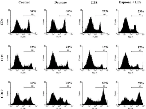
Fig. 2. Dapsone does not alter the subsets of spleen cells. The cells were seeded at a concentration of 2 × 10⁶ cells/mL in 6-well culture plates and treated with 50 µg/mL dapsone and 1 µg/mL LPS. After 2 days, the treated cells were stained for subset markers and analyzed using flow cytometry as explained in the Methods section. The number on the histogram represents the percentage of positive cells.
포사 측정을 위해 annexin V-fluorescein isothiocyanate (FITC)/propidium iodide(PI) kit(Thermo Fisher Scientific, USA)를 사용하였다. 또 다른 세포활성 및 세포사 측정방법 으로 미토콘드리아 막전위를 측정하기 위해, rhodamine 123(Sigma)으로 30분간 염색하였다. 염색된 모든 세포는 FACSCalibur와 CellQuest(Beckton, Dickinson and Company, USA)로 분석하였다.
Enzyme-linked immunosorbent assay(ELISA)와 nitric oxide(NO) 분석
다양한 농도의 dapsone(0, 12.5 및 50 µg/mL)과 1 µg/mL 의 LPS로 3일 동안 처리한 비장세포의 상층액을 회수한 후 ELISA를 실시하였다. CytoSet kit(Thermo Fisher Scientific) 를 이용하여 주요 염증 관련 사이토카인인 interleukin(IL)-6, IL-10, tumor necrosis factor(TNF)-alpha의 함량을 측정하였 다. 또한 NO의 생산량은 Griess reagent(Sigma)를 이용하여 정량하였다.
통계분석
그래프의 결과는 평균값과 표준편차(mean ± SD)로 제시 하였으며, ANOVA 분석을 한 후 Tukey-Kramer multiple comparisons test와 Student’s t-test를 이용하여 유의성을 검 정하였다. 대조군에 대한 실험군의 유의성은 *p < 0.05, **p <
0.01, ***p < 0.001로 표시했다.
결 과
비장세포 생존율에 대한 dapsone의 효과
비장세포의 생존율에 대한 dapsone의 효과를 알아보기 위 해 dapsone과 LPS를 처리하였다(Fig. 1). LPS는 대표적인 염증 유발물질이며 본 연구에서 염증 유사 상황을 유도하기 위해 사용되었다. MTT assay에서 LPS 처리군은 dapsone의 전 농도 범위에서 대조군보다 유의하게 흡광도가 증가하였 으나(p < 0.001), 각 시험군 내에서 dapsone의 농도에 따른 흡광도의 증가 또는 감소는 보이지 않았다. Dapsone은 0–50 µg/mL의 농도에서 LPS 유무와 상관없이 비장세포의 생존율 에 유의한 영향을 주지 않았지만, LPS가 있을 때 dapsone 100 µg/mL은 독성을 보였다.
비장세포의 주요 subset에 대한 dapsone의 영향 비장세포의 subset 구성에 대해 dapsone이 영향을 미치는지 알아보기 위해, LPS가 있을 때와 없을 때 dapsone을 처리하 여 CD4, CD8, CD19의 발현량을 측정하였다. 유세포분석에 서 세 가지 표면 마커의 발현은 대체로 dapsone보다는 LPS 에 의존적이었으며, 예외적으로 dapsone을 단독 처리했을 때 CD19의 발현이 감소하였다(Fig. 2). 예상대로 LPS는 비장세 포에서 CD19의 발현을 증가시켰다. 이러한 결과는 dapsone이 비장세포의 주요 subset, 즉 CD4+, CD8+, CD19+ 림프구의 구성 비율에 부분적으로만 영향을 미치는 것으로 보인다.
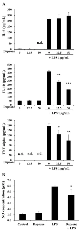
Fig. 3. Dapsone decreases the production of tumor necrosis factor (TNF)-alpha and interleukin (IL)-10 in LPS-treated spleen cells. (A) To check the production of IL-6, IL-10, and TNF-alpha, spleen cells were seeded at a concentration of 4 × 10⁵cells/200 µL/well and treated with the indicated concentrations of dapsone and 1 µg/mL LPS in combination for 3 days. (B) To assay the production of nitric oxide (NO), spleen cells containing adhesive cells were seeded at a concentration of 8 × 10⁵cells/200 µL/well and treated with the 50 µg/mL dapsone and 1 µg/mL LPS in combination for 3 days. *, **, *** indicate p < 0.05, 0.01, 0.001, respectively, compared to LPS alone.
n.d., not detected.
LPS를 처리한 비장세포에서 dapsone의 TNF-alpha와 IL-10 생산 억제
Dapsone이 비장세포의 면역사이토카인 생산에 영향을 주 는지 알아보기 위해 dapsone과 LPS를 처리한 비장세포의 상층액에서 세 가지 사이토카인(TNF-alpha, IL-6, IL-10)을 ELISA로 측정하였다. 측정 결과, dapsone 단독으로는 비장 세포의 세 가지 사이토카인 생산에 영향이 없었다(Fig. 3A).
흥미롭게도 dapsone은 LPS가 있을 때 농도 의존적으로 TNF-alpha와 IL-10의 생산을 동시에 감소시켰으며 IL-6의 생산에는 영향이 없었다. 이러한 결과는 dapsone이 LPS를 처리한 비장세포에서 염증 관련 사이토카인의 생산을 선택 적으로 조절한다는 사실을 의미한다. 또한 다른 기능성 시험 으로 동일한 상층액에서 NO를 정량하였다. NO assay에서 dapsone은 LPS 처리한 비장세포의 NO 생산을 유의하게 감 소시켰다(Fig. 3B).
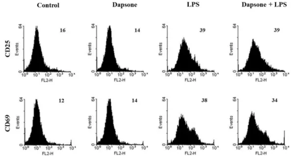
림프구의 활성화 마커의 발현에 대한 dapsone의 효과 비장세포의 활성화 상태에 대해 dapsone이 영향을 주는지 알아보기 위해 림프구의 활성화 마커 발현을 측정하였다. 본 연구에서는 림프구의 세포 표면에서 CD25와 CD69의 발현 을 유세포분석을 통해 확인하였으며 dapsone은 두 가지 활 성화 마커의 발현을 변화시키지 않았다(Fig. 4). 따라서 dapsone은 LPS 유무와 상관없이 비장세포의 활성화 상태에 영향을 주지 않는 것으로 판단된다.
Dapsone 처리한 비장세포의 세포사 측정
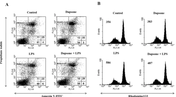
Dapsone이 비장세포에서 세포사를 유도하는지 annexin V- FITC/PI 염색법을 사용하여 정량적으로 측정하였다. Dapsone 은 LPS가 있을 때와 없을 때 모두에서 비장세포의 세포사를
증가시켰다(Fig. 5A). 특히 후기 세포자멸사(annexin V+/
PI+)는 dapsone에 의해 증가하였다. 이러한 효과는 미토콘드 리아 활성 막전위를 측정하기 위해 rhodamine 123으로 염색 하여 확인하였다. 세포는 세포사 전에 활성 막전위가 감소하 는 특징을 보인다. Dapsone은 LPS 존재하에서 비장세포의 미토콘드리아 활성 막전위를 감소시켰다(Fig. 5B). 이 결과를 통해 dapsone이 비장세포의 세포사를 유도한다는 사실을 알 수 있었다.
고 찰
Dapsone은 대표적인 sulfone계 약물 중 하나이며, WHO essential medicine에 포함되어 있다. Dapsone의 가장 보편적 인 적응증은 한센병으로서 다른 질환에도 사용되어왔고 특 히 호중구성 dermatoses와 dermatitis herpetiformis를 포함하 는 피부질환에서 그 사용이 시도되었다 [11, 17]. 작용 기전 은 밝혀지지 않았는데 알려진 것으로는 dihydrofolic acid의 생산을 억제하여 감수성이 있는 세균의 증식을 억제하는 것 이다. 이는 dapsone의 약리작용을 설명하기에 부족하며 dapsone의 항염증 작용을 위한 다른 기전이 있을 것으로 추 정된다 [16]. 최근 연구에서는 myeloperoxidase 촉매반응을 억제하는 것이 dapsone의 항염증 기전의 하나로 여겨진다.
그러나 이전 연구는 면역계의 다양한 세포에 대한 dapsone 의 효과를 설명하기에 한계가 있었다. 따라서 본 연구에서는 dapsone의 항염증 기전, 특히 비장세포의 생존율과 기능에 대한 효과를 알아보고자 하였다.
일부 항염증제가 림프구와 같은 면역세포에 독성을 보이 는 사실에 착안하여 [8] dapsone을 비장세포에 처리한 뒤 MTT assay로 세포활성도를 확인하였고 세포사 분석을 시행
Fig. 4. Effects of dapsone on the expression of lymphocyte activation markers. Spleen cells were cultured and treated as described in Figure 2. The cells were stained for the lymphocyte activation markers CD25 and CD69. The numbers on histograms indicate geometric mean fluorescence intensities.
하였다. MTT assay에서 dapsone은 일정 범위의 농도(0–50 µg/mL)에서 비장세포의 생존율에 영향을 미치지 않았지만 annexin V-FITC/PI 염색에서는 후기 세포자멸사를 유도하였 다. 이러한 차이점은 두 분석 사이의 주요 parameter 차이에 기인한 것으로 보인다. MTT assay가 실험 마지막 시기에서 nicotinamide adenine dinucleotide phosphate(NADP)H 의 존적인 oxidoreductase의 활성도를 측정하고 미토콘드리아 기 능을 평가하여 세포생존율을 측정하는 데 반해 annexin V- FITC/PI 염색은 세포자멸사와 같은 세포사 과정에서 세포 표 면에 발현하는 phosphatidylserine과 annexin V가 결합하므 로 세포사의 과정에 있는 세포를 감지할 수 있다. 또한 dapsone이 비장세포의 세포자멸사를 유도하는 사실은 부분적 으로 과립구감소증과 같은 dapsone에 의한 혈액학적 독성과 연관된다 [4].
TNF-alpha와 IL-10은 면역계에서 대표적인 염증과 항염증 사이토카인이며 [5, 14], IL-6는 다양한 염증 과정에 관련된 다기능 사이토카인이다 [10, 13]. 본 연구를 통해 dapsone이 농도 의존적으로 비장세포에서 TNF-alpha와 IL-10의 생산을 억제한다는 사실을 확인했다. 따라서 dapsone은 비장세포에 서 염증 관련 사이토카인의 생산을 선택적으로 조절하는 것 으로 보이며, 이러한 효과로 dapsone이 항염증 작용을 나타 내는 것으로 보인다. TNF-alpha와 IL-10이 염증성과 항염증 사이토카인으로 반대작용을 보이지만, dapsone에 의해 양쪽 모두 생산이 감소하는 사실은 관련 사이토카인 사이에서 negative feedback 방식의 조절 기전을 추정해 볼 수 있다.
또한 NO는 세포 내의 미생물을 죽일 수 있어 세포성 면역 에도 관여하는 주요 물질인데 [3] 본 연구 결과, dapsone은 LPS에 의해 유도된 NO의 생산을 감소시켰다.
림프구의 활성화 마커로 CD25와 CD69를 사용하였는데 CD25는 IL-2 수용체의 alpha chain으로 림프구의 clonal expansion에서 주요한 역할을 하며 [15], CD69는 림프구의 초기 활성화 마커로 사용된다 [9]. LPS가 있을 때와 없을 때 모두 림프구 활성화 마커의 발현에 대한 dapsone의 영향 은 거의 없었다.
본 연구에서는 dapsone이 비장세포의 세포자멸사를 유도 하고 염증 관련 사이토카인의 생산을 조절한다는 결과를 얻 었다. 이러한 결과는 dapsone이 항균제로서의 사용뿐 아니라 다양한 염증 질환에 사용될 가능성을 보여주어 기초 및 임 상 분야에서 dapsone을 사용하는 데 유용한 정보를 제공할 수 있을 것으로 기대된다.
감사의 글
이 논문은 2015학년도 제주대학교 학술진흥연구비 지원사 업에 의하여 연구되었습니다.
References
1. Anderson R. Enhancement by clofazimine and inhibition by dapsone of production of prostaglandin E2 by human polymorphonuclear leukocytes in vitro. Antimicrob Agents Chemother 1985, 27, 257-262.
2. Booth SA, Moody CE, Dahl MV, Herron MJ, Nelson RD.
Dapsone suppresses integrin-mediated neutrophil adherence function. J Invest Dermatol 1992, 98, 135-140.
3. Chakravortty D, Hensel M. Inducible nitric oxide synthase and control of intracellular bacterial pathogens. Microbes Infect 2003, 5, 621-627.
Fig. 5. Dapsone increases death in spleen cells. (A) The cells were cultured and treated as described in Figure 2. After treatment, the cells were stained with annexin V-fluorescein isothiocyanate/propidium iodide. The quadrants show viable (lower left), early apoptotic (lower right), late apoptotic (upper right), and necrotic (upper left) cells. (B) The cells were stained with rhodamine 123 solution. The numbers in histograms represent geometric mean fluorescence intensities.
4. Coleman MD. Dapsone-mediated agranulocytosis: risks, possible mechanisms and prevention. Toxicology 2001, 162, 53-60.
5. Croft M, Duan W, Choi H, Eun SY, Madireddi S, Mehta A. TNF superfamily in inflammatory disease: translating basic insights. Trends Immunol 2012, 33, 144-152.
6. Joo HG, Goedegebuure PS, Sadanaga N, Nagoshi M, von Bernstorff W, Eberlein TJ. Expression and function of galectin-3, a β-galactoside-binding protein in activated T lymphocytes. J Leukoc Biol 2001, 69, 555-564.
7. Kim MH, Byon YY, Ko EJ, Song JY, Yun YS, Shin T, Joo HG. Immunomodulatory activity of ginsan, a polysaccharide of Panax ginseng, on dendritic cells. Korean J Physiol Pharmacol 2009, 13, 169-173.
8. Kountouras J, Zavos C, Chatzopoulos D. Immunomodulatory benefits of cyclosporine A in inflammatory bowel disease. J Cell Mol Med 2004, 8, 317-328.
9. Marzio R, Mauël J, Betz-Corradin S. CD69 and regulation of the immune function. Immunopharmacol Immunotoxicol 1999, 21, 565-582.
10. Opal SM, DePalo VA. Anti-inflammatory cytokines. Chest
2000, 117, 1162-1172.
11. Plotnikova N, Miller JL. Dermatitis herpetiformis. Skin Therapy Lett 2013, 18, 1-3.
12. Ramos-e-Silva M, Rebello PFB. Leprosy. Recognition and treatment. Am J Clin Dermatol 2001, 2, 203-211.
13. Rincon M. Interleukin-6: from an inflammatory marker to a target for inflammatory diseases. Trends Immunol 2012, 33, 571-577.
14. Sabat R, Grütz G, Warszawska K, Kirsch S, Witte E, Wolk K, Geginat J. Biology of interleukin-10. Cytokine Growth Factor Rev 2010, 21, 331-344.
15. Shipkova M, Wieland E. Surface markers of lymphocyte activation and markers of cell proliferation. Clin Chim Acta 2012, 413, 1338-1349.
16. Van Zyl JM, Basson K, Kriegler A, van der Walt BJ.
Mechanisms by which clofazimine and dapsone inhibit the myeloperoxidase system: a possible correlation with their anti-inflammatory properties. Biochem Pharmacol 1991, 42, 599-608.
17. Wozel G, Blasum C. Dapsone in dermatology and beyond.
Arch Dermatol Res 2014, 306, 103-124.