Article
Risk of necrosis in the adjacent toe after one-toe fillet flap in diabetic foot:
Retrospective study of 107 cases over 5 years
Junhyung Kim, Kyubeom Kim, Jaehoon Choi, Woonhyeok Jeong, Taehee Jo and Sangho Oh
Abstract
Purpose: Fillet flap is a “spare part” concept. This technique allows the defect to be covered without donor site morbidity.
Over the past 5 years, there were 107 diabetic foot cases of one-toe fillet flap in our hospital. After the operation, in some patients, there was necrosis of the adjacent toe that required additional amputation. The aim of our study was to determine the cause of necrosis of the adjacent toe after fillet flap. Methods: The patients were divided into two groups. One group had no necrosis of the adjacent toe (group A) after the operation, and the other group had necrosis of the adjacent toe that required additional amputation after the operation (group B). Then, to confirm the cause of the additional necrosis of the adjacent toe, w
2tests, Fisher’s tests, and logistic regression tests were performed. Results: A total of 107 patients were included, and 48 patients needed additional amputation. The logistic regression test revealed that a fillet flap at the metatarsophalangeal joint (MTPJ), horizontal sutures, and a fillet flap at the second toe were significant risk factors for developing necrosis.
Conclusions: If a fillet flap with a second toe, fillet flap on MTPJ level and horizontal closure after fillet flap is needed, the chance of developing necrosis of the adjacent toe and additional revisional surgery must be communicated preoperatively.
Keywords
amputation, diabetic foot, fillet flap, necrosis, toes
Date received: 21 June 2020; Received revised 22 July 2020; accepted: 2 August 2020
Introduction
Diabetic foot ulcers were defined as any breakdown of the patient’s foot. These foot ulcers are common in patients with diabetes and easily lead to amputation of the patient’s leg.
Currently, the incidence of diabetes is expected to rise, and the incidence of diabetic foot ulcer is elevated.
1The incidence of diabetic foot ulcers in diabetes is 4–10%, and 5% of patients with diabetes are anticipated to have a history of foot ulceration.
2As the risk of diabetic foot ulcers is high, the incidence of diabetic foot ulcers may increase in the future.
3Diabetic foot ulcers are a common complication of diabetes and are caused by various factors, such as poor glucose con- trol, diabetic neuropathy, peripheral vascular disease, struc- tural deformity,
4and reduced pedal soft tissue thickness.
5In addition, patient lifestyle also affects the development of foot
ulcers. These various factors cause foot ulcers in patients with diabetes, and the development of foot ulcers is a complicated process. Therefore, hospitalized patients with diabetic foot ulcers need a multidisciplinary approach determined by a team composed of doctors from endocrinology, vascular surgery, orthopedics, physiatry, and so on.
Department of Plastic and Reconstructive Surgery, Keimyung University Dongsan Hospital, Daegu, South Korea
Corresponding author:
Sangho Oh, Department of Plastic and Reconstructive Surgery, Keimyung University Dongsan Hospital, 1035, Dalgubeol-daero, Dalseo-gu, Daegu, Republic of Korea.
Email: [email protected]
Journal of Orthopaedic Surgery 28(3) 1–7
ª
The Author(s) 2020 Article reuse guidelines:sagepub.com/journals-permissions DOI: 10.1177/2309499020951944 journals.sagepub.com/home/osj
Or thopaedic Surger y
Creative Commons Non Commercial CC BY-NC: This article is distributed under the terms of the Creative Commons
Attribution-NonCommercial 4.0 License (https://creativecommons.org/licenses/by-nc/4.0/) which permits non-commercial use,
reproduction and distribution of the work without further permission provided the original work is attributed as specified on the SAGE and Open
Access pages (https://us.sagepub.com/en-us/nam/open-access-at-sage).
The treatment of diabetic foot ulcers includes tight glu- cose control, physical therapy, dressing with specific mate- rials, and surgical therapy. If the patient’s wound is not severe and the circulation is intact, then conservative treat- ment, such as foam dressing, can be performed. However, if necrosis, infection, or gangrene is found in the patient’s foot, then surgical debridement is needed. After wound preparation, surgery is needed to cover the wound. The surgical options include skin graft, local flap, and free flap.
Recently, most surgeons have chosen a free flap to cover the wound.
6,7However, if patient’s condition, including comorbidity, economical status, and other factors, was not adequate with free flap surgery, our department was to choose a local flap, such as a fillet flap firstly. A fillet flap is a “spare part” concept, which allows the defect to be covered without donor site morbidity.
8The advantage of fillet flaps is simple and the ability to avoid the risks of microsurgery if the patient has peripheral vascular diseases or comorbidities.
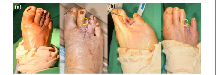
9For dia- betics with many cormorbidities, simple surgery and short duration of treatment is better. When necrosis of only one toe is fully demarcated, the fillet flap is frequently applied. How- ever, after the surgery, we frequently encountered necrosis on the side of the toe. The new complication like necrosis of side toe needed a longer time for diabetic foot ulcer treatment and more surgery. Therefore, in this article, we review our 107 cases of fillet flaps over the course of 5 years to find the cause of necrosis on the side of the toe (Figure 1).
Materials and methods
Brief treatment protocol of diabetic foot ulcer in our institute
The patients’ chief complaints were color change in the toe, pain, or wounds on the foot. Patients often visited plastic
surgery outpatient clinics or endocrinology offices first. If more treatment was needed, then the patient was admitted to our institute. After admission, various departments par- ticipated in developing the patient’s treatment, including doctors from vascular surgery, endocrinology, plastic sur- gery, radiology, nuclear medicine, orthopedics, and so on.
The first step of the treatment process was vascular surgery and endocrinology. Endocrinologists tightly controlled the blood glucose levels during admission and educated the patients on healthy lifestyles after discharge. Vascular sur- geons assessed the status of the patient’s vessels with the ankle–brachial index (ABI), digital artery test, and, if needed, angiography. After the assessment, vascular inter- vention or bypass surgery was performed. In addition, MRI and three-dimensional bone scans were performed to check for the presence of osteomyelitis or abscesses, the extent of the wound. Then, if the foot vessels fully recovered and the patients’ condition, including blood glucose level, was sta- ble, surgical treatment was started with plastic surgery or orthopedics. The orthopedic surgeons treated bony defor- mities, such as Charcot joints, and so on, and plastic sur- geons controlled the soft tissue of the foot with flap or graft surgery. After surgery, postoperative management was started by each department.
Patient selection and ethics
Before the study was conducted, the experimental plan was
approved by the institutional review board of our medical
center. The informed consent of clinical photo in this article
was provided. This study was performed retrospectively,
and patients who underwent a fillet flap primarily by a
single surgeon due to necrosis in only one toe over the
course of 5 years (from 2015 to 2019) were included in our
study. Patient information was obtained through electronic
Figure 1. (a) Preoperative clinical photo of 65 years old female diabetic foot patient with second toe necrosis. (b) Immediate post-
operative clinical photo after second toe fillet flap on metatarsophalangeal joint. (c) Postoperative day (POD) 3 days, new ulceration on
the third toe was occurred. (d) POD 5 days, the third toe ulceration was aggravated. (e) POD 8 days, full layer necrosis with yellow
eschar formation was founded on the third toe medial side. Additional third toe fillet flap was performed on POD 10 days.
medical records, and patients with incomplete medical records were excluded. In addition, we exclude patients who had infections, had a previous history of toe amputa- tion, had osteomyelitis confirmed by magnetic resonance imaging (MRI), and had foot deformities. The foot defor- mities were confirmed using X-ray and clinical photogra- phy by a single surgeon. We obtained the following: patient age; duration of diabetes; necrosis site; preoperative ABI;
previous history of illness, including hypertension, chronic kidney disease, and cardiovascular disease; amputation level; and suture direction.
Hypothesis of our study
Over a period of 5 years, we successfully performed the fillet flap operation for patients with necrosis of one toe, and in our study, we retrospectively review all of these cases. However, we found necrosis in the adjacent toe after a fillet flap; for example, after we treated necrosis in the fifth toe, the fourth toe became necrotic. We eventually realized that most of the necrosis in the adjacent toes empirically occurred at the metatarsophalangeal joint (MTPJ) amputation. Therefore, we hypothesize that MTPJ amputation causes necrosis in the adjacent toe and retro- spectively studied this hypothesis by this procedure with distal amputation. In addition, using medical records, we attempted to identify other risk factors for necrosis in the adjacent toe after a fillet flap.
Study sequence and statistical analysis
We divided the patients into two groups: one group had no necrosis in the adjacent toe (group A) after the operation, and the other group had necrosis in the adjacent toe that required additional amputation after the operation (group B). The statistical analysis is as follows:
1) w
2tests and Fisher’s tests were performed to compare the differences in distribution between the two groups (groups A and B).
2) Logistic regression tests were performed to compare the risk factors of group B with those of group A.
3) The last study was confirmed whether a specific vessel affected the development of necrosis in the adjacent toe. The sequence described as follows:
a) If the flow in the distal toe was not patent, some patients underwent angiography. In angiography, five vessels (superficial femoral artery, popliteal artery, anterior tibial artery, posterior tibial artery, and peroneal artery) were examined.
b) We classified the vessel status into five grades. Then, we divided the vessel status grades into two groups.
Grades 0–2 were combined and called “good,” and grades 3–5 were combined and called “bad”
(Table 1). The rating was graded by vascular sur- geons in our hospital.
c) After the vessel status of the patient was classified, a logistic regression test was performed to assess whether the “bad” status of a vessel specifically affected developing toe necrosis.
Results
The results from “Study sequence and statistical analysis”
section are described as follows:
1) The patient demographics and differences in distri- bution are described in Table 2. As given in Table 2, 107 patients were enrolled, the mean age was 68.72 + 12.12 years, the mean duration of diabetes was 17.3 + 10.72 years, and the mean ABI was 0.8 + 0.43; the number of patients with each condition, including hypertension, chronic kidney disease, and so on, is also described (Table 2). The w
2test and Fisher’s test (Table 2) revealed that there were sta- tistically significant differences in the amputation level and amputation site between the two groups (p < 0.05).
2) The logistic regression test revealed that the amputa- tion at the MTPJ had a statistically significant risk of causing necrosis in the adjacent toe compared with amputation at the proximal or distal interphalangeal joints (PIPJ and DIP, respectively) (p < 0.01). The fillet flaps at the MTPJ carry 131.6 times more risk than fillet flaps at the DIP or PIPJ. Amputation of the second toe had a significantly higher risk than ampu- tations of other toes (p < 0.05). Interestingly, hori- zontal sutures had 6.7 times higher risk of causing necrosis in the adjacent toe than vertical sutures, which was statistically significant. None of the vari- ables had a statistically significant risk of causing necrosis in the adjacent toe (Table 3 and Figure 2).
3) A total of 54 patients underwent angioplasty. After dividing the patients into two groups (“good” and
“bad” groups), the logistic regression test revealed that none of the vessels had a statistically significant risk of causing necrosis in the adjacent toe (p > 0.05, Table 4).
Table 1. Grading the vessel status with angiography.
Grade Description Group
Grade 0 Normal Good
Grade 1 Minimal stenosis
Grade 2 Focal stenosis
Grade 3 Segmental stenosis Bad
Grade 4 Steno-occlusion
Grade 5 Total occlusion
Discussion
The foot is an important and essential structure of the human body. Sensitive changes to the gait and motion of
the foot facilitate smooth walking. In patients with diabetic foot ulcers, the foot ulcer itself is the main cause of ampu- tation.
10During amputation surgery, the remaining toe length is an important factor, especially in the great toe.
After a great toe amputation, foot deformities and foot ulcers can arise and increase the pressure on the foot.
11,12The resulting environment allows diabetic foot ulcers to recur more easily. Therefore, even if an amputation surgery is needed, the remaining toe length is very important.
Diabetes is a disease that requires steady and constant care.
We have treated diabetic foot patients for a long time and have found that treating diabetic patients requires something spe- cial. Although medical treatment is important, the rapport between the doctor and the patient is also very important.
Patients with diabetic complications have many conditions that require treatment. The disease itself is difficult to cure, but patients with chronic complications have poor awareness and require long-term treatments; however, compliance with the treatment is difficult to maintain.
In this article, the study only showed the fillet flap of one toe, but recently, we treated an extensive diabetic foot with a fillet flap. Although difficulties exist in treating the diabetic foot, the treatment method was successful, and the patient was satisfied (Figures 3 and 4). At first, we choose Table 3. Logistic regression test to confirm the risk of developing
necrosis of the adjacent toe.
b Value p Value Exp (B)
Sex (male) 0.359 >0.05
Age 0.011 >0.05
Duration of diabetes 0.008 >0.05
Hypertension 0.317 >0.05
Chronic kidney disease 1.167 >0.05 Cardiovascular disease 0.448 >0.05
Site (right) 0.383 >0.05
Site
Second toe 1.957 <0.05 0.141
Third toe 0.505 >0.05
Fourth toe 0.538 >0.05
Fifth toe 1.330 >0.05
Amputation level (MTPJ) 4.880 <0.001 131.6 Suture direction (horizontal) 1.905 <0.05 6.7
ABI 1.651 >0.05
Angioplasty (yes) 0.230 >0.05
ABI: ankle–brachial index; MTPJ: metatarsophalangeal joint.
Figure 2. (a) Horizontal suture direction after the fillet flap. (b) Vertical suture direction after the fillet flap.
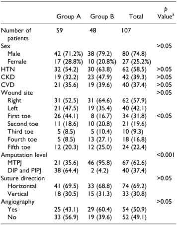
Table 2. Patient demographics.
Group A Group B Total p Value
aNumber of
patients
59 48 107
Sex >0.05
Male 42 (71.2%) 38 (79.2) 80 (74.8) Female 17 (28.8%) 10 (20.8%) 27 (25.2%) HTN 32 (54.2) 30 (63.8) 62 (58.5) >0.05 CKD 19 (32.2) 23 (47.9) 42 (39.3) >0.05 CVD 21 (35.6) 19 (39.6) 40 (37.4) >0.05
Wound site >0.05
Right 31 (52.5) 31 (64.6) 62 (57.9) Left 21 (47.5) 19 (35.4) 40 (42.1)
First toe 26 (44.1) 8 (16.7) 34 (31.8) <0.05 Second toe 11 (18.6) 10 (20.8) 21 (19.6)
Third toe 5 (8.5) 5 (10.4) 10 (9.3) Fourth toe 5 (8.5) 13 (27.1) 18 (16.8) Fifth toe 12 (20.3) 12 (25.0) 24 (22.4)
Amputation level <0.001
MTPJ 21 (35.6) 46 (95.8) 67 (62.6) DIP and PIPJ 38 (64.4) 2 (4.2) 40 (37.4)
Suture direction >0.05
Horizontal 41 (69.5) 33 (68.8) 74 (69.2) Vertical 18 (30.5) 15 (31.3) 33 (30.8)
Angiography >0.05
Yes 25 (43.1) 29 (60.4) 54 (50.9)
No 33 (56.9) 19 (39.6) 52 (49.1)
HTN: hypertension; CKD: chronic kidney disease; CVD: cardiovascular disease; MTPJ: metatarsophalangeal joint; DIP and PIPJ: distal and proximal interphalangeal joint.
a