Tuberc Respir Dis 2013;74:264-268
CopyrightⒸ2013. The Korean Academy of Tuberculosis and Respiratory Diseases. All rights reserved.
The Clinical Assessment of Protease-Activated Receptor-2 Expression in Inflammatory Cells from Peripheral Blood and Bronchoalveolar Lavage Fluid in Idiopathic Pulmonary Fibrosis
Young Sik Park, M.D., Chul-Gyu Yoo, M.D., Ph.D.
Division of Pulmonary and Critical Care Medicine, Department of Internal Medicine and Lung Institute, Seoul National University College of Medicine, Seoul, Korea
Background: Idiopathic pulmonary fibrosis (IPF) is a lethal pulmonary fibrotic disease. In general, the exaggerated activation of the coagulation cascade has been observed during initiation or maintenance of the fibrotic disease.
In our recent study, immunohistochemical expression of protease-activated receptor-2 (PAR-2), which plays a key role in coagulation cascade, was observed in surgical specimen of IPF patients, and associated with poor clinical outcome. The aim of this study was to evaluate the overexpression of PAR-2 in inflammatory cells from peripheral blood and bronchoalveolar lavage fluid in IPF patients.
Methods: From May 2011 to March 2012, IPF patients and controls were enrolled in Seoul National University Hospital. Peripheral blood and bronchoalveolar lavage fluid were collected for analysis of PAR-2 expression. Flow cytometry and reverse transcription polymerase chain reaction were used for PAR-2 receptor and mRNA assessment.
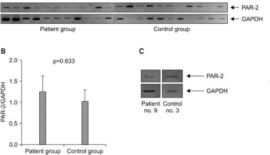
Results: Twelve IPF patients and 14 controls were included in this study. Among them, flow cytometry analysis was conducted from 26 peripheral blood (patient group, 11; control group, 13) and 7 bronchoalveolar lavage fluid (patient group, 5; control group, 2). The expression of PAR-2 receptor was not different between patient and control groups (p=0.074). Among all 24 population, PAR-2 mRNA assessment was performed in 19 persons (patient group, 10; control group, 9). The mRNA expression of PAR-2 was not significant different (p=0.633).
Conclusion: In IPF patients, PAR-2 receptor and mRNA expression were not different from control group.
Key Words: Receptor, PAR-2; Idiopathic Pulmonary Fibrosis; Bronchoalveolar Lavage
Address for correspondence: Chul-Gyu Yoo, M.D., Ph.D.
Division of Pulmonary and Critical Care Medicine, Depart- ment of Internal Medicine and Lung Institute, Seoul National University College of Medicine, 101 Daehak-ro, Jongno-gu, Seoul 110-744, Korea
Phone: 82-2-2072-3760, Fax: 82-2-762-9662 E-mail: [email protected]
Received: Feb. 14, 2013 Revised: Apr. 1, 2013 Accepted: Apr. 1, 2013
CCIt is identical to the Creative Commons Attribution Non-Commercial License (http://creativecommons.org/licenses/by-nc/3.0/).
Introduction
Idiopathic pulmonary fibrosis (IPF) is an inflamma- tory fibrotic lung disease of unknown etiology. Unfor- tunately, IPF is a progressive and irreversible disorder.
In general, median survival of IPF patients is 2.5 to 5 years with or without treatment
1,2. The pathogenesis of
IPF is poorly understood
3. One of the major advances in the pathogenesis of IPF is the shift in current para- digms from inflammation to abnormal wound healing
4. In the wound healing process, coagulation cascade is activated locally from tissue factor dependent extrinsic pathway
5. Recent studies implicated protease-activated receptor-2 (PAR-2) in the fibrosis.
PAR-2 is a G-coupled 7-transmembrane receptor that
is activated by tethered peptide ligand, which is ex-
posed after enzymatic cleavage of the specific site in
the extracellular N-terminal
6. PAR-2 is expressed in vari-
ous tissues and abundant in kidney, pancreas, and gas-
trointestinal tissue than in heart and lungs
7. In each or-
gan, PAR-2 expressions were mainly located in endothe-
lial and epithelial cells
8. Recent studies suggested asso-
ciation of PAR-2 activation with airway inflammation
9-11and pulmonary fibrosis
12. In addition, up-regulation of PAR-2 was observed in lung tissue of IPF patients and possible pathway of the development of pulmonary fib- rosis
13. Recently we conducted PAR-2 expression of sur- gical specimen from IPF patients, and we found the possibility of the association between PAR-2 expression and clinical outcome of IPF
14. So the aim of this study was to evaluate the overexpression of PAR-2 in in- flammatory cells from peripheral blood and bron- choalveolar lavage fluid in IPF patients.
Materials and Methods 1. Study population
We included the patients with IPF in Seoul National University between May 2011 and March 2012. The di- agnosis of IPF was based on the established criteria (20) and clinical diagnosis was judged according to interna- tional guidelines. Peripheral blood and bronchoalveolar lavage fluid were collected after informed consent. The protocol was approved by the Institutional Review Board of the Seoul National University Hospital. All pa- tients gave written informed consent. The study was conducted in accordance with the Declaration of Helsinki.
2. Mononuclear cell isolation
Blood (5 mL) was drawn into heparinized tubes and mononuclear cell isolation was performed within 6 hours from acquisition of specimen. Blood was diluted phosphate buffered saline with equal volume. The mix- ture was layered over 5 mL Ficoll-Paque-Plus solution (density 1.077/mL; Amersham Biosciences, Uppsala, Sweden). The layers of density gradient were separated after centrifugation (2,100 rpm for 20 minutes). Mono- nuclear cells were harvested with micropipette.
3. Flow cytometry
Mononuclear cells (1.0×10
6/mL) were fixed in 4%
paraformaldehyde for 20 minutes at 4
oC and then in- cubated 30 minutes room temperature with CD3 (BD Biosciences, San Jose, CA, USA), CD14 (BD Bioscien-
ces), CD20 (BD Biosciences), PAR-2 (Santa Cruz Biotechnology, Santa Cruz, CA, USA). Each isotype con- trols were used for calibration.
4. Reverse transcription polymerase chain reaction (RT-PCR) and PCR
The total RNA was extracted from the mononuclear cells using RNeasy Mini kit (Qiagen, Valencia, CA, USA), and PCR was followed using commercially avail- able kit (Bioneer, Daejeon, Korea), according to manu- facturere's instructions. Oligonucleotide primers (for- ward: 5'-GTT GAT GGC ACA TCC CAC GTC-3', reverse:
5'-GTA CAG GGC ATA GAC ATG GC-3') were designed for PAR-2. The PCR conditions were as follows; a pre-denaturating for 94
oC for 1 minute, followed by 40 cycles fo denaturation at 94
oC for 1 minute, annealing at 64
oC for 1 minute, and extension at 72
oC for 1 minute. PCR products were separated by electrophore- sis through 1% agarose gel and the signal intensity was analyzed by ImageJ. Glyceraldehyde-3-phosphate de- hydrogenase (GAPDH) controls were used to stand- ardize the quantification of samples.
RT-PCR was conducted to examination the mRNA ex- pression of PAR-2 and GAPDH.
5. Statistical analysis
All data were showed as mean±SD. Student t-test was used for comparison between two groups. SPSS version 19.0 (SPSS Inc., Chicago, IL, USA) was used for statistical analysis and p<0.05 was considered as stat- istically significant difference.
Results 1. Study population
Table 1 shows the clinical characteristics of IPF pa-
tients (n=12) and controls (n=14). Median age was 66 in
IPF group and 63 control group. Pulmonary function test
showed statistically significant different forced vital ca-
pacity (FVC) and forced expiratory volume in one sec-
ond/FVC, because chronic obstructive pulmonary disease
(COPD) patients (n=8) were included in control group.
Figure 1. Distribution of protease-activated recep- tor-2 (PAR-2) and CD14 double positive cells in idi- opathic pulmonary fibrosis patient (A) and control (B).
Table 1. Baseline characteristics of included patients with idiopathic pulmonary fibrosis and controls
Variable Patient (n=12) Control (n=14) p-value
Median age (range), yr 66 (43–80) 63 (37–75) 0.315
Male sex, n (%) 8 (66.7) 13 (92.9) 0.148
Pulmonary function test (n=22)
FVC (% pred) 73 106 0.005
FEV1 (% pred) 82 62 0.178
FEV1/FVC 77.7 43.9 0.001
White blood cell count (n=24) 7,000 7,705 0.686
Neutrophil 3,938.5 3,892.8 0.862
Lymphocyte 1,931.8 2,462.7 0.083
FVC: forced vital capacity; FEV1: forced expiratory volume in one second.