DOI 10.3339/jkspn.2008.12.2.143
1)
INTRODUCTION
Urinary N-acetyl-beta-D-glucosaminidase (NAG), a lysosomal enzyme of 130 kDa mole- cular mass, is normally excreted in low amounts as a result of normal exocytosis pro-
접수 : 2008년 9월 9일, 승인 : 2008년 10월 1일 책임저자 : 이재승, 서울시 서대문구 성산로 250
연세대학교 의과대학 소아과학교실 Tel : 02)2228-2054 Fax : 02)393-9118 E-mail : jsyonse@yuhs.ac
cess[1]. Urinary beta 2-microglobulin(B2M) is a low molecular weight protein that easily pass through the glomerular basement memb- rane, and is re-absorbed and catabolized at the proximal tubules[2].
It has been reported that their excretions are increased in various conditions associated with renal tubular damage, such as diabetes mellitus, nephrotic syndrome, vesicoureteral reflux, heavy metals poisoning, the use of aminoglycosides, valproate, contrast media or
Urinary N-Acetyl-beta-D-Glucosaminidase and beta 2-Microglobulin in Children with Various Renal Diseases
So Jin Yoon, M.D., Jae Il Shin, M.D.
Jae Seung Lee, M.D. and Hyon Suk Kim, M.D.*
The Institute of Kidney Disease, Department of Pediatrics, Laboratory Medicine
*Yonsei University College of Medicine, Severance Children s Hospital, Seoul, Korea
= Abstract =
Purpose : Urinary N-acetyl-beta-D-glucosaminidase(NAG) and beta 2-microglobulin(B2M) is considered to be a marker of tubulointerstitial injury. The aim of this study was to exa- mine the urinary levels of NAG and B2M in children with various renal diseases.
Methods : We studied 21 children(8.9±4.5 years, Male:Female=14:7) and they were divided into three groups: group I(steroid-sensitive nephrotic syndrome-4 patients), group II(various kinds of glomerulonephritis-4 patients), and group III(normal urinalysis or non-glomerular renal diseases-13 patients).
Results : Urinary NAG levels in groups I and II were significantly higher than those in group III(19.4±11.5 and 30.0±30.1 vs. 4.7±3.9, P=0.01), while urinary B2M levels did not differ among the 3 groups, although urinary NAG levels were positively correlated with uri- nary B2M levels(r=0.49, P=0.03). Urinary NAG and B2M levels were all correlated with pro- teinuria(r=0.79, P<0.001 and r=0.68, respectively, P=0.001) serum albumin(r=-0.72, P<0.001 and r=-0.57, respectively, P=0.01) and cholesterol(r=0.58, P=0.006 and r=0.56, respectively, P=
0.013) levels.
Conclusions : Urinary excretions of NAG and B2M are increased in children with steroid- sensitive nephrotic syndrome and various kinds of glomerulonephritis, suggesting tubular dysfunction might be present in these diseases. (J Korean Soc Pediatr Nephrol 2008;
12:143-149)
Key Words : N-acetyl-beta-D-glucosaminidase, Beta 2-microglobulin, Tubulointerstitial in-
jury, Children
other nephrotoxic drugs[3-7]. However, the clinical usefulness of these parameters in children has not been fully elucidated.
In this study, we aimed to evaluate renal tubular function in children with various renal diseases through assessment of the urinary levels of NAG and B2M.
METHODS
We recruited 21 children with various renal diseases(8.9±4.5 years, Male:Female=14:7) from January 2003 to December 2003, and they were divided into three groups: group I(stero- id-sensitive nephrotic syndrome-4 patients), group II(various kinds of glomerulonephritis-4
patients), and group III(normal urinalysis or non-glomerular renal diseases-13 patients).
In Group I, three patients were in a nephro- tic state(proteinuria>40 mg/m2/hr and serum albumin<2.5 g/dL) and one in a non-nephrotic state. Group II included focal segmental glo- merulosclerosis(FSGS), lupus nephritis, acute poststreptococcal glomerulonephritis(APSGN), and glomerulonephritis due to vasculitis. Group III included normal urinalysis, idiopathic he- maturia, hemorrhagic cystitis, lower urinary tract infection, lupus without nephritis, He- noch-Schoenlein purpura without nephritis, nut-cracker syndrome and idiopathic hyper- calciuria(Table 1).
Random urine samples were obtained from
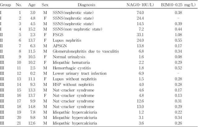
Table 1. Clinical characteristics of children with various renal diseases
Group No. Age Sex Diagnosis NAG(0-10U/L) B2M(0-0.25 mg/L)
I I I I II II II II III III III III III III III III III III III III III
1 2 3 4 5 6 7 8 9 10 11 12 13 14 15 16 17 18 19 20 21
3.0 4.8 4.5 15.2 2.3 13.7 6.3 11.5 10.5 10.2 2.5 0.2 11.1 9.3 13.3 13.7 9.9 14.8 7.8 9.8 12.6
M F M M F F M M F F M M F M M F M M M M M
SSNS(nephrotic state) SSNS(nephrotic state) SSNS(nephrotic state) SSNS(non-nephrotic state) FSGS
Lupus nephritis APSGN
Glomerulonephritis due to vasculitis Normal urinalysis
Idiopathic hematuria Hemorrhagic cystitis Lower urinary tract infection Lupus without nephritis HSP without nephritis Nut-cracker syndrome Nut-cracker syndrome Nut-cracker syndrome Nut-cracker syndrome Idiopathic hypercalciuria Idiopathic hypercalciuria Idiopathic hypercalciuria
74.0 24.4 14.5 7.2 33.1 24.0 13.8 6.8 1.6 2.2 1.8 6.9 1.5 4.0 4.6 4.8 12.6 13.0 1.2 3.1 3.6
0.38
-
0.39
0.44
1.08
0.55
0.17
0.34
0.08
0.29
0.52
-
0.28
0.28
0.17
0.13
0.31
0.29
0.22
0.34
0.26
Abbreviations : NAG, N-acetyl-beta-D-glucosaminidase; B2M, beta 2-microglobulin; SSNS, Steroid sen-
sitive nephrotic syndrome; FSGS, Focal segmental glomerulosclerosis; APSGN, Acute poststreptococcal
glomerulonephritis; HSP, Henoch-Shoenlein purpura
each child. The amount of protein in the urine was assessed as 1+(30 mg/dL), 2+(100 mg/
dL), 3+(300 mg/dL) or 4+(1,000 mg/dL)[8], and hematuria was defined as more than 5 erythrocytes per high-power field(in a sedi- ment of approximately 10 mL of freshly voi- ded urine)[9].
Urinary NAG and B2M were analyzed as parameters for renal tubular function. For the measurement of urinary NAG, spectrophoto- metric assay was performed using chlorophe- nol red-NAG(CPR-NAG) which is hydrolyzed by NAG with the release of chlorophenol red, which can be measured photometrically at 575 nm on a JCA-BM 12. Urinary B2M was mea- sured by radioimmunoassay(RIA).
Statistical analysis was performed using SPSS version 11.0(SPSS Inc, Chicago, IL).
Continuous variables were expressed as mean
± standard deviation. The differences among three groups were analyzed by Kruskal-Wal- lace test. Correlation between two variables was assessed by Spearmans rank correlation test. P<0.05 was considered significant.
RESULTS
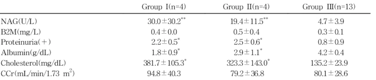
Clinical characteristics of the groups are shown in Table 1. Urinary NAG levels in groups I and II were significantly higher than those in group III(19.4±11.5 and 30.0±30.1 vs.
4.7±3.9, P=0.01), while urinary B2M levels did not differ among the 3 groups(Table 2).
In group I, NAG levels were increased in three of the four patients and B2M in all 3 patients. In group II, two patients with FSGS and lupus nephritis showed significantly in- creased levels of NAG and B2M. In one pa- tient with APSGN, NAG was mildly elevated but B2M was within normal range. In one pa- tient with glomerulonephritis due to vasculitis, however, NAG was normal, but B2M was significantly increased. In group III, two of the four patients with nut-cracker syndrome showed increased level of both NAG and B2M.
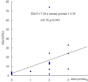
Urinary NAG levels were positively corre- lated with urinary B2M levels(NAG=32.6×
B2M+0.77, r=0.49, P=0.03)(Fig. 1). In 21 pa- tients, urinary NAG levels were correlated with proteinuria (NAG = 7.24 x urinary pro- tein+2.32, r=0.79, P<0.001)(Fig. 2), serum al-
Table 2. Laboratory findings among the three groups
Group I(n=4) Group II(n=4) Group III(n=13) NAG(U/L)
B2M(mg/L) Proteinuria(+) Albumin(g/dL) Cholesterol(mg/dL) CCr(mL/min/1.73 m
2)
30.0±30.2
**0.4±0.0 2.2±0.5
*1.8±0.9
*381.7±105.3
*94.8±40.3
19.4±11.5
**0.5±0.4 2.5±0.6
*2.9±1.1
*323.3±143.0
*79.2±36.8
4.7±3.9 0.3±0.1 0.8±0.9 4.2±0.4 135.2±23.9
80.1±28.6
*P<0.01 in comparison to Group III
**P<0.05 in comparison to Group III
Abbreviation : NAG, N-acetyl-beta-D-glucosaminidase; B2M, beta 2-microglobulin, CCr; Creatinine Clea-
rance
bumin(NAG=-9.18×albumin+44.52, r=-0.72, P
<0.001)(Fig. 3) and serum cholesterol(r=0.58, P=0.006) levels. Urinary B2M levels were also correlated with proteinuria(r=0.67, P=0.001), serum albumin(r=-0.57, P=0.011) and serum cholesterol(r=0.56, P=0.013). However, urinary NAG or B2M did not correlate with serum creatinine (P>0.05) or uric acid levels(P>0.05).
In various renal diseases, such as HSP without nephritis, lupus without nephritis, idi- opathic hematuria, idiopathic hypercalciuria, and cystitis, NAG levels were within normal ranges, but B2M were variably increased.
There was no significant correlation bet- ween CCr and NAG or B2M(P>0.05).
DISCUSSION
The present study demonstrates that urina- ry NAG and B2M are increased in children with SSNS and various kinds of glomerulo- nephritides, suggesting the presence of renal tubular impairment in these patients.
NAG excretion has been considered as a
non-invasive and reliable marker of the tubu- lo-toxicity of proteinuria in the early stage of membranous nephropathy, FSGS and minimal change disease in adult studies[6,10]. In glo- merular diseases, analysis of isoenzymes of NAG demonstrated that the increased urinary excretion of this enzyme is due to an increa- sed release by the renal tubular cells and not to increased filtration across the damaged glo-
Fig. 1. Relationships between urinary NAG and B2M levels.
Fig. 3. Relationships between urinary NAG and serum albumin levels.
Fig. 2. Relationships between urinary NAG and
proteinuria.
merular capillary wall[11].
Regarding childhood nephrotic syndrome, Valles et al. reported that urinary NAG and B2M levels were significantly increased in the steroid-resistant nephrotic syndrome(SRNS) group as compared to the SSNS in remission and controls, and there were no differences between the SRNS group and SSNS in relapse[12]. They concluded that proximal tu- bule cell dysfunction, partially affected by massive albuminuria, might account for the higher values of B2M and NAG excretion in the SRNS patients and urinary B2M and NAG levels are not helpful in identifying histological evidence of structural tubulointerstitial damage in children with SRNS[12]. In our study, there were five patients with nephrotic syndrome (one with FSGS and four with SSNS, and urinary NAG or B2M levels were significantly increased in these patients as compared to those of the control group, which were in ac- cordance with previously reports[4, 12] Fur- thermore, NAG was significantly decreased in non-nephrotic state compared to nephritic sta- te. However, Tsau et al. had reported the contrasting results that there were no signifi- cant changes in urinary NAG levels during heavy proteinuria and after remission, and no correlation was found between urinary protein excretion and NAG in children with nephrotic syndrome[13].
Regarding childhood lupus nephritis, Marks et al. reported that the lupus nephritis group had elevated urinary NAG levels than non- nephritis lupus or normal children[14]. Simi- larly, our study showed that one patient(No.
6) with lupus nephritis had an elevated urina- ry NAG level, while the other with non-neph-
ritis lupus showed a normal NAG level. How- ever, Marks et al pointed that tubular dys- function with elevated urinary NAG levels was present in 2 lupus non-nephritis patients with no evidence of glomerular disease, who eventually developed biopsy-proven lupus ne- phritis later, suggesting evidence of tubular dysfunction in lupus non-nephritis patients might help to identify lupus nephritis prior to the onset of albuminuria[14]. Therefore, the increase of urinary NAG level can be used as an early index for the damage of tubular epi- thelium, and may be useful in detecting or monitoring renal disease in lupus patients.
Group III include normal uinalysis, cystitis and Nut-cracker syndrome etc. together be- cause these diseases usually dont have tubular injury.
We had one patient with HSP without ne- phritis, and urinary NAG level was within normal range. Müller et al. demonstrated that urinary NAG or alpha1-microglobulin levels were highest(>mean +4 SD) in patients with early kidney involvement, intermediately high (>mean +2 SD, <mean +4 SD) in those who developed renal involvement during follow-up, and normal in those with a benign further clinical course. Therefore, urinary NAG levels may be a good prognostic marker for the de- velopment of HSP nephritis[15].
In idiopathic hypercalciuria, tubular impair- ment might occur, especially in patients with urolithiasis or nephrocalcinosis[16]. However, they could not find a direct relationship bet- ween the urinary NAG level and the degree of calcium leakage[16], although the relationship between the urinary NAG and the daily uri- nary calcium excretion was statistically signi-
ficant in an experimental model of hypercal- ciuria[17]. In our study, all three patients with idiopathic hypercalciuria without nephrocalci- nosis showed normal urinary NAG levels, but one of them did increased B2M level.
We also had four patients with nut-cracker syndrome, two of whom(Nos.17 and 18) showed increased levels of NAG and B2M, suggesting left renal venous hypertension might cause the excretion of proteinuria and these urinary enzymes.
It is reported that creatinine correction is rather incorrect because of diurnal variation in NAG level[18].
However, there are some limitations in this study(1) small numbers of patients,(2) a lack of control groups, and(3) a retrospective na- ture of the study.
Nevertheless, we suggest that the measure- ment of NAG or B2M may be a useful and non-invasive examination for assessing a proximal tubular damage in pediatric patients with nephrotic syndrome or the early stages of various kinds of glomerulonephritides.
한 글 요 약
다양한 신장질환 환아들에서 요중 N-Acetyl-beta-D-Glucosamini dase와 beta 2-Microglobulin
연세대학교 의과대학 소아과학교실, 진단검사의학교실*
윤소진·신재일·이재승·김현숙
*목 적 : 요 중 N-acetyl-beta-D-glucosamini- dase(NAG) 와 beta 2-microglobulin(B2M) 은 신세뇨관 간질 손상의 표적으로 생각된다. 이 연구
의 목적은 다양한 신장 질환 환아 에서 요 중 NAG 와 B2M 수치를 검사해 보는 것이다.
방 법 : 우리는 21명의 환아(8.9±4.5세, 남:녀=
14:7)를 조사해서 세 군으로 분류하였다: I군(스테 로이드에 반응하는 신증후군 환아-4명), II군(다양 한 종류의 사구체 신염 환아-4명), III군(정상뇨 또 는 비사구체성 신장 질환 환아-13명).
결 과 : I군과 II군에서의 요 중 NAG 수치는 III 군에서보다 유의하게 높았다.(19.4±11.5와 30.0±
30.1 vs. 4.7±3.9 , P=0.01) 반면에 요 중 NAG 수치와 B2M 수치가 양의 상관 관계에 있음에도 (r=0.49, P=0.03), 요 중 B2M 수치는 세 군에서 차이가 없었다. 요 중 NAG 와 B2M 수치는 모두 단백뇨, 혈중 알부민, 콜레스테롤과 상관관례를 보 였다.
결 론 : NAG 와 B2M 의 배설량은 스테로이드 에 반응하는 신증후군 환아와 다양한 종류의 사구 체 신염 환아에서 증가되어 있었다. 이는 이런 질 환들에서 세뇨관 기능저하를 의미하는 것으로 보 인다.
참 고 문 헌