R E S E A R C H Open Access
Risk factors for postoperative infection of odontogenic cysts associated with
mandibular third molar
Jin-woo Kim, Do-hyun On, Jin-yong Cho and Jaeyoung Ryu *
Abstract
Background: Odontogenic cysts associated with lower third molar are common. The prognosis for surgical treatment is relatively good. However, postoperative infection discourages the clinicians. Hence, we would like to investigate the factors associated with infection after surgical treatment of cysts associated with the mandibular third molar.
Methods: We retrospectively reviewed the medical and radiographic records of 81 patients who were diagnosed with dentigerous cyst or odontogenic keratocyst and underwent cyst enucleation. The factors affecting postoperative infection were divided into host factor, treatment factor, and cystic lesion factor. To identify the factors associated with postoperative infection, we attempted to find out the variables with significant differences between the groups with and without infection.
Results: A total of 81 patients (64 male and 17 female) were enrolled in this study. There was no statistical relationship about the postoperative infection between all variables (gender, smoking, diabetes mellitus, age, bone grafting, related tooth extraction, previous marsupialization or decompression, type of antibiotics, cortical perforation associated with cystic lesion, preoperative infection, preoperative cyst size).
Conclusions: The results of this study suggest that it is not necessary to avoid bone grafts that are concerned about postoperative infection.
Keywords: Odontogenic cysts, Surgical procedures, Surgical wound infection
Background
Intrabony cysts are common lesions of the oral and maxillofacial region [1]. The treatment of cystic lesions associated with mandibular third molar has relatively fa- vorable prognosis [2]. A dentigerous cyst (DC) and odontogenic keratocyst (OKC) are known as common odontogenic cyst in the mandibular third molar region [2 – 4]. In case which the lesions are similar in size and location, the treatment is similar [5]. Enucleation of cys- tic lesion with extraction of associated tooth is usually performed, though decompression or marsupialization can also be performed taking into account the patient
condition, size and location of the lesion, the potential for pathological fractures, and recurrence [5, 6].
Although surgeons prescribe the antibiotics and edu- cated a patient about the importance of no smoking and maintenance of oral hygiene, surgical site infections may occur irrespective of bone grafts. Unexpected infection symptoms after surgery of cysts may result in poor satis- faction of patients and clinicians. If the statistically sig- nificant factors that cause these infections can be identified, treatment predictability can be improved. The purpose of this study was to determine the factors asso- ciated with infection after surgical treatment of cysts as- sociated with the mandibular third molar.
Methods
This study was performed in accordance with the Dec- laration of Helsinki for medical protocols. The regional
© The Author(s). 2020 Open Access This article is distributed under the terms of the Creative Commons Attribution 4.0 International License (http://creativecommons.org/licenses/by/4.0/), which permits unrestricted use, distribution, and reproduction in any medium, provided you give appropriate credit to the original author(s) and the source, provide a link to the Creative Commons license, and indicate if changes were made.
* Correspondence: [email protected]
Department of Oral & Maxillofacial Surgery, Gachon University Gil Medical Center, 21, Namdong-daero 774 beon-gil, Namdong-gu, Incheon 21565, South Korea
Maxillofacial Plastic and Reconstructive Surgery
Kim et al. Maxillofacial Plastic and Reconstructive Surgery (2020) 42:4
https://doi.org/10.1186/s40902-020-00248-5
Ethical Review Board granted approval (GDIRB2020- 041) for the study. We retrospectively reviewed the med- ical and radiographic records of 81 patients who were diagnosed with DC or OKC and underwent cyst enucle- ation between 2012 and 2018. Postoperative infection was defined as obvious pain with formation of swelling and pustulation. Patients with postoperative infection signs were classified as infection group and patients who were not as non-infection group. The factors affecting postoperative infection were divided into host factor, treatment factor, and cystic lesion factor. Age, gender, underlying disease such as diabetic mellitus, and smok- ing of patients were investigated as host factors. Bone graft to the defect after enucleation, type of antibiotics (amoxicillin with clavulanic acid or cephalosporin), mar- supialization or decompression before enucleation, the presence or absence of mesial tooth (lower second molar), and its additional treatment such as extraction, endodontic treatment, or apicoectomy were also investi- gated as treatment factors. Cortical perforation around cyst, size of cystic lesion, preoperative infection, and type of cyst (dentigerous cyst or odontogenic keratocyst) were also evaluated as cystic lesion factors. The size of cystic lesion was calculated using software (INFINITT PACS®, INFINITT Healthcare, Seoul, South Korea) which was measured the longest diameter of the radiolucent lesion.
To identify the factors associated with infection, we attempted to find out the variables with significant dif- ferences between the groups with and without infection.
Then, we tried to find out which detailed treatment or patient factors influenced the outcome of infection.
Results
A total of 81 patients (64 male and 17 female) were en- rolled in this study (Table 1). The mean age was 47.3 years. Postoperative infection occurred in 21 patients.
The average age of the infected patients was 45.3 (22–
77) years old and the infection occurred at average 5 weeks (10–93 days) postoperatively. Infection was oc- curred at 14 patients (20.1%) in DC and 7 patients (50%) in OKC, respectively. There was no statistical signifi- cance between the groups with and without the infection by diagnosis (p = 0.054). In addition, there was no statis- tical relationship between all variables (gender, smoking, diabetes mellitus, age, bone grafting, related tooth ex- traction, previous marsupialization or decompression, type of antibiotics, cortical perforation associated with cystic lesion, preoperative infection, preoperative cyst size). A total of 66 patients did not have tooth extraction related to the cyst (Table 2). No statistical relationship was found between the infection and non-infection groups with regard to the treatment of mesial tooth (ex- traction, endodontic treatment, and apicoectomy of the lesion-related tooth).
Discussion
In this study, we analyzed the factors of infection after surgical treatment of cystic lesion associated with
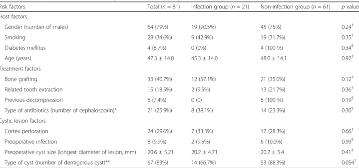
Table 1 Description of risk factors affecting infection after the surgery of cyst enucleation
Risk factors Total ( n = 81) Infection group ( n = 21) Non-infection group ( n = 61) p value
Host factors
Gender (number of males) 64 (79%) 19 (90.5%) 45 (75%) 0.24
†Smoking 28 (34.6%) 9 (42.9%) 19 (31.7%) 0.35
†Diabetes mellitus 4 (6.7%) 0 (0%) 4 (100 %) 0.34
§Age (years) 47.3 ± 14.0 45.3 ± 14.0 48.0 ± 14.1 0.92
‡Treatment factors
Bone grafting 33 (40.7%) 12 (57.1%) 21 (35.0%) 0.12
†Related tooth extraction 15 (18.5%) 2 (9.5%) 13 (21.7%) 0.36
†Previous decompression 6 (7.4%) 0 (0) 6 (100 %) 0.19
§Type of antibiotics (number of cephalosporin)* 21 (25.9%) 8 (38.1%) 14 (23.3%) 0.30
†Cystic lesion factors
Cortex perforation 24 (29.6%) 7 (33.3%) 17 (28.3%) 0.66
†Preoperative infection 8 (9.9%) 2 (9.5%) 6 (10.0%) 0.99
§Preoperative cyst size (longest diameter of lesion, mm) 20.6 ± 5.21 20.2 ± 4.71 20.7 ± 5.4 0.41
‡Type of cyst (number of dentigerous cyst)** 67 (83%) 14 (66.7%) 53 (88.3%) 0.054
††
Chi-square test
§
Fisher ’s exact test
‡
Student ’s t test
*Two kinds of antibiotics were used, amoxicillin with clavulanic acid and cephalosporin
**All subjects were classified as dentigerous cyst or odontogenic keratocyst
Kim et al. Maxillofacial Plastic and Reconstructive Surgery (2020) 42:4 Page 2 of 4
mandibular third molar. Although infection was more likely to occur after surgical treatment of OKC than DC, the results were not statistically significant (p = 0.054).
Furthermore, it could be considered to have a no direct effect on postoperative infection because both diseases are developmental cyst not inflammatory origin. Further analysis about infection occurrence for each diagnosis (DC and OKC) was performed with regard to cortical perforation of the cyst lesion, conditions of mesial tooth, and the presence of bone grafting in patient. There was also no statistical significance. The treatment methods or recurrence may vary depending on the diagnosis, but no significant related factors for postoperative infection were found.
In the case of cyst enucleation, bone grafts are pre- ferred to improve bone healing in the bone defects [7].
On the other hand, studies have shown that bone regen- eration is possible without bone graft [8, 9]. It was re- ported to increase bony density up to 97% in relatively large defect (between 20 and 30 mm in size) and 84% in more large defect (between 30 and 50 mm in size) after one year [8]. Therefore, it may be considered that a bone graft should not be done due to a concern about infec- tion of the graft. However, as with other findings, this study suggests that bone graft can be considered when necessary due to the low correlation between bone graft and infection [10].
Most of the studies focusing on the size reduction and recurrence of cystic lesion have been made for decom- pression procedure before cystectomy. The advantage of decompression procedure of cystic lesion is that the size of the cyst can be reduced to preserve the surrounding anatomical structures and to prevent pathological frac- tures through minimal facial deformation and bone re- generation during healing period [6, 11, 12]. Although decompression have been shown to improve the progno- sis of cyst enucleation in both DC and OKC [13, 14], the results of postoperative infection according to the size of this study were not significant. Although there was no statistical significance, further study is needed to
investigate the relation between decompression and postoperative infection, because there was no postopera- tive infection in 6 patients who underwent decompres- sion procedure before definitive enucleation.
This study found no statistically significant factors as- sociated with postoperative infection of the DC and OKC associated with the mandibular third molar. How- ever, reducing postoperative infection is one of the im- portant things in the surgical treatment. Therefore, it may be necessary to study through an extended popula- tion with a well-designed prospective study.
Conclusions
The results of this study suggest that it is not necessary to avoid bone grafts that are concerned about postopera- tive infection and there is no justification of decompres- sion to reduce the size of cysts only for the prevention of postoperative infection.
Abbreviations
DC: Dentigerous cyst; OKC: Odontogenic keratocyst
Acknowledgements Not applicable
Authors ’ contributions
J-K was a major contributor in writing the manuscript. D-O participated col- lection of data and drafting of the manuscript. J-C analyzed and interpreted the patient data. JR contributed to the conception of the report and critical revising. All authors read and approved the final manuscript.
Funding
We received no funding in support of this study.
Availability of data and materials Not applicable
Ethics approval and consent to participate
This study followed the medical protocols and ethics outlined in the Declaration of Helsinki, and approval was granted by the regional Ethical Review Board (GDIRB2020-041).
Consent for publication Not applicable
Competing interests
The authors declare that they have no competing interests.
Table 2 Comparisons between groups according to the treatment method of cystic lesion-related mesial tooth
Total Infection group Non-infection group p value
Extraction of related tooth 81 2 13 0.36
†Without extraction of related tooth 19 47
Within the patients without extraction of related tooth
Root canal treatment of related tooth 66 11 18 0.24
†Without root canal treatment of related tooth 8 29
Within the patients undergoing root canal treatment of related tooth
Apicoectomy of related tooth 29 0 2 0.51
§Without apicoectomy of related tooth 11 16
†
Chi-square test
§