• 검색 결과가 없습니다.

Synthesis and Biocompatibility of PVA/NaCMC Hydrogels Crosslinked by Cyclic Freezing/thawing and Subsequent Gamma-ray Irradiation

N/A
N/A
Protected

Academic year: 2021

Share "Synthesis and Biocompatibility of PVA/NaCMC Hydrogels Crosslinked by Cyclic Freezing/thawing and Subsequent Gamma-ray Irradiation"

Copied!
7
0
0

로드 중.... (전체 텍스트 보기)

전체 글

(1)

학 술 논 문

161

Synthesis and Biocompatibility of PVA/NaCMC Hydrogels Crosslinked by Cyclic Freezing/thawing and

Subsequent Gamma-ray Irradiation

Ji-Yeon Shin

1

, Heeseok Jeong

2

and Deuk Yong Lee

1

1

Department of Biomedical Engineering, Daelim University, Anyang 13916, Korea

2

Convergence of Institute of Biomedical Engineering and Biomaterials, Seoul National University of Science and Technology, Seoul 01811, Korea

(Manuscript received 15 March 2018 ; revised 28 June 2018 ; accepted 23 August 2018)

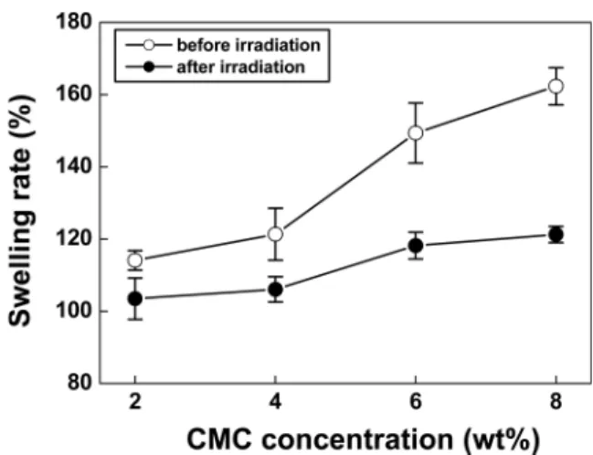
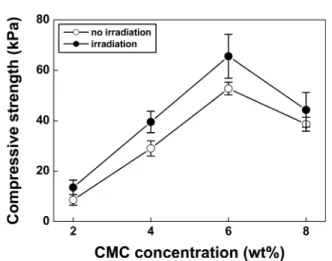
Abstract: Polyvinyl alcohol/sodium carboxymethyl cellulose (PVA/NaCMC) hydrogels were prepared by physical crosslinking (cyclic freezing/thawing) and gamma (γ)-ray irradiation to evaluate the effect of NaCMC concentration (2~8 wt%) on the mechanical properties and the biocompatibility of the PVA/NaCMC hydrogels. The swelling rate of PVA/NaCMC hydrogels regardless of irradiation rose with increasing NaCMC content from 2 wt% to 8 wt%, while the gelation rate was the reverse. As the NaCMC content increased from 2 wt% to 6 wt%, the compressive strength of the hydrogels increased dramatically from 8.5 ± 2.0 kPa to 52.7 ± 2.5 kPa before irradiation and from 13.5 ± 2.9 kPa to 65.5 ± 8.7 kPa after irradiation. When 8 wt% NaCMC was added afterwards, the compressive strength decreased however. The irradiated PVA/NaCMC hydrogels containing 6 wt% NaCMC exhibited the tailored properties of the swelling rate of 118 ± 3.7%, the gelation rate of 71.4 ± 1.3%, the strength of 65.5 ± 8.7 kPa, respec- tively, and no cytotoxicity was observed.

Key words: Polyvinyl alcohol/sodium carboxymethyl cellulose (PVA/NaCMC), Hydrogel, Compressive strength, Cytotoxicity

I. Introduction

With the development of medical technology, the average life span is increased and the living standard have improved. However, traffic accidents, trauma and burn patients are on the rise as well [1- 5]. Transplants of autologous or cultured fibroblasts should be the ultimate treatments of severe burns or trauma. Due to the prolonged procedure, a dressing is needed to protect and heal the wound. Dressings for optimal wound healing do the following: absorb body fluids from the wound, prevent infection, and provide visibility via transparency. Polymer hydrogel is the best material for such aforementioned dressing

properties. It is composed of three-dimensional hydrophilic polymer networks, in which a large amount of water is interposed [1-8].

Polyvinyl alcohol (PVA) is a semi-crystalline polymer whose hydroxyl groups produce inter- and intra- molecular hydrogen bonding. Sodium carboxymethyl cellulose (NaCMC) is a typical ionic-type cellulose ether with multiple carboxyl groups. Double network hydrogels (PVA/NaCMC) can be easily formed due to the good coordination ability, hydrophilicity, and biodegradability of NaCMC [1-5]. PVA/NaCMC hydrogels can be prepared by several methods, such as physical crosslinking by freezing and thawing technique, chemical crosslinking, and radiation- induced crosslinking. The hydrogels prepared by freezing and thawing method exhibited insufficient mechanical strength, but they have been widely used because of their simplicity and lack of toxicity.

Radiation crosslinking has the advantage of being Corresponding Author : Deuk Yong Lee

Department of Biomedical Engineering, Daelim University, Anyang 13916, Korea

TEL: +82-31-467-4835 / FAX: +82-31-467-4432

E-mail: [email protected]

(2)

162

sterilized during crosslinking without exposing to the chemical risks [1-4]. The present study evaluates the effect of NaCMC concentration on the mechanical properties, cytotoxicity, and drug delivery system of the PVA/NaCMC hydrogels by crosslinking the PVA/

NaCMC hydrogels through cyclic freezing/thawing and γ-ray irradiation.

II. Experimental

1. Materials

PVA ([CH

2

CH(OH)]

n

, Mw 85,000~124,000), NaCMC ([C

6

H

7

O

2

(OH)

2

OCH

2

COONa]

n

, Mw 90,000), and MD (metanidazole, C

6

H

9

N

3

O

3

) were purchased from Sigma-Aldrich, USA. All chemicals were used as received without any further purification.

2. Synthesis of hydrogel

3.5 g of PVA was added to 46.5 mL of distilled water and dissolved in an autoclave for 20 min at 121

o

C and 1.2 MPa. 2 g of CMC was added to the aqueous PVA solution and uniformly mixed at 75 rpm using an overhead stirrer. The gas bubbles in the PVA/NaCMC solution were eliminated by using ultrasonic cleaner for 10 minutes. The PVA/NaCMC aqueous solution was then cast on a petri dish and physical crosslinking was carried out by repeating the freezing process for 2 h at -75

o

C and the thawing process for 2 h at room temperature. Subsequent crosslinking was then performed by using the 25 kGy γ-ray irradiation.

3. Characterization (1)Gelation rate (%)

The hydrogel was cut into a size of 5 × 5 mm

2

and immersed in distilled water for 24 h. The specimen was taken out and the surface of the gel was wiped off and dried for 48 h in a vacuum dryer. The gelation rate ( G(%)) was calculated by dividing the weight of the dried hydrogel ( W

d

) by the weight of the polymer initially used ( W

i

), [1,5].

(2) Swelling rate (%)

The hydrogel was cut into a size of 5 × 5 mm

2

and immersed in distilled water for 24 h. The moisture of

the specimen was screened through a 46 µm filter paper using a vacuum pump. The swelling rate ( S(%)) is determined by using the equation, , where W

s

and W

d

represent the weight of the swollen hydrogel and the weight of the dried hydrogel, respectively [1,9-14].

(3) Compressive strength

Compressive strength of PVA/NaCMC hydrogels before and after irradiation was examined by using an Instron 5564 with a crosshead speed of 10 mm/

min. The specimen with a diameter of 15 mm and a thickness of 5 mm was fabricated and the strength was determined at a point where the specimen thickness decreased by 50% [1].

(4) Drug delivery

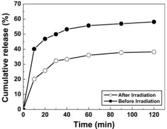
Drug release experiment was performed on antimicrobial drug of MD in a double jacket beaker [15]. The PVA/NaCMC hydrogel with a diameter of 15 mm was added into 300 mL of MD solution with a concentration of 1 mg/L. Drug delivery experiment was performed on MD in a double jacket glass. The change in the adsorption at 319 nm was applied to identify the concentration of MD by using an UV-vis spectrophotometer (V-670, Jasco, Japan) and the concentration of MD was measured as a function of time.

(5) Cytotoxicity

The extract test method was conducted on the PVA/NaCMC hydrogels to evaluate the potential of cytotoxicity on the base of the International Organization for Standardization (ISO 10993-5) [13,14,16]. The PVA/

NaCMC hydrogels were extracted aseptically in single strength Minimum Essential Medium (1X MEM, Dulbecco’s Modified Eagles’s Medium (Gibco) with 10% fetal bovine serum (Gibco) and 1%

penicillin-streptomysin) with serum. The ratio of the PVA/NaCMC hydrogels to extraction vehicle was 0.2 g/mL. The 96-well plate was incubated at a temperature of 37

o

C in a 5% CO

2

atmosphere. The test extracts were maintained in an incubator for 24 h. The test extracts were placed onto three separate confluent monolayers of L-929 (NCTC Clone

G %( ) Wd Wt ---

=

S %( ) (Ws×Wd) Wd --- 100×

=

(3)

163 929, ATCC, USA) mouse fibroblast cells propagated

in 5% CO

2

. For this test, confluent monolayer cells were trypsinized and seeded in 10 cm

2

wells (35 mm dishes) with a micropipette. Simultaneously, triplicates of reagent control, negative control (high density polyethylene film, RM-C), and positive control (polyurethane film, RM- A) were placed onto the confluent L-929 monolayers. All monolayers were incubated for 48 h at 37

o

C in the presence of 5% CO

2

. After incubation, the morphological change of the cell was examined to assess the biological reaction by using the inverted microscope (TS100-F, Nikon, Japan) and the iMark microplate absorbance spectrophotometer (Bio-Rad, USA) [16,17]. Water- soluble tetrazolium salts (WSTs) are a series of other water-soluble dyes for MTT assays, which can provide different absorption spectra of the formed formazans. EZ-cytox yields a water-soluble formazan, which can be read directly. The absorbance of the colored solution is quantified by measuring at a wavelength of 415 nm with the microplate absorbance spectrophotometer [14,16,17]. The value of untreated cell (control sample, only cultured with culture medium) was set as 100% and those of the treated cells were expressed as the percentage of the control sample.

(6) Cell proliferation

Cell counting kit-8 (CCK-8, Dojindo Molecular Technologies, Inc., Japan) was used for the assay of cell proliferation. CCK-8, being nonradioactive, allows sensitive colorimetric assays for the determination of the number of viable cells in cell proliferation [16].

WST is reduced by dehydrogenases in cells to give an orange colored product (formazan), which is soluble in the tissue culture medium. The amount of the formazan dye generated by dehydrogenases in cells is directly proportional to the number of living cells.

The 96-well plate containing 100 mL of cell suspension (5 × 10

3

cells/well) was incubated for 24 h at a temperature of 37

o

C in a 5% CO

2

atmosphere.

The test extracts (10 mL) were added to the plate and maintained for an appropriate length of time (6, 12, 24, 48 h) in an incubator. After adding 10 mL of CCK-8 solution to each well of the plate, the plate was incubated for 2 h. Then, the absorbance of the

colored solution is quantified by measuring at a wavelength of 450 nm with the microplate absorbance spectrophotometer [16].

III. Results and Discussion

The PVA/NaCMC hydrogels having the composition of 7 wt% PVA with NaCMC concentration of 0 to 8 wt% were prepared by repeating freezing/thawing twice and subsequent irradiation with 25 kGy γ-ray.

The swelling rate of PVA/NaCMC hydrogels was determined by measuring the weight change with time after soaking for 24 h in distilled water at room temperature, as shown in Fig. 1. The swelling rate of the PVA/NaCMC hydrogels before and after irradiation rose from 114% to 162% and 103 wt% to 121 wt%

with increasing the NaCMC concentration from 2 wt% to 8 wt%, respectively. Although the swelling rate of the PVA/NaCMC hydrogels before irradiation was always higher than that of irradiated hydrogels, the addition of NaCMC to PVA exhibited similar behavior regardless of γ-ray irradiation. Since the irradiated γ-rays may form radicals capable of crosslinking to the polymer, the 3-D network structure is likely to be increased, leading to the reduction of the volume capable of containing water [1,4,17]. The highest swelling rates of 162% and 121% were observed for the PVA/NaCMC hydrogels containing 8 wt% NaCMC before and after irradiation, respectively, suggesting that higher NaCMC content

Fig. 1. Swelling rate of PVA/CMC hydrogels as a function of

CMC concentration.

(4)

164

was determined to be effective to swelling capability [5]. The FTIR spectra of PVA powder, NaCMC powder, and PVA/6 wt% NaCMC hydrogels before and after irradiation are shown in Fig. 2. PVA is a semi-crystalline polymer whose hydroxyl groups (-OH) produce inter and intra-molecular hydrogen bonding.

C=O, C-O, C-H, and O-H were partially observed for NaCMC due to the presence of the numerous hydroxyl and carboxylic groups (-COOH) in NaCMC [19]. The PVA/NaCMC hydrogels containing 6 wt% of NaCMC exhibited almost similar peaks regardless of irradiation. The spectra showed a main peak at 3200~3400 cm

−1

due to stretching of hydroxyl groups.

And C=O stretching at 1640 cm

−1

, C-H stretching at 1414 cm

−1

, O-H at 1329 cm

−1

, and C-O stretching at

1073 cm

−1

are also visible. The presence of O-H groups at 3200~3400 cm

−1

are likely due to increase in hydrogen bond as a result of crosslinking (cyclic freezing/thawing) and γ-ray irradiation crosslinking [1].

After removing the residual polymers of the PVA/

NaCMC hydrogels by washing in distilled water for 48 h at room temperature, the hydrogels were dried for 48 h at 60

o

C to determine the gelation rate [1,5].

Experimental results (Fig. 3) revealed that the gelation rates of irradiated specimens were always higher than those of the PVA/NaCMC hydrogels before irradiation. The gelation rate of the PVA/

NaCMC hydrogels before and after γ-ray irradiation decreased gradually from 79.6% to 65.3% and from 82.9% to 70.4% with increasing NaCMC content from 2 wt% to 8 wt%, respectively, as depicted in Fig. 3.

The increase in the gelation rate with decreasing the NaCMC content improved the crosslinking so that 3- dimensional network structure inside the hydrogel increased. Then, the volume capable of containing water was decreased, resulting in the reduction in swelling rate. It was found that the gelation rate was inversely proportional to the swelling rate and higher content of NaCMC was detrimental to the gelation rate of PVA/NaCMC hydrogel.

The compressive strength of the PVA/NaCMC hydrogel before and after irradiation is shown in Fig 4. Similar results were observed regardless of irradiation. As the amount of NaCMC increased from Fig. 2. FT-IR spectra of PVA powder, CMC powder, PVA/

6wt% CMC hydrogels before and after irradiation.

Fig. 3. Gelation rates of PVA/CMC hydrogels as a function of CMC concentration.

Fig. 4. Compressive strength of PVA/CMC hydrogels before

and after γ -ray irradiation.

(5)

165

2 wt% to 6 wt%, the strength of the hydrogels before and after irradiation increased dramatically from 8.5 ± 2.0 kPa to 52.7 ± 2.5 kPa and from 13.5 ± 2.9 kPa to 65.5 ± 8.7 kPa, respectively, and then decreased when 8 wt% of NaCMC was added. PVA exhibits excellent mechanical properties but inferior water- soluble properties after crosslinking. On the other hand, NaCMC shows the reverse phenomena (poor strength and excellent swelling rate). In the PVA/

NaCMC composites, the strength and swelling rate are increased at first by CMC addition due to synergic combination of PVA and NaCMC, but the strength and swelling rate are decreased due to higher content of NaCMC (8 wt%). The highest strength was

observed for the PVA/NaCMC hydrogels containing 6 wt% NaCMC probably due to the optimum combination of swelling rate and gelation rate.

A cytotoxicity test of the irradiated hydrogels

containing various NaCMC concentration in the

range of 2 wt% to 8 wt% determines whether the

hydrogels will have a toxic effect on living cells

[15,16]. The test extracts with the irradiated

hydrogels regardless of NaCMC concentration showed

no evidence of causing cell lysis or toxicity, as depicted

in Fig. 5. The irradiated hydrogels containing

NaCMC of 2%, 4%, 6%, and 8% exhibited cell

viability of 112%, 144%, 193%, and 172% compared

to the negative control, respectively, as measured at

Fig. 5. Photographs of cell morphology: (a) positive control, (b) negative control, and the extracts of PVA/ (c) 2 wt%, (d)

4 wt%, (e) 6 wt% and (f) 8 wt% CMC hydrogels from EZ-cytox after exposing with the hydrogel suspensions for 48 h.

(6)

166

a wavelength of 415 nm by using the microplate absorbance spectrophotometer [9-16]. The qualitative morphological grading of cytotoxicity of the irradiated PVA/NaCMC hydrogels was determined to be scale 0.

In addition, the cell proliferation results of the irradiated PVA/NaCMC hydrogels (Fig. 6) suggested that L-929 cells adhered well to the PVA/NaCMC hydrogels and proliferated continuously with increasing time regardless of NaCMC concentration. Therefore, it is conceivable that the crosslinked PVA/NaCMC hydrogels are considered to be clinically safe and effective due to the absence of cytotoxicity and excellent cell proliferation under the condition of this study.

The drug release behavior of MD (antimicrobial drug) is shown in Fig. 7. The drug release rate of the PVA/NaCMC hydrogels increased dramatically from

0 to 10 min at the beginning and then reached the plateau region from 30 min regardless of irradiation.

Although the cumulative drug release rate of the hydrogels was 58%, it decreased down to 38% when the hydrogels were irradiated. It may be due to the formation of 3-dimensional network structure inside the hydrogel.

IV. Conclusions

PVA/NaCMC hydrogels were crosslinked by cyclic freezing/thawing and subsequent γ-ray irradiation to evaluate the influence of NaCMC concentration on the physical properties and the cytotoxicity of the PVA/NaCMC hydrogels. The swelling rate of PVA/

NaCMC hydrogels regardless of γ-ray irradiation increase with raising the NaCMC content from 2 wt% to 8 wt%, however, the gelation rate was the reverse. As the NaCMC content increased from 2 wt% to 6 wt%, the compressive strength of the hydrogels before and after irradiation increased dramatically from 8.5 ± 2.0 kPa to 52.7 ± 2.5 kPa and from 13.5 ± 2.9 kPa to 65.5±8.7 kPa, respectively.

However, the strength decreased with further loading of NaCMC. The γ-ray irradiated PVA/NaCMC hydrogels containing 6 wt% NaCMC exhibited the optimum properties of the swelling rate of 118 ± 3.7%, the gelation rate of 71.4 ± 1.3%, the strength of 65.5 ± 8.7 kPa, respectively, and no cytotoxicity was detected. The crosslinked hydrogels exhibited no evidence of causing cell lysis or toxicity, implying that the PVA/NaCMC hydrogels are clinically safe and effective.

References

[1] S. Jo, Y. Lim, M. Youn, H. Gwon, J. Park, Y. Nho, and H.

Shin, “Fabrication and characterization of PVA/CMC hydro- gels by freezing-thawing technique and gamma-ray irradia- tion,” Polym. Korea, vol. 33, pp. 551-554, 2009.

[2] J.H. Lee, K.R. Park, Y.C. Nho, and T. Son, “Preparation and characterization of hydrogels by radiation,” J. Chitin Chi- tosan, vol. 8, pp. 10-17, 2003.

[3] S. Hwang, S. Ahn, J. Park, S.I. Jeong, H. Gwon, D.Y. Lee, and Y. Lim, “Characterization and preparation of the hydro- gel has excellent release effect of the active ingredients using a radiation crosslinking technology,” J. Radiat. Industry, vol.

9, pp. 199-207, 2015.

Fig. 7. MD release behavior of PVA/6 wt% CMC hydrogels.

Fig. 6. Proliferation of L-929 cells on the irradiated PVA/

CMC hydrogels.

(7)

167 [4] O.J. Lee, J. Kim, H.W. Ju, B.M. Moon, H.J. Park, F.A.

Sheikh, and C.H. Park, “Fabrication and characterization of silk/PVA hydrogels by sonication and freezing-thawing tech- nique,” Polym. Korea, vol. 37, pp. 717-721, 2013.

[5] J. Kim, C.M. Lee, D. Kim, and K. Lee, “Development of aloin loaded PVA/CMC hydrogel for wound healing,”

Polym. Korea, vol. 37, pp. 802-808, 2013.

[6] C.M. Hassan and N.A. Peppas, “Structure and morphology of freeze/thawed PVA hydrogels,” Macromolecules, vol. 33, pp. 2472-2479, 2000.

[7] S. Huang, Z. Yang, H. Zhu, L. Ren, W.W. Tjiu, and T. Liu,

“Poly(vinyl alcohol)/nano-sized layered double hydroxides nanocomposite hydrogels prepared by cyclic freezing and thawing,” Macromol. Res., vol. 20, pp. 568-577, 2012.

[8] M. Lee, H. Bae, S. Lee, N. Chung, H. Lee, S. Choi, S.

Hwang, and J. Lee, “Freezing/thawing processing of PVA in the preparation of structured microspheres for protein drug delivery,” Macromol. Res., vol. 19, pp. 130-136, 2012.

[9] J. Kim, D.Y. Lee, E. Kim, J. Jang, and N. Cho, “Tissue response to implant of hyaluronic acid hydrogel prepared by microbeads,” Tiss. Eng. Regen. Med., vol. 11, pp. 32-38, 2014.

[10] D.Y. Lee, C. Cheon, S. Son, Y. Kim, J. Kim, J. Jang, and S.

Kim, “Influence of molecular weight on swelling and elastic modulus of hyaluronic acid dermal fillers,” Polym. Korea, vol. 39, pp. 976-980, 2015.

[11] C. Chun, D.Y. Lee, J. Kim, M. Kwon, Y. Kim, and S. Kim,

“Effect of molecular weight of hyaluronic acid on viscoelas- tic and particle texturing feel properties of HA dermal bipha- sic fillers,” Biomater. Res., vol. 20, pp. 275281, 2016.

[12] C. Cheon, Y. Kim, S. Son, D.Y. Lee, J. Kim, M. Kwon, Y.

Kim, and S. Kim, “Viscoelasticity of hyaluronic acid dermal fillers prepared by crosslinked HA microspheres,” Polym.

Korea, vol. 40, pp. 600-606, 2016.

[13] S. Song, J. Choi, H. Cho, D. Kang, D.Y. Lee, J. Kim, and J.

Jang, “Synthesis and characterization of porous poly(e- caprolactone)/silica nanocomposites,” Polym. Korea, vol.

39, pp. 323-328, 2015.

[14] Y. Kim, S. Son, C. Chun, J. Kim, D.Y. Lee, H.J. Choi, and T.

Kim, “Effect of PEG addition on pore morphology and bio- compatibility of PLLA scaffolds prepared by freeze drying,”

Biomed. Eng. Lett., vol. 6, pp. 287-295, 2016.

[15] J. Baik, J. Park, J. Jeong, S.I. Jeong, H. Gwon, S. Ahn, Y.

Lim, “Preparation and characterization of poly(vinyl alco- hol) hydrogel contain metronidazole by irradiation,” J. Rad.

Ind., vol. 10, pp. 21-27, 2016.

[16] B. Seol, J. Shin, G. Oh, D.Y. Lee, and M. Lee, “Characteris- tics of PU/PEG hybrid scaffolds prepared by electrospin- ning,” J. Biomed. Eng. Res., vol. 38, pp. 248-255, 2017.

[17] G. Oh, J. Rho, D.Y. Lee, M. Lee, and Y. Kim, “Synthesis and characterization of electrospun PU/PCL hybrid scaffolds,”

Macromol. Res., vol. 26, pp. 48-53, 2018.

[18] K. Park and Y. Nho, “Preparation and properties of PVP/

PEG/carrageenan hydrogels by radiation,” Appl. Chem., vol.

7, pp. 423-426, 2003.

[19] A.J. Al-bermany, B.Y. Kadem, and L.T.H. Kadouri, “Prepa-

ration of study the mechanical properties of CMC/PVA com-

posites by sound waves,” Adv. Phys. Theor. Appl., vol. 15,

pp. 11-20, 2013.

수치

Fig. 1. Swelling rate of PVA/CMC hydrogels as a function of CMC concentration.
Fig. 3. Gelation rates of PVA/CMC hydrogels as a function of CMC concentration.
Fig. 6. Proliferation of L-929 cells on the irradiated PVA/

참조

관련 문서

Fig.. X-ray diffraction patterns for GTD-111 alloys after cyclic hot corrosion.. 염 도포 방법에 따른 부식속도는 SI D,SISDD순이다.염 도포량이 많을수록

Surface morphology and chemical composition of samples were characterized by field emission scanning electron microscopy (FE-SEM), contact angle measurement and

- Mechanical-collimation-based Gamma CT system uses a mechanical collimator to record the unscattered gamma rays events, and to block the scattered events and other

Activated H-Ras expression in human fibroblast cell lines increases the activity of Ku80 to bind injuried DNA, reduces γ-H2AX expression by UV irradiation,

The structure and film optical properties were investigated by X-ray diffraction(XRD), the particle size and thickness were investigated by scanning

출처 : IAEA 발표 자료(Comprehensive inspection exercise at bulk handling facilities, “U-235 Enrichment measurements by gamma-ray spectroscopy”) 13.  Uranium

– Analyze unloading and cyclic loading behavior for both rheological models and for real materials, including cyclic stress‐strain curves, irregular variation of strain with

4.1 SEM images of bare GAC, Ni-NP/GAC composite prepared by the microwave irradiation method, and Ni-NP/GAC composite prepared by the solution plasma process.. EDX