© Copyright
Keimyung University School of Medicine 2015 Received: September 18, 2015
Revised: October 6, 2015 Accepted: October 13, 2015
Corresponding Author: Jae Hyung Park, M.D., Department of Physiology,
Keimyung University School of Medicine, 1095 Dalgubeoldae-ro, Dalseo-gu, Daegu 42601, Korea
Tel: +82-53-580-3878 E-mail: [email protected]
• The authors report no conflict of interest in this work.
Resistin is a recently described novel adipokine that has been suggested to play an important role in the development of insulin resistance and type 2 diabetes by inhibiting insulin receptor signaling in myocytes and adipocytes. In the present study, we evaluated the direct effect of resistin on insulin secretion in pancreatic beta-cells.
After treatment with recombinant resistin for 30 min, changes in glucose uptake, intracellular ATP and calcium levels, and insulin secretion were measured in glucose-stimulated INS-1 cells. The number of insulin granules morphologically docked to the plasma membrane was measured using a total internal reflection fluorescence microscope. Resistin significantly inhibited glucose-stimulated insulin secretion in INS-1 cells. Although resistin had no effects on intracellular glucose uptake, ATP and calcium levels, it caused a significant decrease in the number of docked insulin granules. In addition, the expression of rab3A was decreased after treatment with resistin. These results suggest that resistin can inhibit insulin secretion through inhibition of insulin granule docking via downregulation of rab3A in pancreatic beta-cells. The present finding may also be an important mechanism of resistin for the development of type 2 diabetes.
Key Words: Insulin secretion, Obesity, Rab3A, Resistin, Type 2 diabetes
Department of Physiology, Keimyung University School of Medicine, Daegu, Korea
Jae Hyung Park , M.D.
Resistin Inhibits Insulin Secretion Through Inhibition of Insulin Granule Docking Via Downregulation of Rab3A in Pancreatic Beta-cells
박재형
계명대학교 의과대학 생리학교실
췌장 베타세포에서 rab3A 억제를 통한 Resistin의 인슐린 분비 저해 작용
서 론
지방조직은 온도 변화와 외상으로부터 인체를 보호하고 과도한 에너지를 축적하는 기능을 담당하는 기관으로 알려져 있었다. 최근 지방세포가 다양한 호르몬, 성장인자 및 사이토카인(cytokine)들을 분비하는 또 다른 내분비 기관인 것으로 알려졌다[1].
지방세포에서 분비되는 이러한 물질들을 아디포카인 (adipokines)이라 하며, 인슐린 저항성, 제 2형 당뇨병 및 비만의 다양한 합병증의 병인에 관여하는 것으로 알 려 졌 다 [ 2 ] . 아 디 포 카 인 의 대 표 적 인 예 로 adiponectin, leptin, tumor necrosis factor-alpha, visfatin, retinol-binding protein 4 그리고 resistin이 있으며[1], 이들은 인슐린 민감성뿐만 아니라 췌장 베타세포의 기능을 조절할 수 있는 것으로 알려져 있다[3-6].
Resistin은 비만도에 따라 혈중 농도가 증가하는 대표적인 아디포카인으로, 비만에 의한 인슐린 저항성 증가에 중요한 역할을 하는 것으로 알려져 있다[7].
생쥐에서 resistin은 흰색 지방조직에 대부분 발현되어 있으나, 비만 생쥐의 경우 혈중 resistin 농도도 상당히 높다. 그러므로 resistin은 혈액 순환을 통해 타 조직과 세포에 영향을 줄 수 있다. R e s i s t i n은 근육과 흰색지방에서 인슐린 작용을 억제하여 포도당 흡수를 저해함으로써 당뇨병 발생에 중요한 역할을 할 뿐만 아니라[8-12], 시상하부, 심장 그리고 간에서도 중요한 역할을 하는 것으로 알려져 있다[13-15].
하지만, resistin이 췌장 베타세포에 미치는 영향에 대해서는 거의 알려진 바가 거의 없다. 외인성 resistin이 인슐린 분비를 억제한다는 보고가 있지만 그 기전에 대해서는 잘 알려져 있지 않다[16]. 본 연구에서는 resistin이 췌장 베타세포의 기능에 미치는 영향과 그 기전에 대해 알아보고자 하였다.
재료 및 방법
1. 재료
Fetal bovine serum (FBS)과 RPMI-1640 배지는
Gibco (Carlsbad, CA, USA)에서 구입하였다.
Resistin은 Cayman Chemical (Ann Arbor, MI, USA)에서 구입하였고, 그 외 모든 화합물들은 Sigma- Aldrich (St. Louis, MO, USA)에서 구입하였다.
2. 세포 배양
Rat insulinoma INS-1 세포는 RPMI 1640 배지에 10% FBS, 2 mM L-glutamine, 1 mM sodium pyruvate, 50 μM β-mercaptoethanol, 50 U/ml penicillin과 50 U/ml streptomycin을 첨가하여 5%
CO2 및 37℃ 조건에서 배양하였다.
3. 인슐린 분비측정
I N S-1 세포에서 인슐린 분비량은 효소면역 분석법(Mercodia, Uppsala, Sweden)을 이용하여 측정하였다. INS-1 세포를 35-mm tissue culture dish에 1 × 10⁶ 세포 수/dish의 농도로 배양한 후, 2 mM 포도당을 포함한 Krebs-Ringer phosphate- HEPES 용액 [KRPH 용액: 10 mM phosphate buffer, pH 7.4; 1 mM MgSO4, 1 mM CaCl2, 136 mM NaCl, 4.7 mM KCl, 0.2% bovine serum albumin and 10 mM HEPES, pH 7.6]에 30분간 resistin과 함께 노출시켰다. 다시 INS-1 세포를 16.7 mM 포도당을 포함한 KRPH용액에 30분간 노출시켰다. 30분 후, 세포 배양액을 거둔 후 1000 rpm 조건에서 5분간 원심분리를 하였다. 원심분리 후, 상층액을 모아 -20℃에 보관하였다.
4. 세포 내 2-deoxy-[3H]-포도당 흡수량 측정
INS-1 세포를 35-mm tissue culture dish에 1 × 10⁶ 세포 수/dish의 농도로 배양한 후 2 mM 포도당을 포함한 KRPH 용액에 30분간 resistin과 함께 노출시켰다. 이후 2-deoxy-[3H] 포도당(0.1 mM, 0.5 μCi/ml; PerkinElmer Life and Analytical Science, Waltham, MA, USA)을 세포에 노출시켰다. 10분 후, 0.2 M NaOH를 이용하여 세포를 용해하고 scintillation counter를 이용하여 세포 내 포도당 흡수량을 측정하였다.
5. 세포 내 ATP 농도 측정
INS-1 세포 내 ATP 농도 측정은 luciferase assay (Invitrogen, Carlsbad, CA, USA)를 이용하여 측정하였다. INS-1 세포를 35-mm tissue culture dish에 1 × 10⁶ 세포 수/dish의 농도로 배양한 후, 2 mM 포도당을 포함한 KRPH 용액에 2시간 동안 노출시켰다. 다시 INS-1 세포를 16.7 mM 포도당을 포함한 KRPH용액에 30분간 노출시킨 후 세포를 용해하여 세포 내 ATP 농도를 측정하였다. ATP 농도는 단백질 정량을 통해 표준화하였다.
6. 세포 내 칼슘 농도([Ca2+]i) 측정
[Ca2+]i 변화는 confocal laser microscope (LSM 5 EXCITER; Carl Zeiss, Jena, Germany)를 이용하여 측정하였다. 칼슘을 측정하기 위해 excitation과 e m i s s i o n 파장은 각각 488 n m와 516 n m로 설정하였다. 형광 이미지는 3초 간격으로 모아 LSM5 software (Carl Zeiss)를 이용하여 분석하였다. 세포에 포도당 처리를 위해 1 ml/min 유속과 37℃ 조건의 flow system을 구축하였다. INS-1 세포를 칼슘 민감성 염색약인 fluo-4/acetoxymethyl ester (3 μM;
Molecular Probes, Eugene, OR, USA)와 2 mM 포도당을 포함한 KRPH 용액에 30분간 노출시켰다.
이후 세포를 20분간 염색약이 없는 KRPH 용액에 노출시켜 fluo-4/ acetoxymethyl ester의 esterase cleavage가 발생될 수 있도록 하였다. [Ca2+]i 측정값의 안정화를 확인한 후, 세포에 5분간 16.7 m M의 포도당을 처리하였다. 결과값은 초기 형광 밝기 값(F0)을 기준으로 하여 형광 밝기 값의 변화를 그래프로 표현하였다.
7. Total Internal Reflection Fluorescence Micro- scopy (TIRFM)를 이용한 분석
인슐린-GFP가 발현된 INS-1 세포를 10 mg/ml laminin (Sigma-Aldrich)이 코팅된 high-refractive- index glass coverslip (Olympus, Tokyo, Japan)에 1
× 105 세포 수/dish의 농도로 배양한 후, 2 mM
포 도 당 을 포 함 한 K R P H 용 액 에 2 시 간 동 안 노출시켰다. 다시 INS-1 세포를 16.7 mM 포도당을 포함한 KRPH용액에 30분간 노출시키면서 TIRFM (ix70; Olympus)을 이용하여 세포막에 결합한 인슐린 과립의 수와 인슐린 과립의 분비양상을 측정하였다.
세포막에서 200 nm 거리 내에 있는 과립의 수를 단위면적당 과립의 수로 환산하여 분석하였다.
8. Western blot 분석
INS-1 세포 균질액을 SDS-PAGE에 투입하고 항 rab3A 항체(Abcam, Cambridge, UK)와 항 beta-actin 항체(Sigma-Aldrich)를 이용하여 immunoblot을 시행하였다. Western blot 단백질 밴드는 horseradish peroxidase가 결합된 2차 항체(Abcam)와 화학발광 시약(Amersham Biosciences, Little Chalfont, UK)을 이용하여 검출하였다.
9. 통계학적 분석
결과는 mean ± SEM으로 표현하였고, SPSS version 20.0 (SPSS Inc., Chicago, IL, USA) 을 이용하여 통계학적 분석을 하였다. Area under the curve (AUC)는 Microcal Origin software version 9.1 (Northampton, MA, USA)을 이용하여 분석하였다. 두 그룹 간의 비교는 Student’s two-tailed t-test를 이용하였고, 두 그룹 이상의 비교는 Bonferroni 수정을 가한 A N O VA를 이용하여 분석하였다.
통계학적 유의성은 p < 0.05으로 설정하였다.
결 과
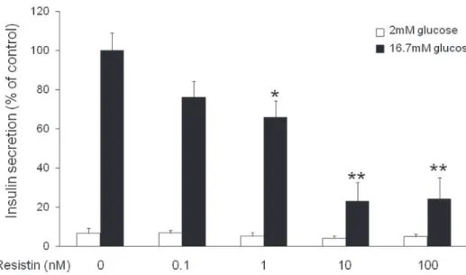
1. Resistin의 포도당 자극성 인슐린 분비 (glucose- stimulated insulin secretion, GSIS) 억제효과
INS-1 췌장 베타세포에서 resistin이 GSIS에 미치는 영향에 대해 조사하였다. Resistin을 30분간 전 처리한 후 포도당을 2 mM 또는 16.7 mM의 농도로 처리하여 인슐린 분비반응을 관찰하였다. Resistin은 INS-1
세포에서 농도 의존적으로 GSIS를 억제하였다(Fig. 1).
저농도 포도당 자극 시 resistin은 인슐린 분비를 감소시키는 경향을 보였으나 유의한 차이는 없었다.
Resistin 농도 1, 10 그리고 100 nM에서 유의한 GSIS 억제효능을 확인하였고, 본 실험에서는 resistin 10 nM 농도를 이용하여 모든 실험을 진행하기로 결정하였다.
Resistin에 의한 세포 내 포도당 흡수량(Fig. 2A)과 ATP 농도(Fig. 2B)의 변화는 관찰되지 않았다. 또한, 포도당 자극에 의한 [Ca2+]i 변화에도 resistin은 아무런 영향을 주지 않았다(Fig. 3A&B). 이러한 결과들을 통해 resistin은 췌장 베타 세포에서 인슐린 분비 기전 중 포도당 흡수, ATP 생성 그리고 [Ca2+]i 증가 기전과는 관계없이 인슐린 분비를 억제하는 것으로 생각된다.
Resistin의 이러한 인슐린 분비억제 효능은 인슐린 분비 기전 중 마지막 과정인 인슐린 분비과립의 세포막 결합과정에 관여할 것으로 예상하였다.
2. Resistin의 rab3A 저해를 통한 인슐린 분비 과립의 세포막 결합 억제 효과
Resistin이 인슐린 분비 기전 중 인슐린 분비과립의
Fig. 1. Effect of resistin on glucose-stimulated insulin secretion in INS-1 cells. Dose- dependent effect of resistin on GSIS levels. The values represent the mean ± SEM, n = 5. *p < 0.05 and **p < 0.01 vs cells treated with 16.7 mM glucose alone.
Fig. 2. Effects of resistin on cellular glucose uptake and ATP levels in INS-1 cells. Cells were stimulated with 10 nM resistin for 30 min, followed by 16.7 mM glucose for 10 min. Cellular glucose (A) and ATP (B) levels were measured in INS-1 cell lysates. The values represent the mean
± SEM, n = 5.
A
B
세포막 결합과정에 관여하는지를 확인하기 위해 TIRFM을 이용하여 인슐린 과립의 분비양상과 세포막 결합(docking) 정도를 확인하였다. 인슐린-GFP가 발현된 INS-1 세포에 resistin을 30분간 전 처리한 후 인슐린 과립의 분비양상을 관찰하였을 때(F i g.
4A&B), 대조군에 비해 1단계 인슐린 분비반응이 유의하게 감소하였다. 2단계 인슐린 분비반응은 resistin에 의해 감소하는 경향을 보였지만 유의한 변화는 없었다(Fig. 4C). 이를 통해 resistin은 1단계 인슐린 분비과정에 관여하는 것으로 생각할 수 있다.
다음으로 resistin이 인슐린 분비과립의 세포막 결합에 미치는 영향을 알아보기 위해 TIRFM를 이용하여 세포막에서 200 nm 거리 내에 있으면서 형태학적으로
세포막에 결합된 과립의 수를 측정하였다(Fig. 5A).
포도당 자극 없이 resistin을 30분간 처리한 후 세포막에 결합된 인슐린 과립의 수를 측정하였을 때, resistin을 처리하기 전에 비해 세포막에 결합된 인슐린 과립의 수가 유의하게 감소하였다(Fig. 5B). 또한, 인슐린 과립의 세포막 결합에 중요한 역할을 하는 것으로 알려진 rab3A 단백질의 발현양이 resistin에 의해 확연하게 감소하였다(Fig. 5C).
고 찰
본 연구에서는 resistin이 인슐린 분비를 억제하는 Fig. 3. Effect of resistin on glucose-stimulated [Ca2+]i increase in INS-1 cells. Cells were stimulated with 10 nM resistin for 30 min, followed by 16.7 mM glucose for 5 min. (A) Representative data from five independent experiments are shown. Solid line, control; dotted line, resistin. (b) The area under the curve (AUC) is depicted as the percentage of the values in the presence of 16.7 mM glucose alone. The values represent the mean ± SEM, n = 5.
효능을 rat insulinoma INS-1 세포에서 확인하였고, 그 기전으로 r e s i s t i n인 인슐린 분비과립의 세포막 결합(docking)에 중요한 역할을 하는 rab3A 단백질의 발 현 을 억 제 함 으 로 써 인 슐 린 분 비 를 저 해 함 을 증명하였다(Fig. 6).
Resistin은 근육세포와 지방세포에서 세포 내 포도당 흡수를 저해함으로써 당뇨병 발생에 중요한 역할을 하는 것으로 알려져 있다[9,11]. 췌장 베타세포에서도 세포 내 포도당 흡수율의 변화는 인슐린 분비에 중요한 인자이다[17]. 본 연구에서 resistin은 췌장 베타세포의
포도당 흡수율에는 아무런 영향을 주지 못하였고, 이를 통해 췌장 베타세포에서 resistin은 포도당 운반체와 포도당 흡수율 결정에 중요한 역할을 하는 glucokinase 활성에 아무런 영향을 주지 못할 것으로 예상하였다.
췌장 베타세포는 식후 고혈당에 반응하여 인슐린을 분비한다. 고농도의 포도당은 베타 세포 내로 흡수되어 세포 내 ATP 농도를 증가시키고, 생성된 ATP는 세포막 ATP-sensitive potassium channel에 결합하여 세포막의 탈분극을 유발한다. 이러한 탈분극은 베타 세포의 전압 의존성 칼슘 통로를 활성화시켜 세포 내 칼슘농도를 Fig. 4. Effect of resistin on the insulin secretion phase in INS-1 cells. Cells were infected with adenovirus carrying the insulin-GFP fusion protein. The cells were preincubated in 2 mM glucose buffer with or without 10 nM resistin for 30 min. At 30 sec after acquisition of the image, the cells were stimulated with 16.7 mM glucose for 20 min. The histogram of the fusion events at 30-sec intervals in a cell-surface area of 200 μm2 was generated (A, B). The data are representative of four independent experiments. (C) The sum of the fusion events in each phases induced by glucose stimulation is presented. The values represent the mean ± SEM, n = 4. *p <
0.05 vs control values.
증가시키고 이를 통해 세포 내 인슐린 과립의 분비를 유발한다[17]. 본 연구에서는 인슐린 분비량을 결정하는 중요한 인자인 세포 내 ATP 농도와 칼슘 농도 변화에 resistin이 미치는 영향을 조사하였다. Resistin은 베타세포 내 ATP 농도와 칼슘 농도 변화에 아무런 영향을 주지 못하였다.
최근 resistin은 췌장 베타 세포주인 RINm5F 세포에서 인슐린 분비를 억제한다고 보고된 바 있다 [16]. 하지만 그 기전에 대해서는 자세히 알려져 있지 않다. 본 연구에서는 resistin이 rab3A 억제를 통해 인슐린 분비를 저해함을 최초로 증명하였다. 인슐린 분비 과립은 세포질에서 세포막으로 targeting 과정을 통해 이동하고 docking, priming 그리고 fusion 과정을
통해 세포막에 결합하여 분비된다[18]. Rab3는 rab family의 small G 단백으로 분비과립의 targeting과 docking 과정에서 중요한 역할을 하는 것으로 알려져 있고, 특히 rab3A와 rab3A-연관 단백질들은 칼슘 의존적 과립분비의 docking에 중요한 역할을 하는 것으로 알려져 있다[19-21]. Rab3A 발현 및 활성에 영향을 미치는 것으로 알려진 물질로는 heregulin beta- 1[22], alpha-synuclein[23], rabconnectin-3[24], growth inhibitory factor[25], rabin3[26] 등이 알려져 있으나, 췌장 베타세포에서 rab3A 발현 및 활성에 영향을 미치는 물질은 아직까지 알려진 바가 없다. 본 연구에서 resistin이 rab3A의 발현을 억제함으로써 인슐린 분비과립의 세포막 결합을 저해하고 이를 통해 인슐린 Fig. 5. Effect of resistin on the number of membrane-docked insulin granules in INS-1 cells. Cells were infected with adenovirus carrying the insulin-GFP fusion protein. The cells were preincubated in 2 mM glucose buffer with or without 10 nM resistin for 30 min. (A) Representative data from four independent experiments are shown.
Scale bar, 10 μm. (B) The number of docked granules was measured in a cell-surface area of 200 μm2. The values represent the mean ± SEM, n = 4. **p < 0.01 vs control values. White bars, 2 mM glucose. (C) The expression of rab3A was detected by Western blot analysis of whole cell lysates.
A
B C
분비를 저해함을 처음으로 밝혔다.
인체에서는 동물 실험에서와는 달리 지방세포에서 resistin의 발현이 낮고 오히려 대식세포와 같은 면 역 세 포 들 에 발 현 이 많 이 되 어 있 는 것 으 로 알려져[27], 인체에서의 resistin의 역할은 마우스에서의 역할과 다를 수도 있다. 하지만 인체 resistin 유전자의 polymorphism이 인슐린 저항성과 깊은 연관성이 있는 것으로 알려져 있으며[28], 비만 또는 당뇨병 환자의 혈중 resistin 농도가 증가되어 있어[29-30], resistin이 인간의 인슐린 저항성 및 당뇨병 발생에도 중요한 역할을 할 것으로 생각된다.
간, 근육, 지방세포, 심장 그리고 시상하부 등 다양한 장기에 대한 resistin의 다양한 효능이 알려져있지만, 아직까지 세포막의 resistin 수용체에 대해서는 알려진 바가 없다. 본 연구에서도 췌장 베타세포의 resistin 수용체에 대해서는 알아보지 못하였고 앞으로 추가적인
연구가 필요하겠다. 또한, resistin을 췌장 베타세포에 장기간 처리하였을 때 세포 증식 및 생존에 미치는 영향에 대해서도 추가적인 연구가 필요하겠다. 본 연구를 통해 resistin이 비만에 의한 인슐린 저항성 증가 뿐만 아니라 췌장 베타세포의 기능을 저해함으로써 제 2형 당뇨의 발생에 중요한 역할을 함을 증명하였다. 제 2형 당뇨의 병인인 resistin의 중요성과 그 기전을 밝힘으로써 resistin을 이용한 새로운 당뇨병 치료제 개발에 도움이 될 수 있겠다.
요 약
체중 증가 시 지방세포에서 분비가 증가되는 아디포카 인인 resistin은 간, 근육 그리고 지방세포에서 인슐린 저항성 증가에 중요한 역할을 하는 것으로 알려져 있다. 본 Fig. 6. Proposed mechanism to explain the effect of resistin on insulin secretion in pancreatic beta-cells. KATP: ATP-sensitive potassium channel, Mito: mitochondria, ΔΨ: membrane depolarization, VDCC: voltage-dependent calcium channel.
연구에서는 resistin이 췌장 베타세포에 미치는 영향에 대해 조사해보았고, 그 결과 r e s i s t i n은 인슐린 분비과립의 세포막 결합에 중요한 역할을 하는 rab3A를 억제함으로써 인슐린 분비과립의 세포막 결합을 저해하고 최종적인 인슐린 분비를 억제함을 증명하였다.
참 고 문 헌
1. Ahima RS. Adipose tissue as an endocrine organ.
Obesity (Silver Spring) 2006;14 Suppl 5:242S-9S.
2. Antuna-Puente B, Feve B, Fellahi S, Bastard JP.
Adipokines: the missing link between insulin resistance and obesity. Diabetes Metab 2008;34:2-11.
3. Brown JE, Thomas S, Digby JE, Dunmore SJ. Glucose induces and leptin decreases expression of uncoupling protein-2 mRNA in human islets. FEBS Lett 2002;513:189- 92.
4. Brown JE, Onyango DJ, Dunmore SJ. Resistin down- regulates insulin receptor expression, and modulates cell viability in rodent pancreatic beta-cells. FEBS Lett 2007;581:3273-6.
5. Trayhurn P, Wood IS. Signalling role of adipose tissue:
adipokines and inflammation in obesity. Biochem Soc Trans 2005;33:1078-81.
6. Rabe K, Lehrke M, Parhofer KG, Broedl UC.
Adipokines and insulin resistance. Mol Med 2008;14:741-51.
7. Steppan CM, Bailey ST, Bhat S, Brown EJ, Banerjee RR, Wright CM, et al. The hormone resistin links obesity to diabetes. Nature 2001;409:307-12.
8. Muse ED, Obici S, Bhanot S, Monia BP, McKay RA, Rajala MW, et al. Role of resistin in diet-induced hepatic insulin resistance. J Clin Invest 2004;114:232-9.
9. Fan HQ, Gu N, Liu F, Fei L, Pan XQ, Guo M, et al.
Prolonged exposure to resistin inhibits glucose uptake in rat skeletal muscles. Acta Pharmacol Sin 2007;28:410-6.
10. Rajala MW, Obici S, Scherer PE, Rossetti L. Adipose-
derived resistin and gut-derived resistin-like molecule- beta selectively impair insulin action on glucose production. J Clin Invest 2003;111:225-30.
11. Sanchez-Solana B, Laborda J, Baladron V. Mouse resistin modulates adipogenesis and glucose uptake in 3T3-L1 preadipocytes through the ROR1 receptor.
Mol Endocrinol 2012;26:110-27.
12. Jorgensen SB, Honeyman J, Oakhill JS, Fazakerley D, Stockli J, Kemp BE, et al. Oligomeric resistin impairs insulin and AICAR-stimulated glucose uptake in mouse skeletal muscle by inhibiting GLUT4 translocation. Am J Physiol Endocrinol Metab 2009;297:E57-66.
13. Graveleau C, Zaha VG, Mohajer A, Banerjee RR, Dudley-Rucker N, Steppan CM, et al. Mouse and human resistins impair glucose transport in primary mouse cardiomyocytes, and oligomerization is required for this biological action. J Biol Chem 2005;280:31679-85.
14. Steppan CM, Wang J, Whiteman EL, Birnbaum MJ, Lazar MA. Activation of SOCS-3 by resistin. Mol Cell Biol 2005;25:1569-75.
15. Niederwanger A, Kranebitter M, Ciardi C, Tatarczyk T, Patsch JR, Pedrini MT. Resistin impairs basal and insulin-induced glycogen synthesis by different mechanisms. Mol Cell Endocrinol 2007;263:112-9.
16. Nakata M, Okada T, Ozawa K, Yada T. Resistin induces insulin resistance in pancreatic islets to impair glucose-induced insulin release. Biochem Biophys Res Commun 2007;353:1046-51.
17. Straub SG, Sharp GW. Glucose-stimulated signaling pathways in biphasic insulin secretion. Diabetes Metab Res Rev 2002;18:451-63.
18. Seino S, Shibasaki T. PKA-dependent and PKA- independent pathways for cAMP-regulated exocytosis. Physiol Rev 2005;85:1303-42.
19. Takai Y, Sasaki T, Shirataki H, Nakanishi H. Rab3A small GTP-binding protein in Ca(2+)-dependent exocytosis. Genes Cells 1996;1:615-32.
20. Huang CC, Yang DM, Lin CC, Kao LS. Involvement
of Rab3A in vesicle priming during exocytosis:
interaction with Munc13-1 and Munc18-1. Traffic 2011;12:1356-70.
21. Lin CC, Huang CC, Lin KH, Cheng KH, Yang DM, Tsai YS, et al. Visualization of Rab3A dissociation during exocytosis: a study by total internal reflection microscopy. J Cell Physiol 2007;211:316-26.
22. Vadlamudi RK, Wang RA, Talukder AH, Adam L, Johnson R, Kumar R. Evidence of Rab3A expression, regulation of vesicle trafficking, and cellular secretion in response to heregulin in mammary epithelial cells.
Mol Cell Biol 2000;20:9092-101.
23. Dalfo E, Ferrer I. Alpha-synuclein binding to rab3a in multiple system atrophy. Neurosci Lett 2005;380:170- 5.
24. Kawabe H, Sakisaka T, Yasumi M, Shingai T, Izumi G, Nagano F, et al. A novel rabconnectin-3-binding protein that directly binds a GDP/GTP exchange protein for Rab3A small G protein implicated in Ca(2+)-dependent exocytosis of neurotransmitter.
Genes Cells 2003;8:537-46.
25. Liu Y, Ren H, Wu C, Bai S, Zhang X, Ru B.
Attenuation of zinc-induced neuronal death by the interaction of growth inhibitory factor with Rab3A in rat hippocampal neurons. Neurosci Lett 2004;358:149-
52.
26. Brondyk WH, McKiernan CJ, Fortner KA, Stabila P, Holz RW, Macara IG. Interaction cloning of Rabin3, a novel protein that associates with the Ras-like GTPase Rab3A. Mol Cell Biol 1995;15:1137-43.
27. Savage DB, Sewter CP, Klenk ES, Segal DG, Vidal- Puig A, Considine RV, et al. Resistin / Fizz3 expression in relation to obesity and peroxisome proliferator-activated receptor-gamma action in humans. Diabetes 2001;50:2199-202.
28. Pizzuti A, Argiolas A, Di Paola R, Baratta R, Rauseo A, Bozzali M, et al. An ATG repeat in the 3’-untranslated region of the human resistin gene is associated with a decreased risk of insulin resistance. J Clin Endocrinol Metab 2002;87:4403-6.
29. Youn BS, Yu KY, Park HJ, Lee NS, Min SS, Youn MY, et al. Plasma resistin concentrations measured by enzyme-linked immunosorbent assay using a newly developed monoclonal antibody are elevated in individuals with type 2 diabetes mellitus. J Clin Endocrinol Metab 2004;89:150-6.
30. Azuma K, Katsukawa F, Oguchi S, Murata M, Yamazaki H, Shimada A, et al. Correlation between serum resistin level and adiposity in obese individuals.
Obes Res 2003;11:997-1001.