․교신저자: 전상윤 광주시 남구 월산로 141번지 동신대학교 광주한방병원 한방내과 TEL: 062-350-7207 FAX: 062-350-7141 E-mail: [email protected]
․이 논문은 2013년도 동신대학교 일반대학원 한의학 박사학위 논문임.
고지방 식이로 유발된 고지혈증 동물 모델에서 枸杞子加味方의 효과 연구
안가영, 조재준, 신민구, 전상윤 동신대학교 한의과대학 내과학교실
Study of the Effects of Gugijagami-bang in a Hyperlipidemic Animal Model Induced with a High-Fat Diet
Ga-young An, Jae-joon Joe, Min-koo Shin, Sang-yun Jeon
Dept. of Internal Medicine, College of Korean Medicine, Dong-Shin University
ABSTRACT
Objectives:
This study was undertaken to investigate the effects of Gugijagami-bang (GGB) in a hyperlipidemic animal model induced by a high-fat diet using diverse biological methods.
Methods:
This study was to determine whether fractionated GGB extracts inhibit reactive oxygen species (ROS) and nitric oxide (NO) in RAW 264.7 cells. Hyperlipidemia was induced by a high-fat diet fed for 6 weeks. Total cholesterol, LDL cholesterol, HDL cholesterol, triglyceride, liver function and histologic change of liver were measured after oral administration of GGB.
Results:
1. DPPH scavenging bow performance was increased in a concentration-dependent manner by GGB.
2. Compared to the control group, NO production (%) and ROS production (%) were decreased significantly by GGB.
3. Total-cholesterol, LDL-cholesterol, triglyceride were decreased significantly by GGB.
4. HDL cholesterol increased more than the control group, but not significantly.
5. In histopathologic examination, fatty liver (hepatic steatosis) was inhibited, almost no rounds of fat were observed in the liver.
Conclusions:
GGB would appear effective in the prevention and treatment of atherosclerosis, ischemic heart disease, other cardiovascular diseases caused by hyperlipidemia.
Key words:
Gugijagami-bang, hyperlipidemia, RAW 264.7 cell, antioxidant activity
Ⅰ. 서 론
현대사회는 서구화된 식생활 습관으로 탄수화물, 지방, 단백질의 과다 섭취와 운동이 부족해짐에 따
라 혈액 내 콜레스테롤 및 지방 축적이 증가되고
있다
1. 특히 체내에 과잉 축적된 지방은 생체 내의
에너지 대사에 직접적으로 영향을 주어 열량소비
저하, 지방산화의 억제 및 혈액 내 유리지방산의
과잉
2, 혈중 지질과산화의 촉진, 중성지방의 증가,
당대사의 이상, 인슐린의 분비와 감수성의 약화 등
3,
생체 내 대사산물의 생성과 기능에 이상을 초래하
고, 이로 인해 아니라 심장질환, 등의 각종 성인병
및 만성 질환을 유발한다
4. 특히 고지혈증은 죽상
경화증을 일으키고, 이는 우리나라 주요 사망원인 인 심혈관과 뇌혈관 질환의 주요 위험인자가 되고 있다
1.
한의학에서 고지혈증은 痰飮, 瘀血, 氣滯, 寒凝, 虛勞 등의 범주에 속하며
5, 嗜食肥甘厚味의 外因과 肝脾腎 機能失調의 內因으로 인해 濕濁, 火熱, 脾 胃濕熱 등의 원인에 의해 발생하는 것으로 인식하 고 있다
6.
고지혈증에 대한 한의학적 연구로는 정
7의 加味 溫膽湯, 황
8의 茵蔯胃苓湯, 차
9의 降脂通脈飮, 유
10의 夏枯草散 박
11의 加味地黃湯 등 처방과 茵蔯蒿
12, 枳實
13, 白何首烏
14, 丹蔘
15등 약재를 이용하여 고지 혈증에 대한 효과를 실험적으로 증명하고, 이에 대 한 기전을 밝히려는 연구가 이루어지고 있다. 특히 枸杞子는 고지혈증에 대한 실험 연구가 활발히 연 구되고 있다
16-18.
枸杞子加味方은 현재 동신대학교 한방병원에서 고지혈증에 활용되고 있는 처방으로 滋補肝腎 益 精明目하는 枸杞子를 군약으로 하여 生地黃, 丹蔘, 牧丹皮, 甘菊, 山査肉, 石菖蒲, 麥門冬, 黃連, 梔子, 柴胡, 黃芩, 防風 등으로 구성되어 있다.
임상적으로 혈중 지질이 감소하는 효과를 나타 내는 枸杞子加味方에 대하여 실험적으로 규명하고, 기전을 밝히고자 연구하게 되었다.
이에 저자는 枸杞子加味方 추출물을 Raw 264.7 cell을 이용하여 세포독성, DPPH 소거능, No 생성 억제, ROS 활성효과를 측정하고, 고지혈증 질환 모델동물인 6주령의 수컷 ApoE(k/o) 생쥐를 이용 하여 간 및 신 독성, 혈중 인자 등을 관찰하여 유 의한 결과를 얻었기에 보고하는 바이다.
Ⅱ. 실 험
1. 재 료 1) 약 재
본 실험에 사용한 枸杞子加味方 (Gugijagami-bang, GGB)의 구성 약물은 대전대학교 부속한방병원에
서 구입하여 정선한 후 사용하였다. 한첩 분량은 아래와 같다(Table 1).
Herbal
name Scientific name Volume (g) 枸杞子 Lycii Fructus 20
生地黃 Rehmanniae Radix 8 丹 蔘 Salvia miltiorrhiza Bunge 8
牧丹皮 Moutan Cortex 6
甘 菊 Chrysanthemi Flos 6 山査肉 Crataegi Fructus 6 石菖蒲 Acori Graminei Rhizoma 6
麥門冬 Liriopis Tuber 6
黃 連 Coptidis Rhizoma 4 梔 子 Gardeniae Fructus 4 夏枯草 Prunellae Spica 4 柴 胡 Bupleuri Radix 4 黃 芩 Scutellariae Radix 4 防 風 Saposhnikovia Radix 4 Total amount 90 Table 1. The Compositions of Gugijagami-bang (GGB)
2) 세 포
실험에 사용된 RAW 264.7 세포는 한국세포주은 행(Korea)에서 구입하였다.
3) 동물 및 사육조건
실험동물은 고지혈증 질환 모델 동물인 6주령
수컷 ApoE(k/o) 생쥐를 미국 Jackson Lab.으로부
터 수입하여 사용하였다. 분양 받은 실험동물은 2
주간 일반 고형사료(Table 2. AIN-76A diet Test
Diet Co, USA)로 적응시킨 후 체중 변화가 일정하
고 건강한 10주령의 생쥐를 선별하여 4마리씩 분리
한 후 0.15% cholesterol, 21% fat으로 조성된 western
식이사료(Table 3. Research Diet, USA)와 물을 자
유롭게 공급하면서 항온(25±2 ℃), 항습(50±5%) 및
12시간 간격의 광주기(light on 07:00-19:00)로 명
암이 조절되는 SPF(Specific Pathogen Free) 실험
실에서 실험하였다.
Diets ingredient Concentration (g/kg)
Casein 200
DL-Methionine 3 Corn Starch 150
Sucrose 500
Cellulose 50
Con Oil 50
AIN-76 Mineral Mix 35 AIN-76A Vitamin Mix 10 Choline Bitartrate 2
Ethoxyquin 0.01 Table 2. Composition of AIN-76A Diet
Diets ingredient Concentration (g/kg) Casein, 80 Mesh 195
L-Cystine 3
Corn Starch 50
Maltodextrin 10 100
Sucrose 341
Cellulose 50
Milk Fat, Anhydrous 200
Corn Oil 10
Mineral Mix S 10001 35 Calcium Carbonate 4 Vitamin Mix V10001 10
Choline Bitartrate 2 Cholesterol 1.5 Ethoxyquin 0.04 Table 3. Composition of Western Diet
4) 시 약
Dulbecco's phosphate buffered saline, RPMI 1640, Collagenase A, Penicillin, pyrogallol, Ethylenediaminetetraacetic acid (ETDA), 3-(4,5- Dimethyl -2-thiazolyl)-2,5-diphenyl-2H-tetrazolium bromide(MTT), Sodium citrate, N-2-hydroxylethyl -piperazine-N-2-ethane sulfonic acid(HEPES), lipopolysaccaride(LPS), 2,7-dichloro-dihydro fluorescin diacettate(DCFH-DA), diethyl pyrocarbonate(DEPC), 2,2Diphenyl-1-picrylhydrazyl(DPPH)는 sigma사(USA)
제품을, Dimethyl sulfoxide(DMSO)는 Sowa chemical 사(Japan) 제품을, Ethylacrtate 는 Junsei사(Japan) 제품을, Potassium Phosphate Monobasic(KH
2PO
4) 는 Yakuri사(Japan) 제품을, RNAzolB M-MLV RT, dNTPs mix, RNase inhibitor Takara사(Japan) 제품 을, First Strand cDNA Synthesis kit는 Pharmacia 사(USA) 제품을, DNase I는 Life Technologies사(USA) 등을 사용하였고, 이 밖에 일반 시약은 특급 시약을 사용하였다.
5) 기 기
본 연구에 사용된 기기는 High performance liquid chromatography(HPLC. Shimadzu Japan), Ice-maker (Vision, Korea), Centrifuge(Hanil Co., Korea), Rotary vaccum evaporator(Büchi 461, Switzerland), Deep freezer(Sanyo Co., Japan), Autoclave(Sanyo, Japan), Ultrasonic cleaner(Branson Ultrasonics Co., USA), ELISA reader(Molecular Divice., USA), Roller Mixer (Gowon scientific technology Co., Korea), 한약유 출기(DWP-1800T, 대웅, Korea), Spectrophotometer (UV-2450, Shimazu, Japan), cell cytometery(FACS, Beckman Co., USA), Balance(Cass, korea), 생화학 기(AU400, Olimpus, USA), 감마카운터기(WIZARD 1470, Finland) 등을 사용하였다.
2. 방 법 1) 시료 추출
GGB 2첩(180 g)에 멸균 증류수 1.5 L를 넣고 전탕 한 후, 여과하여 증류장치로 농축하였다. 농 축액을 동결 건조기를 이용하여 완전 건조한 다음 얻어진 분말(수득률 17.8%, 32.1 g)을 냉동 보관 (-80 ℃) 하면서 실험 농도로 희석하여 사용하였다.
2) 시료 HPLC 패턴 확인
GGB 추출물 30 mg을 멸균 증류수 1 ml에 녹여
0.45 μm membrane filter로 여과 후 이 중 20 μl를
HPLC 시료로 사용하였다. HPLC는 Shimadzu(Japan)
사의 system controller(CBM-20A), pump(LC-20AD),
column oven(CTO-20A), UV/VIS detector(SPD-20A)
를 사용하였으며, column은 ACE 5 C18(250×4.6 mm, 5 μm)을 사용하였다. 이동상은 water(A)와 acetonitrile(B) 로 gradient elution system을 적용시켜 0%(5분 B), 0-30%(35분 B), 30-100%(10분 B)로 설정하였다.
유속은 1.0 μl/min 이었으며 column 온도는 40 ℃를 유지하였다. UV wavelength는 254 nm로 설정하여 분석하였다.
3) In vitro 실험 (1) 세포독성 측정
Raw 264.7 cells은 96well plates에 10
4cells/well 로 분주하여 24시간 동안 배양 한 후, GGB를 각각 50, 100, 200 ug/ml의 농도로 처리하여 24시간 동안 배양하였다. 배양 후, 10 ul의 WST solution을 첨가 한 후 CO
2배양기(37 ℃, 5% CO
2)에서 30분 반응 시 킨 후, 450 nm에서 흡광도의 변화를 측정하여 대 조군에 대한 세포 생존율을 백분율로 표시 하였다.
(2) 항산화 활성 측정
① 2,2-diphenyl-1-picrylhydrazyl(DPPH) 소거능 측정 150 mM DPPH/EtOH 150 μl에 GGB를 25, 50, 100, 200 μg/ml 농도로 희석하여 100 μl씩 첨가한 후 37 ℃에서 30분간 반응시켰다. 이를 ELISA reader 를 이용하여 wave length 517 nm에서 흡광도를 측 정한 후 아래의 방법으로 계산하였다.
DPPH 소거능 (%) =
( 대조군의 흡광도-GGB처리군의 흡광도 )×100 대조군의 흡광도
② Total Nitric oxide 생성 억제 효과 측정 NO의 농도는 배양액 내의 nitrite 농도를 Griess Reagent System을 이용하여 측정하였다. Raw 264.7 cells은 96 well plates에 1×10
4cells/100 μl로 분주하여 24시간 동안 배양 한 후, GGB을 각각 50, 100, 200 μg/ml 농도로 처리하고, LPS 1 μg/μl을 처리하여, 다시 24시간 동안 배양하였다. N
1buffer 50 μl를 각 well에 처리한 후, 10분간 상온에서 암소 반응 후 540 nm에서 흡광도를 측정하였다. Nitrite standard의 농도별 표준곡선을 이용하여 배양액의 NO 농도를 결정하였다.
③ Reactive oxygen species(ROS) 활성 측정 Raw 264.7 cell 내에서 reactive oxygen species (ROS)를 측정하기 위하여 2',7'-Dichlorodihydrofluorescin diacetate(DCFH-DA)를 이용하였다. 48 well plate 에 Raw 264.7 cell를 2×10
5/well/400 μl씩 첨가하고 LPS 1 μg/ml 및 GGB 50, 100, 200 μg/ml 농도를 처리한 후 37 ℃, 5% CO
2배양기에서 24시간 배양 하였다. 배양 종료 후 DCFH-DA 10 μM을 처리하 여 15분 동안 빛이 차단된 상온에서 배양한 후 차 가운 PBS를 넣어 1,200 rpm에서 5분간 원심 분리 한 다음 상청액을 제거하고 다시 PBS 400 μl를 부 유시켜 유세포 분석기(Flow cytometer)를 이용하 여 형광강도의 세기에 따른 ROS를 측정하였다.
4) In vivo 실험 (1) 약물투여
각 실험군은 정상군, 대조군 그리고 GGB 50, 100, 200, 400 투여군으로 나누어 정상군은 일반 사료와 식수를 충분히 공급하였다 대조군과 GGB 투여군은 Table 3의 조성으로 만들어진 고콜레스테롤 사료와 식수를 충분히 공급하였으며, GGB 투여군은 고콜레 스테롤 사료 투여 2주 후부터 400 mg/kg/0.5 ml의 양으로 정해진 시간(오전 11시)에 매일 투여하였다.
(2) 간 및 신 독성 측정
실험 종료 후 혈액을 채취 해 혈청을 분리하여 Alanine aminotransferase(ALT), Alkaline Phosphatase (AST), 요소질소(BUN) 등의 검사를 실시하였다.
(3) 혈중 지질 인자 측정
실험 종료 후, ApoE(k/o) 생쥐에서 심장 천자를 통하여 채혈한 후 Total cholesterol 및 HDL(High- density lipoprotein), LDL(Low-density lipoprotein), triglyceride 함량을 바이오톡스텍(오창, Korea)에 측정 의뢰하였다.
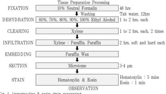
(4) Hematoxyline & Eosin 염색
각 실험군 별로 적출한 간 조직 10% 중성 포르 말린에 48시간 고정하여 고정이 완료된 각 조직들 은 흐르는 수돗물에서 12시간 수세하여 조직 내 고 정액을 완전 제거하였다. 조직의 탈수를 위해 60%
에서부터 100% 알코올에 이르기까지 농도 상승 순으
로 통상의 방법에 따라 탈수하고, xylene에 투명과정 을 거친 다음 파라핀 블럭을 제작하였다. 제작된 블 럭은 박절기(microtome)를 이용하여 3~4 μm 두께로
절편을 만들어 탈 파라핀 및 함수 과정을 거친 다음 hematoxyline & eosin(H&E) 일반 염색을 실시하여 광학현미경상에서 관찰 및 사진 촬영 하였다(Fig. 1).
Tissue Preparation Processing
FIXATION 10% Neutral Formalin 48 hrs
Washing Tab water, 12hrs DEHYDRATION 60%, 70%, 80%, 90%, 100% Ethyl Alcohol 1 to 2 hrs, each
CLEARING Xylene 1 to 2 hrs, each, 2 times
INFILTRATION Xylene : Paraffin, Paraffin 2 hrs, soft and hard each
EMBEDDING Paraffin Wax
SECTION Microtome 3-4 μm
STAIN Hematoxylin & Eosin Hematoxylin : 5 mins Eosin : 1 min
OBSERVATION Fig. 1. Hematoxyline & eosin stain processing.
3. 통계처리
본 실험에서 얻은 결과를 ANOVA multi t-test (JAVA, Bonferroni Ver 1.1)로 분석하여 p값을 구 하였다. 각 대조군을 정상군과, 실험군을 대조군과 비교하여 p<0.05 일 때 유의성이 있는 것으로 판정 하였다.
Ⅲ. 실험결과
1. HPLC 패턴 확인
GGB 메탄올 추출물을 HPLC로 pattern 분석한 결과 254 nm에서 retention time이 16.7분, 22.7분, 24.1분, 26.4분대에 peak를 확인할 수 있었다(Fig. 2).
5.0 10.0 15.0 20.0 25.0 30.0 35.0 40.0 45.0 min
0 100 200 300 400 mV
Detector A Ch2:254nm
Fig. 2. HPLC chromatogram of water extract of GGB.
Column; ACE 5 C18 (250×4.6 mm, 5 μm), sample injection volume; 5 μl, mobile phase; H2O(A)/ACN(B) gradient elution : 0% (5min B), 0~30% (35min B), 30~100% (20min B), flow rate; 1.0 ml/min, detector; PDA detector
2. In vitro 실험
1) 세포독성에 미치는 영향
정상군의 세포생존율 100±0%로 했을 때, GGB의 50, 100, 200(μg/ml) 농도에서는 각각 132±5, 130±6, 130±6(%)로 나타났다(Fig. 3).
Fig. 3. Effects of GGB on the cell viability of RAW 264.7 cells.
Cells were treated with 50, 100, 200 μg/ml of Caesalpinia sappan Lignum for 24hrs. Cell viability was determined using the WST assay. The results are expressed as mean±SD from three independent experiments.
2) 항산화 활성 측정 (1) DPPH 소거활성 측정
DPPH의 소거 활성은 25, 50, 100, 200(μg/ml) 농도에서 각각 8.2±4.5, 22.5±5.0, 44.3±5.2, 76.5±3.4 (%)로 나타나 농도 의존적으로 소거활성이 증가 하는 것으로 나타났다(Fig. 4).
Fig. 4. Scavenging activity of GGB on DPPH free radical.
GGB were reacted with DPPH for 30 minutes at 37 ℃, and the absorbance at 517 nm due to DPPH radical was determined. The results are the mean±SD of three independent experiments.
(2) NO 생성억제효과
대조군을 100.0±8.7%로 나타냈을 때, 정상군은 27.7±12.7%, GGB50은 101.8±3.1%, GGB100은 86.8
±7.5%, GGB200은 62.5±3.1%로 나타나 GGB100과 GGB200에서 유의성 있는(** p<0.01, *** p<0.001) 감소를 나타내었다(Fig. 5).
Fig. 5. NO production rate of GGB.
NO production rate of GGB at final concentration 50, 100, 200 (μg/ml). The results are the mean±SD of three independent experiments. Normal : Raw 264.7 cells, Control : Raw 264.7 cells and LPS (2 μg/ml), GGB50 : Raw 264.7 cells and LPS and GGB 50 μg/ml, GGB100: Raw 264.7 cells and LPS and GGB 100 μg/ml, GGB200 : Raw 264.7 cells and LPS and GGB 200 μg/ml. The results are expressed as mean±S.D from three independent experiments. Significant value was calculated by compared with control group by ANOVA multi t-test (** p<0.01, *** p<0.001).
3) ROS 활성 측정
대조군을 100.0±0%로 나타냈을 때, 정상군은 34±18%,
GGB50은 90±9%, GGB100은 86±12%, GGB200은
75±8%로 나타나 대조군에 비해 실험군에서 농도
의존적으로 감소하였으며, 특히 GGB200에서 유의
성 있는(* p<0.05) 감소를 나타내었다(Fig. 6).
Fig. 6. The inhibitory effect of GGB on reactive oxygen species (ROS).
Raw 264.7 cells were incubated with GGB for 24hr at 37 ℃. The cells were stressed with LPS for 30 min and were labeled with DCFH-DA for 15 min. Normal : Raw 264.7 cells, Control : Raw 264.7 cells and LPS (2 μg/ml), GGB50 : Raw 264.7 cells and LPS and GGB 50 μg/ml, GGB100 : Raw 264.7 cells and LPS and GGB 100 μg/ml, GGB200: Raw 264.7 cells and LPS and GGB 200 μg/ml. The results are expressed as mean±S.D from three independent experiments.
Significant value was calculated by compared with control group by ANOVA multi t-test (* p<0.05).
3. In vivo 실험 1) 간 독성 평가
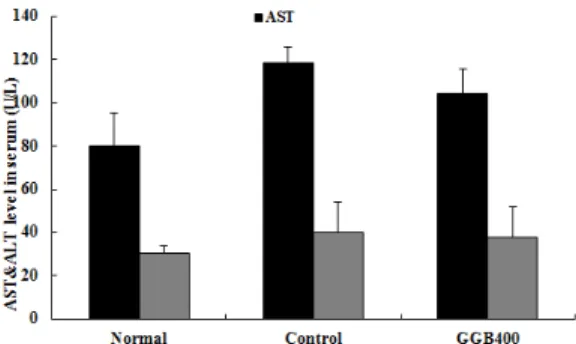
AST는 정상군이 80.3±15.0(I.U/L), 대조군이 118.5
±15.0(I.U/L), GGB400이 104.3±11.4(I.U/L)으로 나 타났으며, ALT는 정상군이 30.2±3.7(I.U/L), 대조군 이 40.0±14.1(I.U/L), GGB400이 37.7±14.0(I.U/L) 으로 나타났다(Fig. 7).
Fig. 7. Effect of GGB on the ALT and AST in ApoE
(k/o) hyperlipidemia mice.
Normal : normal mouse, Control : Hyperlipidemic diet and normal saline (0.2 ml/day) treated group, GGB400 : Hyperlipidemic diet and GGB (400 mg/0.2 ml/day) treated group, Values represent the means±SD of 4 mouse.
2) 신 독성 평가
BUN은 정상군이 25.0±3.3(I.U/L), 대조군이 20.3
±4.0(I.U/L), GGB400이 19.5±3.0(I.U/L)으로 나타 났다(Fig. 8).
Fig. 8. Effect of GGB on the ALT and AST in ApoE (k/o) hyperlipidemia mice.
Normal : normal mouse, Control : Hyperlipidemic diet and normal saline (0.2 ml/day) treated group, GGB400 : Hyperlipidemic diet and GGB (400 mg/0.2 ml/day) treated group, Values represent the means±SD of 4 mouse.
3) 혈중 지질 인자 측정
(1) 총 콜레스테롤에 미치는 영향
정상군이 116.8±7.6(mg/dl), 대조군이 1929.0±12.9
(mg/dl), GGB400은 1575.5±132.9(mg/dl)으로 나타나
GGB400에서 대조군에 비하여 유의성 있는(** : p<0.01)
감소를 나타내었다(Fig. 9).
Fig. 9. Effect of GGB on the Total-cholesterol level in ApoE (k/o) hyperlipidemia mice.
Normal : normal mouse, Control : Hyperlipidemic diet and normal saline (0.2 ml/day) treated group, GGB400 : Hyperlipidemic diet and GGB (400 mg/0.2 ml/day) treated group, Values represent the means±SD of 4 mouse. Statistical significance is based on the difference when compared with control (** p<0.01).
(2) LDL cholesterol에 미치는 영향
정상군이 13.3±4.1(mg/dl), 대조군이 1541.7±69.3 (mg/dl), GGB400은 1219.3±57.3(mg/dl)으로 나타나 GGB400에서 대조군에 비하여 유의성 있는(*** : p<0.001) 감소를 나타내었다(Fig. 10).
Fig. 10. Effect of GGB on the LDL-cholesterol level in ApoE (k/o) hyperlipidemia mice.
Normal : normal mouse, Control : Hyperlipidemic diet and normal saline (0.2 ml/day) treated group, GGB400 : Hyperlipidemic diet and GGB (400 mg/0.2 ml/day) treated group, Values represent the means±SD of 4 mouse. Statistical significance is based on the difference when compared with control (*** p<0.001).
(3) HDL cholesterol에 미치는 영향
정상군이 99.8±11.3(mg/dl), 대조군이 305.0±84.7
(mg/dl), GGB400은 314.0±72.7(mg/dl)으로 나타나 GGB400에서 대조군에 비하여 증가하였으나 유의 성은 나타나지 않았다(Fig. 11).
Fig. 11. Effect of GGB on the HDL level in ApoE (-/-) hyperlipidemia mice.
Normal : normal mouse, Control : Hyperlipidemic diet and normal saline (0.2 ml/day) treated group, GGB400 : Hyperlipidemic diet and GGB (400 mg/0.2 ml/day) treated group, Values represent the means±SD of 4 mouse.
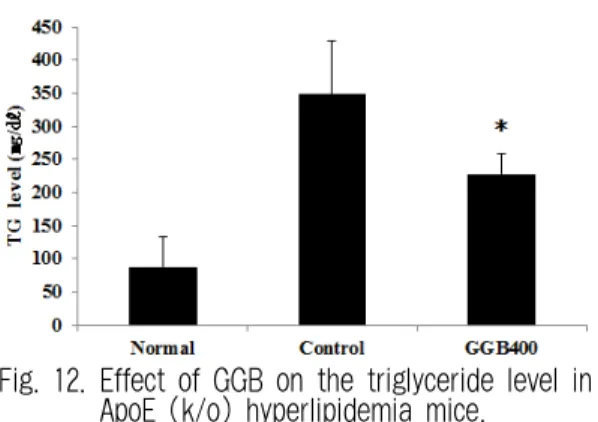
(4) Triglyceride에 미치는 영향
정상군이 88.3±44.5(mg/dl), 대조군이 348.0±80.2 (mg/dl), GGB400은 228.3±29.5(mg/dl)으로 나타나 GGB400에서 대조군에 비하여 유의성 있는(* : p<0.05) 감소를 나타내었다(Fig. 12).
Fig. 12. Effect of GGB on the triglyceride level in ApoE (k/o) hyperlipidemia mice.
Normal : normal mouse, Control : Hyperlipidemic diet and normal saline (0.2 ml/day) treated group, GGB400 : Hyperlipidemic diet and GGB (400 mg/0.2 ml/day) treated group, Values represent the means±SD of 4 mouse. Statistical significance is based on the difference when compared with control (* p<0.05).
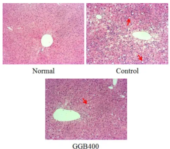
4) 조직 변화에 미치는 영향 (1) 간조직에 미치는 영향
실험 종료 후 간을 적출하여 염색한 결과 대조 군에서는 간세포에서 지방질 축적에 의한 광범위 한 지방공포인 흰색 원형조직이 많이 분포하여 전 형적인 지방간 모습(화살표) 병변 소견이 나타나 있으며 세포핵이 줄어든 반면, GGB 400에서는 대 조군에 비하여 지방간(hepatic steatosis)이 억제되 어 간에 원형의 지방(fat)이 사라진 상태가 관찰되 었다(Fig. 13).
Fig. 13. Representative microscope photographs of liver stained with hematoxylin and eosin.
Normal : normal mouse, Control : Hyperlipidemic diet and normal saline (0.2 ml/day) treated group, GGB400 : Hyperlipidemic diet and GGB (400 mg/0.2 ml/day) treated group