Copyrightⓒ 2008, The Korean Academy of Oral Biology
217
International Journal of Oral Biology
Bicuculline Methiodide (BMI) Induces Membrane Depolarization of The Trigeminal Subnucleus Caudalis Substantia Gelatinosa Neuron in Mice Via Non-GABA
AReceptor-Mediated Action
Hua Yin
1, Seon Ah Park
1, Soon Jeong Choi
2, Janardhan P. Bhattarai
1, Soo Joung Park
1, Bong Jik Suh
2, Seong Kyu Han
1*
1Department of Oral Physiology, 2Department of Oral Medicine, School of Dentistry & Institute of Oral Bioscience, Chonbuk National University, Jeonju
(Received December 8, 2008 ; Revised December 10, 2008 ; Accepted December 12, 2008 )
Bicuculline is one of the most commonly used GABAA receptor antagonists in electrophysiological research.
Because of its poor water solubility, bicuculline quaternary ammonium salts such as bicuculline methiodide (BMI) and bicuculline methbromide are preferred. However, a number of studies have shown that BMI has non-GABAA receptor- mediated effects. The substantia gelatinosa (SG) of the trigeminal subnucleus caudalis (Vc) is implicated in the processing of nociceptive signaling. In this study, we investigated whether BMI has non-GABA receptor- mediated activity in Vc SG neurons using a whole cell patch clamp technique. SG neurons were depolarized by application of BMI (20µM) using a high Cl- pipette solution.
GABA (30-100µM) also induced membrane depolarization of SG neuron. Although BMI is known to be a GABAA receptor antagonist, GABA-induced membrane depolari- zation was enhanced by co-application with BMI. However, free base bicuculline (fBIC) and picrotoxin (PTX), a GABAA and GABAC receptor antagonist, blocked the GABA- induced response. Furthermore, BMI-induced membrane depolarization persisted in the presence of PTX or an antagonist cocktail consisting of tetrodotoxin (Na+ channel blocker), AP-5 (NMDA receptor antagonist), CNQX (non- NMDA receptor antagonist), and strychnine (glycine receptor antagonist). Thus BMI induces membrane depolarization by directly acting on postsynaptic Vc SG
neurons in a manner which is independent of GABAA receptors. These results suggest that other unknown mechanisms may be involved in BMI-induced membrane depolarization.
Key words : Bicuculline methiodide, substantia gelatinosa neuron, trigeminal subnucleus caudalis, whole cell recording
Introduction
The plant alkaloid bicuculline was first shown in 1970 (Curtis et al., 1970) and is supplied as either the free base bicuculline (fBIC) or methyl derivatives. Because of the difficulty to use fBIC itself, quaternary ammonium salts such as bicuculline methiodide (BMI), methochloride and methobromide are preferred (Seutin et al., 1999).
Bicuculline is the most commonly used GABA
Areceptor antagonist in electrophysiological field (Bracci et al., 1996;
Cazalets et al., 1994; Cowley et al., 1995; Jovanovic et al., 1999). However, a number of studies have shown that these derivatives have non-GABA
Areceptor-mediated effects.
For example, BMI blocks a apamin-sensitive Ca
2+-activated potassium (SK) current underlying the afterhyperpolari- zation on dopaminergic neurons (Johnson and Seutin, 1997;
Seutin et al., 1997), nucleus accumbens neurons (Shi and Rayport, 1994) and in various brain regions (Debarbieux et al., 1998). In addition, bicuculline can induce membrane depolarization, prolong Ca
2+-dependent action potentials, block nicotinic responses and acetylcholinesterase activity, and albeit at higher concentrations than those needed to block GABA
Areceptors (Olsen et al., 1976; Heyer et al.,
*Corresponding author: Seong-Kyu Han, Department of Oral Physiology, School of Dentistry & Institute of Oral Bioscience, Chonbuk National University, Jeonju. Tel.: +82-63-270-4030, Fax.: +82-63-270-4004, E-mail: skhan@chonbuk.ac.kr
1981; Zhang and Feltz, 1991).
The substantia gelatinosa (SG) of trigeminal brainstem subnucleus caudalis receives many thin-myelinated A δ- fiber and unmyelinated C primary afferent fiber inputs and is implicated in the processing of nociceptive information (Sessle, 1996). Park et al. (1999) reported the bicuculline- resistant and Cl
--dependent GABA responses in the rat spinal dorsal horn. However, little is known about non- GABAergic action of BMI on the SG neurons of Vc in mice.
In this study, we investigated the effects of BMI on the SG neurons of Vc by whole cell patch clamp technique.
Materials and Methods
All experiments were approved by the Experimental Animal Care and Ethics Committee of Chonbuk National University. Mice (Damul Science, Suwon, Korea) were housed under 12:12 light:dark cycles with free access to food and water. Immature (8-17 days) ICR mice were decapitated and the brains were rapidly removed and placed in the ice-cold bicarbonate-buffered artificial cerebrospinal fluid (ACSF) of the following composition (in mM): 126 NaCl, 2.5 KCl, 2.4 CaCl
2, 1.2 MgCl
2, 11 D-glucose, 1.4 NaH
2PO
4and 25 NaHCO
3(pH 7.4, bubbled with 95 % O
2and 5 % CO
2). Coronal slices (150 to 200 µm thickness) were then cut in ice-cold ACSF using a vibratome (Microm, Germany). Usually brain slices obtained near obex (1-2 mm), the most rostral part of Vc, were used. The slices were allowed to recover in oxygenated ACSF for at least 1 hour at room temperature.
The brain slices were transferred to the recording chamber, held submerged, and continuously perfused with ACSF at a rate of 4-5 ml/min. The slices were viewed with an upright microscope (BX51WI, Olympus, Tokyo, Japan) and Nomarski differential interference contrast optics. Patch pipettes were pulled from thin-wall borosilicate glass- capillary tubing (PG52151-4, WPI, Sarasota, USA) on a Flaming Brown puller (P-97; Sutter Instruments Co., Novato, CA). The pipette solution was passed through a disposable 0.22 µm filter and contained (in mM): 130 KCl, 5 NaCl, 0.4 CaCl
2, 1 MgCl
2, 10 HEPES, 1.1 EGTA (pH 7.3 with KOH). The whole cell recordings were performed using an Axoclamp 2B amplifier (Axon Instruments, Foster City, CA). The tip resistance of the electrode was 4-6 M
Ω. The junction potential between the patch pipette and bath solution was nulled before giga-seal formation. Membrane potential changes were sampled online using a Digidata 1322A interface (Axon Instruments, USA) connected to an IBM PC. Acquisition and subsequent analysis of the acquired data were performed using the Clampex9 software (Axon Instruments, USA). All experiments were carried out at room temperature.
Tetrodotoxin (TTX, 0.5 µM), 6-cyano-7-nitroquinoxaline- 2,3-dione (CNQX, 20 µM), d,l-2-amino-5-phosphonopen-
tanoic acid (AP-5, 50 µM), picrotoxin (PTX, 50 µM) and strychnine (20 µM) were applied to demonstrate direct effects of BMI on recorded neurons (Han et al., 2007, 2008).
All values were expressed as mean ± S.E.M. Student’s paired t-test was used to assess the membrane potential changes induced by GABA alone, BMI alone, GABA with BMI and fBIC. The level of significancy was set at p<0.05.
Results
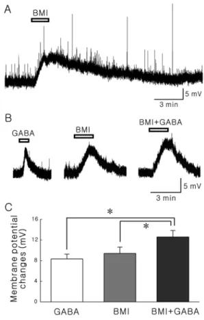
The SG (lamina II) of the Vc was clearly visible as a translucent band, just medial to the spinal trigeminal tract and travels along the lateral edge of the slice. Most of neurons tested were responded to the bath application of BMI (20 µM) with membrane depolarization (14.8 ± 1.54 mV, n=13, Fig. 1A). The mean resting membrane potential of neurons depolarized by BMI was -63.2 ± 2.24 mV (n=13). To examine whether BMI blocks GABA-mediated response on the SG neurons, we applied GABA with or without BMI. Not only GABA (30 µM) alone but also BMI
Fig. 1. BMI-induced membrane depolarization is not mediated by GABAA receptors. A, A representative trace showing membrane depolarization by bath application of 20µM BMI. B, Representa- tive traces by application of 30µM GABA, 20 µM BMI, and GABA with BMI in the same neuron, respectively. C, Comparison of mean membrane potentials induced by GABA alone, BMI alone and GABA with BMI. Asterisks represent significant difference (p<0.05, paired t-test).
(20 µM) alone induced a membrane depolarization.
Interestingly, when 30 µM GABA was applied in the presence of 20 µM BMI, GABA-induced membrane depolarization was still observed (Fig. 1B). The membrane potential changes by GABA with BMI were substantially larger than those of BMI alone or GABA alone (paired t- test, P<0.05). Fig. 1C shows the mean membrane potential changes by GABA alone (8.32 ± 0.93 mV), BMI alone (9.40 ± 1.22 mV) and GABA with BMI (12.6 ± 1.27 mV).
To confirm whether GABA-induced membrane depolari- zation is mediated by the activation of GABA
Areceptor, we applied picrotoxin (PTX), another GABA
Aas well as GABA
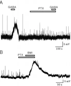
Creceptor antagonist. PTX completely blocked the GABA-induced membrane depolarization. However, PTX alone had no effect on the membrane potential at the employed dose (Fig. 2A). Moreover, BMI-induced mem- brane depolarizations were persisted in the presence of PTX (Fig. 2B, n=4). To examine whether quaternary ammonium salt could be involved in the BMI-induced membrane depolarization, we tested free base bicuculline (fBIC). Fig.
3A shows a representative trace showing depolarization by GABA (100 µM). When fBIC was applied on the same neuron used in Fig. 3A, fBIC completely blocked GABA- induced membrane depolarization in 4 of 5 neurons tested (Fig. 3B) and partially blocked in 1 of 5 tested. Fig. 3C shows the mean membrane potential change by GABA alone and GABA in the presence of fBIC (n=5).
To check whether BMI acts on the SG neurons directly, we applied BMI in the presence of antagonist mixture (AM)
Fig. 2. A. A representative trace showing a complete blocking of GABA (100µM)-induced membrane depolarization by PTX (50 µM). B, A voltage trace showing BMI (20 µM)-induced mem- brane depolarization in the presence of PTX (50µM). Note that PTX failed to induce membrane depolarization.Fig. 3. A, A representative trace showing depolarization by GABA (100µM) at -52.2 mV. B, A trace showing GABA effect in the presence of 20µM free base bicuculline (fBIC, GABAA receptor antagonist, 20µM). Note that GABA-induced membrane depolar- ization shown in Fig 3A was completely blocked by bicuculline. C, Bar graph showing mean membrane potential changes by GABA alone and GABA in the presence of free base bicuculline. * repre- sents p<0.05 by paired t-test).
Fig. 4. A sample trace showing BMI-induced membrane depolar- ization in the presence of antagonist mixture (AM) including tetro- dotoxin (TTX, a Na+ channel blocker, 0.5µM), AP-5 (NMDA receptor antagonist, 50µM), CNQX (non-NMDA receptor antago- nist, 20µM) and strychnine (glycine receptor antagonist, 20 µM).
including tetrodotoxin (TTX, a Na
+channel blocker), AP-5 (NMDA receptor antagonist), CNQX (non-NMDA receptor antagonist) and strychnine (glycine receptor antagonist).
BMI-induced membrane depolarization was still observed in the presence of AM in 7 neurons tested (Fig. 4).
Discussion
Here, we report that 1) BMI itself induced membrane depolarization and failed to block GABA-induced res- ponse; 2) GABA-induced membrane depolarization was completely blocked by PTX, an antagonist for both GABA
Aand GABA
Creceptor; 3) free base bicuculline (fBIC) failed to induce membrane depolarization and GABA-induced membrane depolarization was blocked by fBIC; 4) BMI- induced membrane depolarizations were persisted in the presence of PTX and antagonist mixture (AM) including CNQX, AP-5, strychnine and TTX.
GABA can act on ionotropic GABA
Aand GABA
Creceptors, and metabotropic GABA
Breceptor (reviewed by Bormann, 2000). Ionotropic GABA
Aand GABA
Creceptors are both directly coupled with chloride channels. However, they are pharmacologically and physiologically different.
For instance, GABA
Areceptors are blocked by both bicuculline and PTX (Johnston, 1996), but GABA
Creceptors are bicuculline resistant chloride channel and partially PTX-sensitive (Feigenspan et al., 1993; Qian and Dowling, 1993).
In this study, both GABA and BMI induced membrane depolarization, and when GABA in the presence of BMI was applied, membrane depolarization was not suppressed but enhanced (Fig. 1). Since BMI is a well-known GABA
Areceptor antagonist, similar pattern of response by GABA and BMI may sound strange. In addition, the membrane potential change by co-application of GABA and BMI was bigger than that of BMI alone or GABA alone (Fig. 1C).
Whereas, GABA-induced membrane depolarization was completely blocked by PTX (Fig. 2). These results suggest that there may be a BMI-resistant and PTX-sensitive GABA response, which is similar with previous result in the rat spinal dorsal horn by Park et al. (1999). However, BMI itself induced membrane depolarization, it was difficult to apply GABA in the presence of BMI. Therefore, it was hardly concluded that GABA-induced membrane depolarization was blocked by BMI or not.
Here, BMI, but not fBIC, induced membrane depolar- zation in the majority of the neurons tested. These results indicate that action mechanism of BMI could be different from that of fBIC. A number of studies have suggested the non-GABAergic action of BMI on dopaminergic neuron in various area such as ventral tegmental area and the substantia nigra pars compacta by blocking apamin- sensitive SK current (Seutin et al., 1997), by enhancing the NMDA dependent burst firing (Johnson and Seutin, 1997),
and by blocking nicotinic responses in pituitary cells (Zhang and Feltz, 1991). We also demonstrate here that BMI- induced depolarizing effects were maintained in the presence of excitatory and inhibitory amino acid neurotransmitter receptor antagonist mixture (AM) combined with TTX (Fig. 4). These results imply that BMI can directly act on the postsynaptic SG neuronal dendrite or soma.
Taken together, these results demonstrate that BMI- induced membrane depolarizations which act on the postsynaptic SG neuron directly are not mediated by GABA
Areceptor. These data suggest that the action of BMI is different from that of free base bicuculline and other unknown mechanisms could be involved in the BMI- induced depolarization. Further studies are needed for precise mechanism underlying the BMI-induced membrane depolarization on the SG neurons of Vc.
Acknowledgment
This study was supported by the Korea Science and Engineering Foundation (KOSEF) grant funded by the Korea government (F01-2006-000 -10243-0).
References
Bormann J. The 'ABC' of GABA receptors. Trends Pharmacol Sci. 2000;21:16-9.
Bracci E, Ballerini L, Nistri A. Spontaneous rhythmic bursts induced by pharmacological block of inhibition in lumbar motoneurons of the neonatal rat spinal cord. J Neurophysiol.
1996;75:640-7.
Cazalets JR, Sqalli-Houssaini Y, Clarac F. GABAergic inacti- vation of the central pattern generators for locomotion in isolated neonatal rat spinal cord. J Physiol. 1994;474:173-81.
Cowley KC, Schmidt BJ. Effects of inhibitory amino acid antagonists on reciprocal inhibitory interactions during rhythmic motor activity in the in vitro neonatal rat spinal cord. J Neurophysiol. 1995;74:1109-17.
Curtis DR, Duggan AW, Felix D, Johnston GA. GABA, bicuculline and central inhibition. Nature. 1970;226:1222-4.
Debarbieux F, Brunton J, Charpak S. Effect of bicuculline on thalamic activity: a direct blockade of IAHP in reticularis neurons. J Neurophysiol. 1998;79:2911-8.
Feigenspan A, Wässle H, Bormann J. Pharmacology of GABA receptor Cl- channels in rat retinal bipolar cells.
Nature. 1993;361:159-62.
Han SK, Herbison AE. Norepinephrine suppresses gonadotropin- releasing hormone neuron excitability in the adult mouse.
Endocrinology. 2008;149:1129-35.
Han SK, Park JR, Park SA, Chun SW, Lee JC, Lee SY, Ryu PD, Park SJ. Noradrenaline inhibits substantia gelatinosa neurons in mice trigeminal subnucleus caudalis via alpha(2) and beta adrenoceptors. Neurosci Lett. 2007;411:92-7.
Heyer EJ, Nowak LM, Macdonald RL. Bicuculline: a convulsant with synaptic and nonsynaptic actions. Neurology.
1981;31:1381-90.
Johnson SW, Seutin V. Bicuculline methiodide potentiates NMDA-dependent burst firing in rat dopamine neurons by blocking apamin-sensitive Ca2+-activated K+currents. Neurosci Lett. 1997;231:13-6.
Johnston GA. GABAc receptors: relatively simple transmitter- gated ion channels? Trends Pharmacol Sci. 1996;17:319-23.
Jovanovic K, Petrov T, Stein RB. Effects of inhibitory neurotransmitters on the mudpuppy (Necturus maculatus) locomotor pattern in vitro. Exp Brain Res. 1999;129:172-84.
Olsen RW, Ban M, Miller T. Studies on the neuropharmacological activity of bicuculline and related compounds. Brain Res. 1976;102:283-99.
Park JS, Higashi H, Nagata K, Yoshimura M. Bicuculline- resistant, Cl- dependent GABA response in the rat spinal dorsal horn. Neurosci Res. 1999;33:261-8.
Qian H, Dowling JE.: Novel GABA responses from rod- driven retinal horizontal cells. Nature. 1993;361:162-4.
Sessle, B.J. Mechanisms of trigeminal and occipital pain. Pain Rev. 1996;3:91–116.
Seutin V, Johnson SW. Recent advances in the pharmacology of quaternary salts of bicuculline. Trends Pharmacol Sci.
1999;20:268-70.
Seutin V, Scuvée-Moreau J, Dresse A. Evidence for a non- GABAergic action of quaternary salts of bicuculline on dopaminergic neurones. Neuropharmacology 1997;36:1653-7.
Shi WX, Rayport S. GABA synapses formed in vitro by local axon collaterals of nucleus accumbens neurons. J Neurosci.
1994;14:4548-60.
Zhang ZW, Feltz P.: Bicuculline blocks nicotinic acetylcholine response in isolated intermediate lobe cells of the pig. Br J Pharmacol. 1991;102:19-22.