Rapid Determining for Subtypes and Pandemic Type of Swine Influenza Virus by Diagnostic One-step RT-PCR
Gwang Il Kim1, Jee In Kim2,3, Jin-Hyeap Kwon1, Yoo Hong Min1, Joo Il Kang5, Chang-Ho Lee4, Sung-Hee Kim6 and Jae-Hwan Lim1,2*
1Department of Biological Sciences , Andong National University, Andong 36729, Korea
2Institute of Vaccine Biotechnology, Andong National University, Andong 36729, Korea
3Department of Biochemistry and Cell Biology, School of Medicine, Kyungpook National University, Daegu 41566, Korea
4Gyeongbuk Institute for Bioindustry, Andong 36728, Korea
5Bionics, Seoul 04782, Korea
6Department of Animal Disease Control & Quarantine, Animal and Plant Quarantine Agency, Kimcheon 39660, Korea Received January 15, 2018 /Revised May 2, 2018 /Accepted May 21, 2018
Swine influenza virus (SIV) causes one of the most common diseases of the pig population, and its subtypes are determined by hemagglutinin (HA) and neuraminidase (NA). Recently, the SIV subtype diagnosis has been developed. The method using antigen-antibody reaction rather than PCR was mainly used because of the large change in the ribonucleotide sequences of SIV. Here, we have devel- oped 10 diagnostic primer sets through multi-nucleotide sequences alignment of spreaded SIV since 2008 in Korea and then optimized the reaction of the one-step RT-PCR for rapid determination of SIV subtype. In addition, specific primers were designed to early determine the pandemic SIV by detecting unique M sequences proven in highly infectious and virulent subtypes of the influenza H1N1 (pH1N1). Here, some of the SIVs spread in Korea from 2008 to 2014 have been tested to determine the subtypes and pandemic potential of SIV. All diagnostic primer sets were found to be able to accu- rately determine the SIV subtype and to detect the pandemic SIV. In conclusion, it was confirmed that the optimized one-step RT-PCR analysis using these primer sets is useful for rapid diagnosis of SIV subtypes. These results can be used for development of SIV subtype diagnostic kit to early detect be- fore virulent SIV spreads do.
Key words : Diagnostic one-step RT-PCR, pandemic H1N1, swine influenza virus
*Corresponding author
*Tel : +82-54-820-5797, Fax : +82-54-820-7729
*E-mail : [email protected]
This is an Open-Access article distributed under the terms of the Creative Commons Attribution Non-Commercial License (http://creativecommons.org/licenses/by-nc/3.0) which permits unrestricted non-commercial use, distribution, and reproduction in any medium, provided the original work is properly cited.
Journal of Life Science 2018 Vol. 28. No. 5. 555~562 DOI : https://doi.org/10.5352/JLS.2018.28.5.555
Introduction
Swine influenza virus belongs to orthomyxoviridae and influenza A virus, and is divided into subtypes based on two proteins HA (H1-H16) and NA (N1-N9) on the virus surface [22]. SIV causes one of the common respiratory ill- nesses in pig populations. These can quickly spread to the world and have a great impact on the socioeconomic envi- ronment [18]. In 2009, there was a pandemic H1N1 virus resulting from antigenic shift between animal and human origin of SIV [2, 12, 14, 22]. It spread throughout the world and infected some 500,000 people [5, 12, 24]. Therefore, rapid
and sensitive detection techniques are needed to control epi- demic and epidemic diseases. Conventional subtype diag- nostic methods use the rapid influenza diagnostic test (RIDT) based on immunochromatography to detect viral antigens. This is considered to be the best and quickest diag- nostic method for influenza virus diagnosis [13]. However, this method has difficulties in classifying influenza A sub- types and has been performed mainly for classification of influenza A and B virus types because of low sensitivity and requirement of high amount of SIV [1, 7, 20].
Therefore, reverse transcriptase chain reaction (RT-PCR) techniques that sensitively detect influenza virus have been developed in recent years [3, 4, 15, 21]. PCR-based diagnostic methods can detect type A influenza viruses using highly specific primers and can distinguish other subtypes of influ- enza A virus [8, 16]. Recently, M protein has been focused as a target protein to discriminate between pandemic or en- demic SIV. However, pandemic SIV determination by the method based on antibody-antigen response is quite difficult
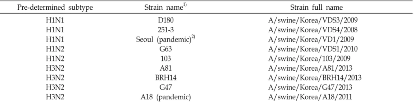
Table 1. SIV strain name and suggested subtypes used in this study
Pre-determined subtype Strain name1) Strain full name
H1N1 H1N1 H1N1 H1N2 H1N2 H3N2 H3N2 H3N2 H3N2
D180 251-3 Seoul (pandemic)2)
G63 103 A81 BRH14
G47 A18 (pandemic)
A/swine/Korea/VDS3/2009 A/swine/Korea/VDS4/2008 A/swine/Korea/VD1/2009 A/swine/Korea/VDS1/2010 A/swine/Korea/103/2009 A/swine/Korea/A81/2013 A/swine/Korea/BRH14/2013 A/swine/Korea/G47/2013 A/swine/Korea/A18/2011
1)The strains of SIV were named and pre-determined those subtypes by APQA (Animal and Plant Quarantine Agency, South Korea).
2)The strain ‘Seoul’ was used in this study with the isolated and tested strain by APQA, Korea.
because M2 protein is pore protein, which is less exposed than HA and NA proteins out of the viral surface. Therefore, the detection method for particular pandemic M sequence by RT-PCR and one-step RT-PCR is developed together with designed specific primers for M sequence [9, 10, 16].
The aim of this study is to rapidly diagnose SIV subtypes and pandemic SIV using specific primers and an optimized one-step RT-PCR analysis system. We designed 10 diag- nostic primer sets to identify the types of SIV using multiple nucleotide sequence alignment of SIV appeared from 2008 to 2014. We have also successfully determined the virulent subtype pH1N1 of the SIV by detecting a unique M sequence.
Ultimately, we are aiming at early detection of pre-spread SIV by developing step RT-PCR based on unique fragments of the HA, NA and M2 genes of influenza A virus.
Materials and Methods
Viral RNA and primers
Viral RNAs (Table 1) were provided and tested at the Animal and Plant Quarantine Agency (APQA) and stored in -80℃ until synthesis cDNA. Those 9 strains of SIV sub- types were named by APQA as follows; D180, 251-3 and Seoul (pandermic) for H1N1 strains, G63, 103 for H1N2 strains, A81, BRH14, G47, A18 (pandermic) for H3N2. The primers for subtyping of SIV were designed by multiple alignment of HA, NA and M gene sequences. The alignment was performed using by ClustalW2 (http://www.geno- me.jp/tools/clustalw/) with SIV nucleotide sequences from the Influenza Research Database (http://www.fludb.org/).
cDNA synthesis and RT-PCR
Reverse transcription (RT) for complementary DNA syn- thesis was performed using iScript™ cDNA Synthesis Kit
(BIO-RAD, Hercules, CA, USA) according to the manu- facturer’s instructions. SIVs were subtyped by conventional RT-PCR targeting the HA, NA and M gene using TaKaRa Ex Taq™ (TaKaRa, Otsu, Japan) with 2 ng of SIV RNA templates. Conventional RT-PCR cycling conditions were as follows: pre melting for 5 minutes at 94℃; 35 cycles of dena- turation for 30 seconds at 94℃, annealing for 30 seconds 53~56℃ and extension for 30 seconds at 72℃; and a final extension of 5 minutes at 72℃.
One step RT-PCR for virus subtyping
One-step RT-PCR targeting the HA, NA and M gene was performed using QIAGEN® One-Step RT-PCR Kit (Qiagen, Hilden, Germany). The PCR mixture consists of QIAGEN One-Step RT-PCR 5ⅹ buffer 10 μl, 2 μl of QIAGEN One-Step RT-PCR Enzyme mix, 2 μl of dNTP Mix, 5 pmole each pri- mer, 2 ng of RNA templates and RNase-free water. Cycling conditions for one step RT-PCR were as follows: reverse transcription for 30 minutes at 50℃, initial PCR activation for 15 minutes at 95℃; 35 cycles of denaturation for 30 sec- onds at 94℃, annealing for 30 seconds 50~68℃ and ex- tension for 60 seconds at 72℃; and a final extension of 10 minutes at 72℃. In this study, diagnostic One-Step RT-PCR with modified cycling condition was also developed and performed with constructing a mixture of Labopass™
M-MuLV reverse transcriptase and Labopass™ G-Taq poly- merase from Cosmo genetech (Korea) with optimized buffer.
Results
Construction of specific primers for HA and NA gene of Swine Influenza A Virus
In order to develop primer set for subtyping of SIVs, we multiple-aligned with complete nucleotide sequences of
Table 2 GeneBank accession numbers of SIVs and segments lengths of each target genes
Subtype Strain name NCBI Taxon.
ID #
GeneBank accession # / Segment length (bp)
HA NA M
H1N1
A/swine/Korea/CY01-04/2012 A/swine/Korea/CY01-05/2012 A/swine/Korea/CY01-06/2012 A/swine/Korea/CY11-01/2011(H1N1) A/swine/Korea/CY11-02/2011(H1N1) A/swine/Korea/SCJ28/2010(H1N1) A/swine/Korea/SCJ33/2010(H1N1) A/swine/Korea/SCJ41/2010(H1N1) A/swine/Korea/SCJ42/2010(H1N1)
1281708 1281709 1281710 1281711 1281712 762450 762451 762452 762453
KC471369/1701 KC471377/1701 KC471385/1701 KC471345/1701 KC471353/1701 HM189557/1701 HM189558/1701 HM189559/1701 HM189560/1701
KC471371/1410 KC471379/1410 KC471387/1410 KC471347/1410 KC471355/1410 HM189445/1410 HM189446/1410 HM189447/1410 HM186448/1410
KC471372/982 KC471380/982 KC471388/982 KC471348/982 KC471356/982 HM189475/977 HM189476/977 HM189477/977 HM189478/976
H1N2
A/swine/Korea/CY03-11/2012(H1N2) A/swine/Korea/CY0423-12/2013 A/swine/Korea/CY0423-33/2013(H1N2) A/swine/Korea/CY12-03/2011(H1N2) A/swine/Korea/VDS1/2010
1281717 1337100 1337101 1281718 1038687
KC471425/1701 KF142495/1778 KF142503/1732 KC471361/1701 JN043428/1701
KC471427/410 KF142497/1466 KF142505/1410 KC471363/410 JN043437/1410
KC471428/982 KF142498/1027 KF142506/2007 KC471364/982 JN043433/971
H3N1
A/swine/Korea/CY02-07/2012 A/swine/Korea/CY02-08/2012(H3N1) A/swine/Korea/CY03-12/2012(H3N1) A/swine/Korea/CY03-13/2012(H3N1)
1281713 1281714 1281715 1281716
KC471393/1701 KC471401/1701 KC471433/1701 KC471441/1701
KC471395/1410 KC471403/1410 KC471435/1410 KC471443/1410
KC471396/982 KC471404/982 KC47136/982 KC47144/982
H3N2
A/swine/Korea/A18/2011
A/swine/Korea/CY02-09/2012(H3N2) A/swine/Korea/CY02-10/2012(H3N2) A/swine/Korea/CY03-14/2012(H3N2) A/swine/Korea/CY03-15/2012(H3N2) A/swine/Korea/CY03-16/2012(H3N2) A/swine/Korea/CY03-17/2012(H3N2) A/swine/Korea/CY03-18/2012(H3N2) A/swine/Korea/CY03-19/2012(H3N2) A/swine/Korea/D79/2011
A/swine/Korea/KSB/2012 A/swine/Korea/PL01/2012
1224818 1281719 1281720 1281721 1281722 1281723 1281724 1281725 1281726 1224819 1224820 1357697
JX501999/1701 KC471409/1701 KC471417/1701 KC471449/1701 KC471457/1701 KC471465/1701 KC471473/1701 KC471481/1701 KC471489/1701 JX502000/1701 JX501998/1701 KF382729/1701
JX502005/1410 KC471411/1410 KC471419/1410 KC471451/1410 KC471459/1410 KC471467/1410 KC471475/1410 KC471483/1410 KC471491/1410 JX502006/1410 JX502004/1410 KF382731/1410
JX502002/982 KC471412/982 KC471420/982 KC471452/982 KC471460/982 KC471468/982 KC471476/982 KC471484/982 KC471492/982 JX502003/982 JX502001/982 KF382732/982 H1N1, H3N1, H1N2, H3N2 swine influenza A virus and
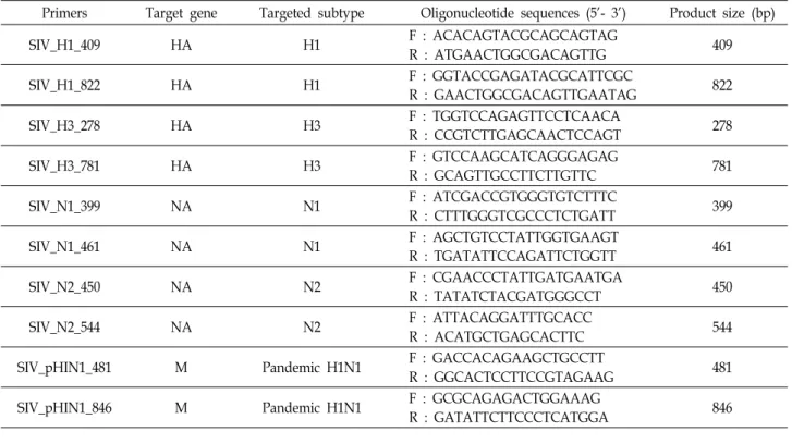
pandemic pH1N1 SIV that occurred from 2008 to 2014 in South Korea (Table 2). As a result, we found specifically con- served sequences among SIV subtypes and developed com- plementary primer sets. In order to determine the H sub- types, we aligned and compared HA sequences in H1N1 and H1N2 in one set and H3N1 & H3N2 SIVs in another set and then constructed H1 primers, H1_409, H1_822 and H3 primer, H3_278, H3_781. The NA sequences of H1N1 &
H3N1 and H1N2 & H3N2 SIVs also compared in the same manner to design N1 primers, N1_399 and N1_461 and N2 primers, N2_450 and N2_544. Finally, to determine pan- demic pH1N1 A virus, we multiple aligned the matrix M gene sequences and identified specific conserved sequences.
And then the detection primers, pH1N1_ 481 and pH1N1_846, were developed (Table 3).
Subtyping and pandemicity identification of Swine Influenza A Virus spread in Korea
By multiple sequences alignment, we designed 4 sets of primers which were able to discriminate H1 and H3 subtype.
Two specific H1 primers, H1_409, H1_822, were clearly used for detection for H1N1 and H1N2 subtypes of SIV named D180, 251-3, Seoul and 103 which had been occurred in South Korea. Also, SIVs named A81, BRH14, G47 and A18 were determined as H3 subtype by using two H3 primers, H3_278, H3_781 (Fig. 1A, Fig. 1B). But SIV named G63 was able to be determined as a H1 subtype by showing a weak band in H1 detection PCR using only with H1_409 primer (Fig. 1A). In the same manner, the N1 and N2 forms of all SIVs were distinguished by RT-PCR with four sets of NA primers. N1 specific band sizes of 399 bp and 461 bp were confirmed by PCR reactions on the SIV N1 subtype using
Table 3. List of primers used in this study
Primers Target gene Targeted subtype Oligonucleotide sequences (5'- 3') Product size (bp)
SIV_H1_409 HA H1 F : ACACAGTACGCAGCAGTAG
R : ATGAACTGGCGACAGTTG 409
SIV_H1_822 HA H1 F : GGTACCGAGATACGCATTCGC
R : GAACTGGCGACAGTTGAATAG 822
SIV_H3_278 HA H3 F : TGGTCCAGAGTTCCTCAACA
R : CCGTCTTGAGCAACTCCAGT 278
SIV_H3_781 HA H3 F : GTCCAAGCATCAGGGAGAG
R : GCAGTTGCCTTCTTGTTC 781
SIV_N1_399 NA N1 F : ATCGACCGTGGGTGTCTTTC
R : CTTTGGGTCGCCCTCTGATT 399
SIV_N1_461 NA N1 F : AGCTGTCCTATTGGTGAAGT
R : TGATATTCCAGATTCTGGTT 461
SIV_N2_450 NA N2 F : CGAACCCTATTGATGAATGA
R : TATATCTACGATGGGCCT 450
SIV_N2_544 NA N2 F : ATTACAGGATTTGCACC
R : ACATGCTGAGCACTTC 544
SIV_pHIN1_481 M Pandemic H1N1 F : GACCACAGAAGCTGCCTT
R : GGCACTCCTTCCGTAGAAG 481
SIV_pHIN1_846 M Pandemic H1N1 F : GCGCAGAGACTGGAAAG
R : GATATTCTTCCCTCATGGA 846
A
B
C
D
Fig. 1. Determination of HA and NA type for SIV subtyping by conventional PCR. Subtyping for SIV HA and NA by PCR amplifica- tion with designed primers. A, Conventional PCR was performed with specific H1 primer sets, H1-409, H1_822, based on conserved sequence. All H1 subtype of SIV was determined by PCR except strain G63. B, The H3 subtypes of SIV were also determined by PCR with designed H3 primers, H3_278 and H3_781, without any misidentification. Also, PCR amplifica- tions for NA subtyping were performed with N1 and N2 primers. C, Conventional PCR for N1 subtying was performed with specific N1 primer sets, N1_399 and N1_461 based on conserved sequence. d, N2 subtype of SIVs was determined by using N2 primer, N2_450 and N2_544. Negative control (N.C.) indicates the result of PCR reaction without template.
A
B
Fig. 2. Identification of pandemic SIV strain. A, Conventional PCR was performed with specific primer sets based on conserved sequence of pandemic strain. The pandemic SIV were amplified by selected primers, pH1N1_481 and pH1N1_846. B, The pandemic strain were determined without mis-detection by One-step RT-PCR with specific primer, pH1N1_481.
A
B
C
D
Fig. 3. Identification of SIV subtype by One-step RT-PCR with selected primers. SIV subtyping with amplification of HA and NA genes by One-step RT-PCR. A, B, One-step RT-PCR was performed with H1 subtype-specifically de- signed primers, H1_409 and H1_822 and with H3 specif- ic primer sets, H3_278 and H3_781. c, d, The SIV con- tained N1, N2 were amplified and determined using pri- mers, N1_399 and N2_450. However, strain ‘D180, de- termined as H1N1 by conventional PCR was not de- tected by selected primers H1_822 and N1_399.
primers N1_399 and N1_461 designed through N1 subtype conserved sequence analysis. Also, determination PCR using N2 subtype-specific N2_450 and N2_544 primers revealed specific bands representing for N2 subtype (Fig. 1C, Fig. 1D).
In the case of G63 subtype determination, a weak N2 sub- type-specific band was observed in NA subtyping PCR as seen in H1 subtype determination (Fig. 1D). As indicated by the weak band in N1 detection PCR using both N1_399 and N1_461 primers, the SIV named D180 was determined as N1. However, these results demonstrate that the designed HA and NA subtype specific primers are suitable for the diagnosis of SIV subtypes.
In addition, to determine the virulent viruses called pH1N1, we used specifically designed primers based on highly conserved matrix M gene among in pandemic SIV subtypes. Apparently, PCR diagnoses using two designed primers, pH1N1_481 and pH1N1_846 were clearly dis- tinguished the pH1N1 viruses from other SIVs (Fig. 2A).
Rapid determination of SIV subtypes by developed One-step RT-PCR
We conducted a diagnostic PCR using the QIA One-Step RT-PCR Kit and sensitive primers selected based on pre- vious results using conventional PCR. All viruses were clear- ly identified by the marked bands as HA and NA subtypes and then pandemic SIV also was distinguished from other SIVs (Fig. 3, Fig. 2B). However, as in the previous two-step RT-PCR, the SIV of D180 was insufficient to determine the subtype. Therefore, we have also developed one-step RT- PCR with optimized buffering components. Here, the most sensitive and specific primers identified by conventional RT-PCR methods were selected for diagnostic one-step RT-PCR analysis (Fig. 4). However, in the H1N2 SIV, G63, which has never been reported as an epidemic, this diag- nostic PCR reaction shows a specially amplified band that is pandemic. In addition, one-step RT-PCR with BRH14, G47 and A81 identified as H3N2 also showed that these SIVs
Fig. 4.Diagnosis of pandemic strain and identification of subtypes from SIV by One-step RT-PCR with specific primer sets. Analysis of SIV for subtyping and diagnosis of pandemic strain was performed by modified One-step RT-PCR using optimized buffers and optimized conditions. All primer sets were well-constructed for determination of SIV subtypes corresponding to specific amplification. However, PCR reaction for pandemic SIV determination with primer 7 showed exact PCR band even in three out of seven SIVs known as non-pandemic. (M: size marker, 1: N1_399, 2: N2_450, 3: H1_409, 4: H1_822, 5: H3_278, 6: H3_781, 7: pH1N1M_481, open triangle: pandemic strain, asterisk: nonspecific band, see Discussion).
had an M protein with pandemic features.
As a result of the one-step RT-PCR developed, all SIVs spread in Korea have been identified as precise subtypes such as H1N1, H1N2, H3N1 and H3N2, and have dis- tinguished pandemic SIVs with specific M proteins. Also subtyping for SIV G63 and D180 were determined to its cor- responding subtype showing high band intensities. Overall, these results, using the developed diagnostic one-step RT- PCR analysis, suggest that spread SIV can be rapidly identi- fied as a subtype of the SIV, as well as identifying potential pandemicity.
Discussion
The emergence of novel influenza virus is occurred by genetic reassortment among different influenza viruses co-infection in the same host or mutative variation of HA and NA gene [3]. Occasionally, it may lead to high infective virus such as pandemic H1N1 in 2009. The pH1N1 virus is representative fatal influenza virus having mixed genomes of swine, avian and human influenza virus. In particular, it is characterized by being easily transmitted from pig to
pig [4, 8, 16]. In order to prevent the spread of pandemic or endemic viruses, the important point is timely diagnosis and treatment. Generally, RT-PCR based on differences among nucleotide sequences and RIDT based on antigenic detection are highly sensitive and specific methods [1, 6, 11].
Recently, quantitative RT-PCR assays are mostly used for rapid detection of influenza. Comparing these two methods, RIDT is less sensitive than RT-PCR assay [15, 17, 19, 23].
On the other hand, for the detecting subtype signature by PCR method, target conserved sequences of HA, NA and M genes are required to develop the specific primers by mul- tiple alignments of full genome sequences [6, 9, 10, 16].
In this study, we aimed to develop the primer sets and one-step RT-PCR assay method for rapidly and specifically determining subtypes of Swine Influenza A viruses. First, conventional two-step RT-PCR was performed, and the sub- type of SIV was determined by electrophoresis. With the ex- ception of only one SIV ‘G63’ showing a low intensity band in the H1 subtyping, subtypes of other SIVs are clearly dis- tinguishable by designed primers. In this regard, there are possibilities that ‘G63’ has a large numbers of variations in nucleotide sequences or ‘G63 is a pandemic virus but is not
proved as an epidemic because of efficient prevention control. Second, the primers for SIV subtype diagnosis used by selecting from the results of conventional RT-PCR might be very sensitive in one-step RT-PCR assay. However, G63, BRH14, G47 and A81 were recently proved to have a M pro- tein derived from pandemic H1N1(unpublished data). As a result, all samples are determined clearly. In other words, developed one-step RT-PCR analysis system using opti- mized the buffer solution and the developed primer sets was shown to be highly efficient at accurately detecting SIV, de- termining subtypes, and distinguishing the pandemicity.
Taken together, the developed and optimized one-step RT-PCR using the specific primers described above makes it possible to diagnose the subtypes and pathogenicity of SIV. Also, this efficient diagnostic assay can be assisted to detect SIV subtypes rapidly and prevent the virulent SIV infection.
Acknowledgment
This work supported by a grant from 2015 Research Funds of Andong National University.
References
1. Balish, A., Garten, R., Klimov, A. and Villanueva, J. 2013.
Analytical detection of influenza A (H3N2) v and other A variant viruses from the USA by rapid influenza diagnostic tests. Influenza Other Respir. Viruses 7, 491-496.
2. Brookes, S. M., Núñez, A., Choudhury, B., Matrosovich, M.
and Essen, S. C., et al. 2010. Replication, pathogenesis and transmission of pandemic (H1N1) 2009 virus in non-im- mune pigs. PLoS One 5, e9068.
3. Choi, Y. K., Goyal, S. M., Kang, S. W., Farnham, M. W. and Joo, H. S. 2002. Detection and subtyping of swine influenza H1N1, H1N2 and H3N2 viruses in clinical samples using two multiplex RT-PCR assays. J. Virol. Methods 102, 53-59.
4. Fereidouni, S. R., Starick, E., Grund, C., Globig, A., Metten- leiter, T. C., Beer, M. and Harder, T. 2009. Rapid molecular subtyping by reverse transcription polymerase chain re- action of the neuraminidase gene of avian influenza A viruses. Vet. Microbiol. 135, 253-260.
5. Garten, R. J., Davis, C. T., Russell, C. A., Shu, B. and Lin- dstrom, S., et al. 2009. Antigenic and Genetic characteristics of the early isolates of Swine-origin 2009 A (H1N1) Influenza Viruses circulating in humans. Science 325, 197-201.
6. Harmon, K., Bower, L., Kim, W. I., Pentella, M. and Yoon, K. J. 2010. A matrix gene–based multiplex real‐time RT‐
PCR for detection and differentiation of 2009 pandemic H1N1 and other influenza A viruses in North America.
Influenza Other Respir. Viruses 4, 405-410.
7. Hawkes, M., Richardson, S. E., Ipp, M., Schuh, S., Adachi, D. and Tran, D. 2010. Sensitivity of rapid influenza diag- nostic testing for swine-origin 2009 a (H1N1) influenza virus in children. Pediatrics 125, e639-e644.
8. Hiromoto, Y., Uchida, Y., Takemae, N., Hayashi, T., Tsuda, T. and Saito, T. 2010. Real-time reverse transcription-PCR assay for differentiating the pandemic H1N1 2009 influenza virus from swine influenza viruses, J. Virol. Methods 170, 169-172.
9. Ji, M. J., Cho, B. K., Cho, Y. S., Choi, Y. J. and Kwon, D., et al. 2013. Development of a specific and rapid diagnostic method for detecting influenza A (H1N1) pdm09 virus in- fection using immunochromatographic assay. Osong Public Health Res. Perspect. 4, 342-346.
10. Kang, X. P., Jiang, T., Li, Y. Q., Lin, F. and Liu, H., et al.
2010. A duplex real-time RT-PCR assay for detecting H5N1 avian influenza virus and pandemic H1N1 influenza virus.
Virol. J. 7, 113.
11. Kaul, K. L., Mangold, K. A., Du, H., Pesavento, K. M., Nawrocki, J. and Nowak, J. A. 2010. Influenza A subtyping:
seasonal H1N1, H3N2, and the appearance of novel H1N1.
J. Mol. Diagn. 12, 664-669.
12. Khanna, M., Kumar, B., Gupta, A. and Kumar, P. 2012.
Pandemic influenza A H1N1 (2009) virus: lessons from the past and implications for the future. Indian J. Virol. 23, 12-17.
13. Kim, Y. K., Uh, Y., Chun, J. K., Kim, C. and Kim, H. Y.
2010. Evaluation of new hemagglutinin-based rapid antigen test for influenza A pandemic (H1N1) 2009. J. Clin. Virol.
49, 69-72.
14. Lange, E., Kalthoff, D., Blohm, U., Teifke, J. P. and Breithaupt, A., et al. 2009. Pathogenesis and transmission of the novel swine-origin influenza virus A/H1N1 after ex- perimental infection of pigs. J. Gen. Virol. 90, 2119-2123.
15. Lee, C. S., Kang, B. K., Lee, D. H., Lyou, S. H., Park, B.
K., Ann, S. K., Jung, K. and Song, D. S. 2008. One-step multi- plex RT-PCR for detection and subtyping of swine influenza H1, H3, N1, N2 viruses in clinical samples using a dual priming oligonucleotide (DPO) system. J. Virol. Methods 151, 30-34.
16. Lorusso, A., Faaberg, K. S., Killian, M. L., Koster, L. and Vincent, A. L. 2010. One-step real-time RT-PCR for pan- demic influenza A virus (H1N1) 2009 matrix gene detection in swine samples. J. Virol. Methods 164, 83-87.
17. Miarka, M., Horban, A., Maliszewska, H., Biliński, P. and Prus-Kowalczuk, W. 2014. A clinical utility of a strip test for influenza A/B and comparison with detection by RT PCR. Acta Biochim. Pol. 61, 485-487.
18. Meiners, C., Loesken, S., Doehring, S., Starick, E., Pesch, S., Maas, A., Noe, T., Beer, M., Harder, T. and Grosse Beilage, E. 2014. Field study on swine influenza virus (SIV) infection in weaner pigs and sows. Tierarztl. Prax. Ausg. G Grosstiere Nutztiere 42, 351-359.
19. Nougairede, A., Ninove, L., Zandotti, C., de Lamballerie, X. and Gazin, C., et al. 2010. Point of care strategy for rapid diagnosis of novel A/H1N1 influenza virus. PLoS One 5, e9215.
초록:진단용 one-step RT-PCR을 통한 돼지 인플루엔자 바이러스의 아형 및 pandemic 유형에 대 한 신속한 결정
김광일1․김지인2,3․권진협1․민유홍1․강주일5․이창호4․김성희6․임재환1,2*
(1안동대학교 생명과학과, 2안동대학교 백신산업연구소, 3경북대학교 의과대학 생화학세포생물학교실, 4경북바이
오산업연구원, 5코스모진텍, 6국립농림축산검역본부)
Swine influenza virus (SIV)는 돼지 개체군에서 가장 흔한 질병을 일으키는 바이러스 중 하나이며 그 subtype 은 hemagglutinin (HA)와 neuraminidase (NA)에 의해 결정됩니다. 최근 SIV subtype 진단 방법이 개발되고 있으 나 SIV의 리보뉴클레오타이드 서열의 많은 변이로 인해 PCR 보다는 항원-항체 반응을 이용하는 방법이 주로 사 용되고 있다. 본 연구에서는 SIV 하위 유형의 신속한 결정을 위하여 2008년 이후 국내에서 발생한 SIV의 다중 염기서열 정렬을 통하여 10개의 subtype 진단 프라이머 세트를 개발하고 이를 이용한 one-step RT-PCR 반응을 최적화하였다. 또한 감염력이 높고 독성이 있는 인플루엔자 H1N1 (pH1N1)의 아형에서 확인된 독특한 M 유전자 서열을 검출함으로써 pandemic SIV를 조기에 결정하도록 특이적 프라이머를 설계하였다. 2008년부터 2014년까지 한국에서 발생한 9종의 SIV RNA를 활용하여 SIV의 아형 및 pandemic 가능성을 결정하기 위해 시험 분석한 결과 모든 진단 프라이머 세트는 SIV 아형을 정확하게 결정하였으며 pandemic SIV를 검출할 수 있는 것으로 확인되었 다. 결과적으로 이들 프라이머 세트를 이용한 최적화된 one-step RT-PCR 분석이 SIV 아형의 신속한 진단에 유용 하다는 것이 확인하였다. 이러한 결과는 SIV 하위 유형 및 pandemic SIV가 확산되기 전에 조기 발견을 위한 키트 로 개발될 수 있음을 시사한다.
20. Su, L. C., Chang, C. M., Tseng, Y. L., Chang Y. F., Li, Y.
C., Chang, Y. S. and Chou, C. 2012. Rapid and highly sensi- tive method for influenza A (H1N1) virus detection. Anal.
Chem. 84, 3914-3920.
21. Tsushima, Y., Uno, N., Sasaki, D., Morinaga, Y., Hasegawa, H. and Yanagihara, K. 2015. Quantitative RT-PCR evalua- tion of a rapid influenza antigen test for efficient diagnosis of influenza virus infection. J. Virol. Methods 212, 76-79.
22. Vincent , A., Awada, L., Brown, I., Chen, H. and Claes, F., et al. 2014. Review of influenza A virus in swine world-
wide: a call for increased surveillance and research.Zoonoses Public Health 61, 4-17.
23. Vincent, A. L., Lager, K. M., Faaberg, K. S., Harland, M.
and Zanella, E. L., et al. 2010. Experimental inoculation of pigs with pandemic H1N1 2009 virus and HI cross‐re- activity with contemporary swine influenza virus antisera.
Influenza Other Respir. Viruses 4, 53-60.
24. World Health Organization. 2009. Influenza (seasonal).
Available at http://www.who.int/ mediacentre/factsheets/
fs211/en/index.html