Anti-proliferative and Pro-apoptotic Activities by Pomace of Schisandra chinensis (Turcz.) Baill. and Schizandrin
Hyun-Ji Kim
1, Yu-Mi Seo
1, Eun-Ju Lee
1, Chungwook Chung
1, Hwa-Jung Sung
2, Ho-Yong Sohn
2, Jong-Yi Park
3and Jong-Sik Kim
1*
1Department of Biological Sciences, Andong National University, Andong 36729, Korea
2Department of Food and Nutrition, Andong National University, Andong 36729, Korea
3Gyeongbuk Institute for Bio Indudustry, Andong 36728, Korea
Received October 19, 2017 /Revised February 5, 2018 /Accepted February 12, 2018
Schisandra chinensis (Turcz.) Baill. (omija) is often used in Chinese medicine to treat various human dis- eases, and is known to possess various bioactive components such as schizandrin and gomisin A. In the present study, we prepared ethanol extracts of pomace of Schisandra chinensis (PSC) and inves- tigated their effects on cell viability and expression changes of pro-apoptotic genes such as ATF3, NAG-1 and p21 in human colorectal cancer HCT116 cells. PSC significantly reduced cell viability in a dose-dependent manner, and also dramatically induced the expression of ATF3, NAG-1 and p21 genes, with resveratrol used as a positive control. We also assessed the effects of pure compound schizandrin (SZ) derived from Schisandra chinensis on cell viability and expression of pro-apoptotic genes such as ATF3, NAG-1 and p21. The results showed that SZ also decreased cell viabilities in a dose-dependent manner and increased the expression of ATF3, NAG-1 and p21 genes. In addition, apoptosis was de- tected in SZ-treated HCT116 cells, which was confirmed with PARP cleavage. PARP cleavage was re- covered in part by the transfection of NAG-1 siRNA. The results indicate that NAG-1 is one of the genes responsible for apoptosis induced by SZ. Overall, our findings may contribute to understanding the molecular mechanisms of anti-proliferative and pro-apoptotic activities mediated by PSC and SZ.
Key words : Anti-proliferation, apoptosis, NAG-1, pomace of Schisandra chinensis, schizandrin
*Corresponding author
*Tel : +82-54-820-5798, Fax : +82-54-820-7705
*E-mail : [email protected]
This is an Open-Access article distributed under the terms of the Creative Commons Attribution Non-Commercial License (http://creativecommons.org/licenses/by-nc/3.0) which permits unrestricted non-commercial use, distribution, and reproduction in any medium, provided the original work is properly cited.
Journal of Life Science 2018 Vol. 28. No. 4. 415~420 DOI : https://doi.org/10.5352/JLS.2018.28.4.415
서 론
암은 고령화 사회로 인해 사망률 및 발병률이 점차 증가하 고 있는 추세이며, 특히 우리나라의 대장암 발병률이 남자와 여성에서 3위이며 대장암 사망률은 4위를 차지한다[2016년 통 계청 자료]. 대장암의 발병 원인은 낮은 확률의 유전적인 요인 과 동물성지방과 같은 식생활의 서구화에 의해 나타나는 것으 로 알려져 있다[20]. 최근 대장암 외에도 여러 가지 암을 예방 하기 위해 약초 및 채소 등 천연 재료를 소재로 항암 성분을 찾는 연구가 활발하게 이루어지고 있으며[4], 화학적 암 예방 식품 소재는 암 치료에 쓰이는 약물과 유사하여 암세포의 증 식 및 전이 등을 차단을 통해 항암 작용을 나타내는 것으로 보고 되었다[14, 16]. 대표적으로 포도 껍질에서 분리한 resver- atrol을 비롯하여 식물에서 분리한 다양한 파이토케미칼 등이 있으며 여러 종류의 암에 대한 예방 물질로 알려져 있다[12,
16].
오미자(Schsandra chinensis (Turcz.) Baill.)는 오미자과에 속 하는 낙엽성 덩굴식물인 오미자 나무의 붉은 색 과실을 말하 며, 사람이 느낄 수 있는 단맛, 쓴맛, 신맛, 매운맛 및 짠맛의 총 다섯 가지 맛을 가지고 있다고 하여 붙여진 이름이다[5, 15]. 오미자는 고대 중국에서 천식, 기침, 불면증의 치료 의약 으로 사용되었으며, 심장, 폐, 신장 관련 질병의 치료나 완화에 도 주로 사용한다[10, 19, 23]. 또한, 열매를 착즙하여 음료로 제조하기도 하는데 오미자 착즙 후 박 추출물은 항산화 활성 에 효과가 있다고 알려져 있다[8]. 오미자의 주요 약리 성분으 로 schizandrin, schizandrol, schizandran, gomisin류 등의 li- gnans이 함유되어 있으며, 이와 관련된 생리적 기능으로는 혈 압강하작용, 콜레스테롤 저하, 항산화 효과, 항균활성, 항염증 효과 및 항암 효과 등이 있다고 보고되었다[1, 6]. 특히, 오미자 유래 생리활성 중 schizandrin의 경우 간의 해당 작용을 강화 하여 간염을 개선하는데 도움을 준다고 알려져 있다[5]. 또한, 오미자는 암세포 증식 억제에 효능이 있다고 보고되었으나 [15], 세포 사멸 및 항암 활성에 대한 기전 연구는 아직 미흡한 실정이다.
따라서, 본 연구에서는 대장암 세포주인 HCT116에서 오미
자 박 에탄올 추출물(PSC) 및 오미자 유래 생리활성물질인
schizandrin에 의한 암세포 항 성장 활성 및 작용기전을 연구
Table. 1 Sequences of oligonucleotide primers of ATF-3, NAG-1, p21 and GAPDH genes
Gene GeneBank
Sequences
Name Acc No.
NAG-1 NM_004864 F : 5’-CTCTCAGATGCTCCTGGTGT-3’
R : 5’-GAATCTTCCCAGCTCTGGTT-3’
ATF-3 NM_004024 F : 5’-TGGTGTTTGAGGATTTTGCT-3’
R : 5’-ATTTCTTTCTCGTCGCCTCT-3’
p21 NM_078467 F : 5’-CGATGGAACTTCGACTTTGT-3’
R : 5’-GTCCACATGGTCTTCCTCTG-3’
GAPDH NM_008084 F : 5'-TGCACCACCAACTGCTTA-3'
R : 5'-GGATGCAGGGATGATGTT-3‘
하였다. 이러한 연구는 오미자와 오미자 박의 항암활성과 작 용기전을 이해하는데 도움을 줄 것으로 기대된다.
재료 및 방법
오미자 박 추출물 제조 및 시약
본 실험 재료인 오미자 박은 2016년 문경 농가에서 수확한 오미자를 사용하였으며, 오미자 가공 공장에서의 착즙기를 이 용하여 착즙한 직후의 박(수분함량 60.5±3.3%)으로, 4℃의 냉 장조건에서 실험실로 옮겨 사용하였다. 오미자 박 추출물 조 제를 위해 오미자 박 15 kg에 20배의 95% ethanol을 가하여, 상온에서 24시간 추출하였으며, 상기 추출을 3회 반복하였다.
이후 추출액은 filter paper (Whatman No. 2)로 거른 후 감압 농축 (Eyela Rotary evaporator N-1000, Tokyo Rikakikai Co., Ltd. Japan)하여 분말로 조제하였다. 또한 schizandrin (Cat.
No. SML0054)과 resveratrol (Cat. No. R5010)의 경우 Sigma 사(St. Louis, MO, USA)에서 구매하여 사용하였다. 오미자 박 추출물(PSC), schizandrin 및 resveratrol은 dimethyl sulf- oxide (DMSO, Sigma, USA)에 녹여 사용하였다.
동물세포배양
인간 대장암 세포주 HCT116는 American Type Culture Collection (ATCC, Fredrick, MD, USA)에서 구입하였다. 세포 주 배양은 10% fetal bovine serum (FBS, Gibco, Grand Island, NY, USA), 1% penicillin-Streptomycin이 첨가된 Dulbecco’s Modified Eagle Medium (DMEM, Gibco)을 사용하였다. 배양 은 37℃, 5% CO
2조건의 배양기에서 실시하였다.
세포 생존율 연구
오미자 박 추출물, schizandrin 및 resveratrol이 세포 성장 에 미치는 영향을 확인하기 위해 다양한 조건에서 대장암 세 포주 HCT116에 처리한 후 cell viability assay를 수행하였다.
96 well palate에 1×10
5cells/well의 세포를 접종한 후, 24시간 동안 배양하였다. 시료를 24시간 동안 처리하고 MTT 용액을 각 well당 20 μl씩 첨가하여 37℃, 5% CO
2incubator에 2시간
반응시킨 후 배지를 제거하고 각 well 당 유기용매인 DMSO를 100 μl 분주하였다. 그 후 NanoQuant Plate
TM(Tecan Trading AG, Switzerland)를 사용하여 540 nm에서 흡광도를 측정하였 다. 실험 결과는 다섯 개의 well을 독립적으로 수행한 값의 평균을 Sigma plot program 10.0을 이용하여 분석하여 그래프 로 나타내었다.
Reverse Transcription-polymerase Chain Reaction (RT-PCR)
대장암 세포주인 HCT116 cell을 60 mm dish에 well 당 1×10
6cell을 분주한 뒤 24시간 동안 배양한 후 시료를 조건에 따라 처리하였다. 시료를 처리 후 24시간 동안 반응시킨 뒤 RNeasy mini kit (Qiagen, Valencia, CA, USA)를 이용하여 total RNA를 추출하였다. 추출한 total RNA 3 μg을 주형으로 PrimeScript
TMRT-PCR Kit (TaKaRa, Japan)을 이용하여 cDNA를 합성하였다. 합성된 cDNA를 주형으로 특이적인 oli- gonucleotide primer (Table 1)를 이용하여 PCR 과정을 수행 하였다. 최종적인 PCR product는 전기영동 후 Gel Image Analysis System (CoreBio, Seoul, Korea)을 이용하여 band를 관찰하였다.
Western blot analysis
대장암 세포주인 HCT116 cell을 60 mm dish에 well 당
1×10
6cell을 분주한 뒤 24시간 동안 배양한 후 시료를 조건에
따라 처리하였다. 24시간 후 수확한 세포는 4X RIPA (Cell sig-
naling, Beverly, MA, USA) 용액을 처리하여 sonication 실시
후 총 단백질을 분리하였다. 추출한 단백질은 Bradford assay
(Bio-Rad, Hercules, CA, USA)을 이용하여 정량 하였다. 총
15 μg의 단백질을 4-12% acrylamide gel에서 전기영동 한 후,
전기적으로 membrane에 transfer 시켰다. 1차 항체는 NAG-1
(34 kDa), PARP (116 kDa), Cleaved PARP (89 kDa), ACTIN
을 사용하였고, 2차 항체는 HRP-conjugated rabbit antibody
와 HRP-conjugated mouse antibody를 사용하였다. 모든 항체
는 Santa Cruz사(Santa Cruz, CA, USA) 혹은 Cell signaling사
(USA)에서 구입하였다. 1차 항체는 4℃, overnight 처리하였
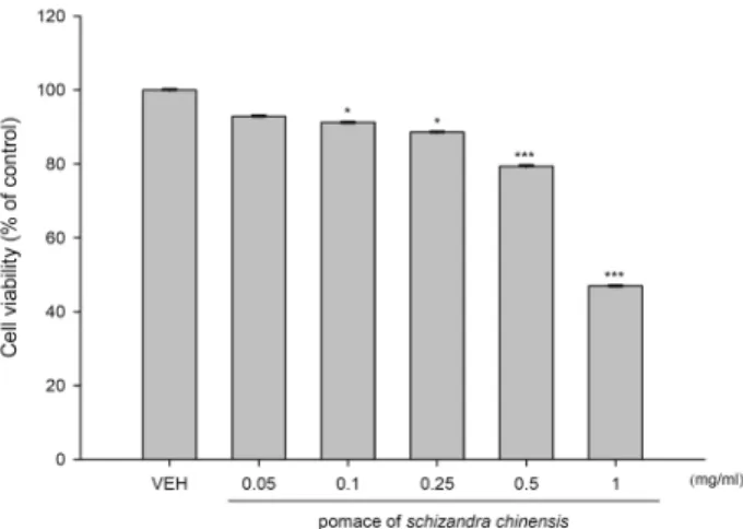
Fig. 1. Effects of ethanol extracts of pomace of Schizandra chinesis (PSC) on cell viabilities in HCT116 cells. HCT116 cells were plated at 1×10⁵ cells/well in a 96-well plate and incubated with five different concentrations of PSC for 24 hr. And then, cell viability was measured using MTT solution. *p<0.05, ***p<0.001.
Fig. 2. Up-regulation of pro-apoptotic genes by pomace of Schi- zandra chinesis (PSC). HCT116 cells were treated with 0.5 mg/ml PSC or 50 μM resveratrol (RES, positive control) for 24 hr. And then, total RNAs were prepared from each sample-treated cells. Total RNA was used for RT-PCR with ATF3, NAG-1 and p21 gene specific primers.
고, 2차 항체를 1시간 동안 반응시켰다. 그 후 enhanced chem- iluminescence kit (ECL
TM, Amersham, Pittsburgh, PA, USA) 을 이용하여 반응시킨 후 C-DIGIT (LI-COR, Lincoln, NE, USA)를 이용하여 확인하였다.
siRNA transfection
대장암 세포주인 HCT116를 약 24시간 동안 배양한 후 se- rum free DMEM에 Lipofectamine 2000 reagent (Invitrogen, Carlsbad, CA, USA) 10 μl와 10 pmole NAG-1 siRNA (Bioneer, Daejeon, Korea) 6 μl를 혼합하여 transfection시켰다. 대조구 는 β-gal siRNA (Bioneer)를 사용하였다. 4시간 후 FBS가 첨가 된 DMEM 배지로 교환시킨 뒤 24시간 동안 추가적인 배양을 시켜준 후 시료를 조건에 따라 처리하였다. 24시간 transfec- tion 후, 수확한 단백질은 Western blot analysis에 의해 단백질 발현을 확인하였다.
통계처리
모든 실험은 최소한 3회 이상 반복실험을 실시 하였으며 실험결과는 평균±표준편차로 나타내었고, 각 실험결과의 유 의성 검토는 시료가 포함되지 않은 대조구와 비교하여 stu- dent’s t-test에 의해 판정하였으며 p<0.05일 때 유의성이 있다 고 판단하였다.
결과 및 고찰
오미자 박 추출물이 세포성장에 미치는 영향
오미자 박 에탄올 추출물(PSC)이 대장암 세포주인 HCT116 세포 증식에 미치는 영향을 확인하기 위해 cell viability assay 를 수행하였다. 즉, 오미자 박 추출물을 0.05, 0.1, 0.25, 0.5, 1 mg/ml 농도로 24시간 동안 처리한 후, 재료 및 방법에 기술한 방법으로 MTT 용액을 이용하여 세포 생존율을 측정하였다.
그 결과, Fig. 1에서 보는 바와 같이 처리한 PSC의 농도가 증가 됨에 따라 세포 생존율이 감소하는 것으로 확인 되었다. 이 결과를 근거로 추후 실험을 위한 농도는 0.5 mg/ml의 농도로 정하여 진행하였다.
오미자 박에 의한 ATF-3, NAG-1, p21 유전자 발현 증가 대장암 세포주인 HCT116 세포주에서 오미자 박 에탄올 추 출물(PSC) 처리에 따른 ATF-3, NAG-1, p21과 같은 pro-apop- totic 유전자의 발현 변화에 미치는 영향을 RT-PCR로 확인하 였다. 즉, 오미자 박 에탄올 추출물을 0.5 mg/ml 농도로 24시 간 동안 처리하고, vehicle로는 DMSO를 처리하였다. 한편, 양 성대조구로서 포도껍질에 풍부한 파이토케미칼인 resveratrol 을 50 μM 농도로 처리하였다. Resveratrol은 대장암 세포주 HCT116 세포에서 세포생존율 감소는 물론 ATF3, NAG-1, p21 을 포함한 다양한 pro-apoptotic 유전자의 발현을 증가시키는
것으로 보고되었다[2, 4, 13]. Fig. 2에서 보는 바와 같이 양성대
조구로 사용한 resveratrol과 동일하게, 0.5 mg/ml PSC를 처
리한 경우에도 ATF-3, NAG-1 그리고 p21 유전자의 발현이
증가되었다. ATF3 (Activating transcription factor 3) 유전자
는 ATF/CREB subfamily의 하나로서, 다양한 생리활성 물질
에 의해 발현이 증가되며 세포사멸을 유도하는 것으로 보고되
었다[11, 21]. 또한 ATF3의 발현은 대장암 세포에서 항암 활성
과 관련되어 암 예방을 위한 분자적 지표로 사용된다고 보고
되었다[7]. NAG-1 (Non-steroidal anti-inflammatory gene-1)
유전자는 TGF-β superfamily의 하나로서 암 성장에 중요한
역할을 한다고 알려져 있다[9, 22]. 또한 NAG-1의 발현은 대장
암, 간암 등 다양한 세포주에서 세포사멸 및 세포 성장억제와
관련이 있다고 보고되었다[17, 18].
Fig. 3. Effects of schizandrin and resveratrol on cell viabilities in HCT116 cells. HCT116 cells were plated at 1×10⁵ cells/well in a 96-well plate and incubated with three different concentrations of schizandrin and 50 μM re- sveratrol (positive control) for 24 hr. And then, cell via- bility was measured using MTT solution. *p<0.05
Fig. 4. Up-regulation of pro-apoptotic genes by schizandrin tre- atement. HCT116 cells were treated with three different concentrations (0.1, 0.25, and 0.5 mM) of schizandrin or 50 μM resveratrol (RES, positive control) for 24 hr. And then, total RNAs were prepared from each sample-treat- ed cells. Total RNA was used for RT-PCR with ATF3, NAG-1 and p21 gene specific primers.
A
B
Fig. 5. Induction of apoptosis by schizandrin treatment and re- covery of apoptosis by NAG-1 siRNA transfection. (A) HCT116 cells were incubated with three different con- centrations (0.1, 0.25, and 0.5 mM) of schizandrin and treated cells were collected for Western blot analysis us- ing PARP, cleaved PARP, and ACTIN antibodies. (B) Either
β
-gal siRNA or NAG-1 siRNA was transfected into HCT116 cells. After 24 hr transfection, cells were treated with DMSO (vehicle) or 0.5 mM schizandrin. After 24 hr treatment, cells were collected for Western blot analy- sis using PARP, cleaved PARP and ACTIN antibodies.오미자 유래 생리활성 물질인 schizandrin이 세포생존율 에 미치는 영향
오미자 유래 생리활성 물질인 schizandrin (SZ)이 대장암 세포 HCT116 성장에 미치는 영향을 연구하기 위하여 cell via- bility assay를 수행하였다. 즉, SZ를 0.1, 0.25, 0.5 mM의 농도 로 HCT116 세포에 24시간 동안 처리하고, 양성 대조구로 re- sveratrol 50 μM을 처리한 후 cell viability assay를 수행하였 다. 그 결과, Fig. 3에서 보는 바와 같이, schizandrin의 처리 농도가 증가됨에 따라 세포 생존율이 감소됨을 확인하였다.
추가 연구를 위한 schizandrin 처리농도는 0.1, 0.25, 0.5 mM의 농도로 정하여 유전자 및 단백질 발현을 확인하였다.
Schiznadrin에 의한 NAG-1, ATF-3, p21 유전자 발현 증가
오미자 유래 생리활성 물질인 schizandrin (SZ)에 의해 ATF3, NAG-1, p21과 같은 pro-apoptotic 유전자의 발현 변화 를 확인하기 위하여 RT-PCR을 이용하였다. 즉, 0.1, 0.25, 0.5 mM의 농도로 SZ를 HCT116 세포를 24시간 동안 처리한 후 유전자의 발현을 확인하였으며, 양성 대조구로는 50 μM re- sveratrol을 처리하였다. 그 결과, 0.25 mM SZ을 처리한 군부 터 ATF-3, NAG-1, p21 유전자의 발현이 증가되는 것을 확인하 였다(Fig. 4). 이러한 결과는 오미자 유래의 순수물질 중의 하 나인 schizandrin이 항암 유전자의 발현을 증가시킬 수 있음을 보여주는 것이다.
Schizandrin에 의한 apoptosis 유도 및 NAG-1 siRNA transfection에 의한 세포사멸의 회복
Schizandrin (SZ)에 의한 세포생존율 감소의 원인이 apop-
tosis와 관련이 있는지 확인하기 위하여, apoptosis의 지표 중 하나인 Poly (ADP-ribose) polymerase (PARP)의 cleavage를 확인하였다. Apoptosis 유도시 PARP 단백질은 89 kDa으로 절단되는 특성을 가지고 있다. 즉, SZ를 0.1, 0.25, 0.5 mM의 농도로 HCT116 세포에 24시간 동안 처리한 후, 절단된 PARP 단백질을 Western blot으로 분석하였다. 그 결과, Fig. 5A에서 보는 바와 같이, 0.1 mM SZ 처리군부터 PARP단백질의 cleav- age를 확인할 수 있었다. 이러한 SZ에 의한 apoptosis의 유도 가 SZ에 의해 발현이 유도되는 NAG-1의 발현 증가와의 관련 성을 확인하기 위해 NAG-1 siRNA를 이용해 증명하였다. 즉, 대조구인 β-gal siRNA (10 pmole)와 NAG-1 siRNA (10 pmole) 를 각각 transfection 한 후, DMSO와 0.5 mM의 SZ를 처리하 였다. 그 결과, Fig. 5B에서 보는 바와 같이, NAG-1 siRNA를 transfection 한 경우 대조구에 비해 NAG-1의 발현 감소는 물 론 cleaved PARP가 감소됨을 확인하였다. 이러한 결과는 SZ 에 의해 유도되는 apoptosis 현상이 SZ에 의해 증가되는 NAG- 1의 발현과 직접적인 관련이 있음을 시사한다.
종합적으로, 본 연구의 결과는 대장암 세포주 HCT116에서 오미자 박 에탄올 추출물(PSC)과 오미자 유래의 순수물질인 schizandrin에 의한 항암 활성과 작용기전을 제시한다.
감사의 글
본 연구는 2016년도 산업통상자원부 바이오테라피산업기 반구축사업(과제번호 N0001805)에 의해 수행되었으며, 이에 감사드립니다.
References
1. Cho, Y. J., Ju, I. S., Kim, B. C., Lee, W. S., Kim, M. J., Lee, B. G., An, B. J., Kim, J. H. and Kwon, O. J. 2007. Biological activity of Omija (Schizandra chinensis Baillon) extracts. J.
Kor. Soc. Appl. Biol. Chem. 50, 198-203.
2. Frank, G., Bottone, Jr. and Brenda, A. M. 2011. The dietary compounds resveratrol and genistein induce activating tran- scription factor 3 while suppressing inhibitor of DNA bind- ing/differentiation-1. J. Med. Food 14, 584-593
3. Guon, T. G. and Chung, H. S. 2014. Effects of Nelumbo nuci- fera root extract on proliferation and apoptosis in HT-29 hu- man colon cancer cells. J. East Asian Soc. Dietary Life. 24, 20-27.
4. Hu, X., Wang, J., Xia, Y, Simayi, M, Ikramullah, S., He, Y., Cui, S., Li, S. and Wushouer, Q. 2016. Resveratrol induces cell cycle arrest and apoptosis in human eosinophils from asthmatic individuals. Mol. Med. Rep. 14, 5231-5236.
5. Kim, B. H., Cho, D. U., Han, K. S. and Bae, Y. L. 2011.
Efficiency analysis of Schisandra tea using image & acoustic signal processing. Journal of Korea Academia Industrial Coop- eration Society 12, 2975-2981.
6. Kim, J. S. and Choi, S. 2008. Physicochemical properties and
antioxidative activities of Omija (Schizandra chinensis Bailon). Kor. J. Food Nutr. 21, 35-42.
7. Kim, K. J., Lee, J., Park, Y. and Lee, S. H. 2015. ATF3 medi- ates anti-cancer activity of trans-10, cis-12-conjugated lino- leic acid in human colon cancer cells. Biomol. Ther. (Seoul).
23, 134-140.
8. Kim, M. S., Sung, H. J., Park, J. Y. and Sohn, H. Y. 2017.
Evaluation of anti-oxidant, anti-microbial and anti-thrombo- sis activities of fruit, seed and pomace of Schizandra chi- nensis Baillon. J. Life Sci. 27, 131-138.
9. Lim, J. H., Park, J. W., Min, D. S., Chang, J. S., Lee, Y. H., Park, Y. B., Choi, K. S. and Kwon, T. K. 2007. NAG-1 up-reg- ulation mediated by EGR-1 and p53 is critical for querce- tin-induced apoptosis in HCT116 colon carcinoma cells.
Apoptosis 12, 411-421.
10. Min, H., Park, E., Hong, J., Kang, Y., Kim, S., Chung, H., Woo, E., Hung, T. M., Youn, U. J. and Kim, Y. S. 2008.
Antiproliferative effects of dibenzocyclooctadiene lignans isolated from Schisandra chinensis in human cancer cells.
Bioorg. Med. Chem. Lett. 18, 523-526.
11. Park, G. H., Song, H. M. and Jeong, J. B. 2017. Kahweol from coffee induces apoptosis by upregulating activating transcription factor 3 in human colorectal cancer cells.
Biomol. Ther. (Seoul). 25, 337-343.
12. Saul, R. B., Javier, F., Ignacio, G. R., Claudio, J. V. and Felipe, L. 2017. New Insights toward colorectal cancer chemo- therapy using natural bioactive compounds. Front. Pharmacol.
8, 1-22.
13. Seung, J. B., Leigh, C. W. and Thomas, E. E. 2002. Resveratrol enhances the expression of non-steroidal anti-inflammatory drug-activated gene (NAG-1) by increasing the expression of p53. Carcinogenesis 23, 425-434.
14. Shin, E. J., Chung, S. W. and Hwang, J. T., 2017. Effect of γ-oryzanol on proliferation and apoptosis of AGS human gastric carcinoma cell. KSBBJ 32, 83-89.
15. Suh, W. S., Park, S. Y., Min, B. S., Kim, S. H., Song, J. H.
and Shim, S. H. 2014. The anti-proliferative effects of com- pounds isolated from Schisandra chinensis. Kor. J. Food Sci.
Technol. 46, 665-670.
16. Sun, S. Y., Hail, N. Jr. and Lotan, R. 2004. Apoptosis as a novel target for cancer chemoprevention. J. Natl. Cancer Inst.
96, 662-672.
17. Wilson, L. C., Baek, S. J., Call, A. and Eling, T. E. 2003.
Nonsteroidal anti‐inflammatory drug‐activated gene (NAG‐1) is induced by genistein through the expression of p53 in colorectal cancer cells. Int. J. Cancer 105, 747-753.
18. Yang, M. H., Kim, J., Khan, I. A., Walker, L. A. and Khan, S. I. 2014. Nonsteroidal anti-inflammatory drug activated gene-1 (NAG-1) modulators from natural products as an- ti-cancer agents. Life Sci. 100, 75-84.
19. Yi, H., Chen, Y., Liu, J., Zhang, J., Guo, W., Xiao, W. and Yao, Y. 2016. Extraction and separation of active Ingredients in Schisandra chinensis (Turcz.) Baill and the study of their antifungal effects. PLoS One 11, e0154731.
20. Yoon, Y. S., Yu, C. S., Jung, S. H., Choi, P. W., Han, K.
R., Kim, H. C. and Kim, J. C. 2007. Characteristics of color-
초록:오미자 박 추출물 및 schizandrin에 의한 암세포 항성장 및 세포사멸 활성
김현지
1․서유미
1․이은주
1․정정욱
1․성화정
2․손호용
2․박종이
3․김종식
1*
(1국립안동대학교 생명과학과, 2국립안동대학교 식품영양학과, 3경북바이오산업 연구원)
오미자는 다양한 인간 질환을 치료하기 위한 한약재로 사용되어 왔으며, schizandrin과 gomisin A와 같은 다양 한 생리활성물질을 함유하고 있는 것으로 알려져 있다. 본 연구에서는 오미자 박으로부터 에탄올 추출물(PSC)을 제조하고, 이들이 대장암 세포인 HCT116의 세포 생존율에 미치는 영향과 ATF3, NAG-1, p21와 같은 pro-apop- totic 유전자의 발현 변화에 미치는 영향을 연구하였다. 오미자 박 에탄올 추출물의 처리는 농도 의존적으로 암 세포생존율을 감소시켰으며, 세 가지 pro-apoptotic 유전자의 발현을 모두 증가시켰다. 또한, 오미자 유래의 순수 물질인 schizandrin도 세포 생존율을 농도의존적으로 감소시켰으며, ATF3, NAG-1, p21 유전자의 발현을 증가시켰 다. 게다가, schizandrin을 처리한 세포에서 PARP cleavage를 확인함으로써 apoptosis가 일어남을 확인하였다. 이 러한 PARP cleavage는 NAG-1 siRNA의 transfection에 의해서 회복됨을 확인하였다. 이러한 결과는 schizandrin 에 의해 유도되는 apoptosis와 NAG-1의 발현증가가 직접적인 관련이 있음을 나타낸다. 종합적으로, 본 연구결과 는 오미자 박 추출물과 schizandrin에 의해 매개되는 항암 활성과 암세포 사멸현상을 이해하는데 도움을 줄 것으 로 사료된다.
ectal cancer detected at the health promotion center. J. Kor.
Soc. Coloproctol. 23, 321-326.
21. Zhang, C., Gao, C., Kawauchi, J., Hashimoto, Y., Tsuchida N. and Kitajima, S. 2002. Transcriptional activation of the human stress-inducible transcriptional repressor ATF3 gene promoter by p53. Biochem. Biophys. Res. Commun. 297, 1302- 1310.
22. Zhang, Z., Wu, L., Wang, J., Li, G., Feng, D., Zhang, B.,
Li, L., Yang, J., Ma, L. and Qin, H. 2014. Opposing effects of PI3K/Akt and Smad-dependent signaling pathways in NAG-1-induced glioblastoma cell apoptosis. PloS One 9, e96283.
23. Zhu, H., Zhang, L., Wang, G., He, Z., Zhao, Y., Xu, Y., Gao, Y. and Zhang, L. 2016. Sedative and hypnotic effects of su- percritical carbon dioxide fluid extraction from Schisandra chinensis in mice. J. Food Drug Anal. 24, 831-838.