The Neurotransmitter Pathway of Itching
Jeong Won Jo and Chi-Yeon Kim*
Department of Dermatology, College of Medicine, Gyeongsang National University, 79 Gangnam-ro, Jinju 52727, Korea Received May 18, 2017 /Revised May 26, 2017 /Accepted May 29, 2017
It was common that the classification of itching was classified into four categories according to the neurophysiological mechanisms of pruritoceptive itching, neuropathic itching, neurogenic itching and psychogenic itching. Recently it was classified by clinical criteria. The neurotransmission pathway of itch is divided into histamine-dependent pathway and histamine-independent pathway. Different re- ceptors and neuropeptides act on each itch mediator. Itch mediators such as histamine, BAM8-22, and chloroquine are transmitted through the histamine-dependent pathway. Cowhage spicule, protease, and TSLP (Thymic stromal lymphopoietin) have been reported to be related to the histamine-in- dependent pathway. These itch mediators, receptors, and neuropeptides are the targets of treatment for itching. Although itching and pain are typical noxious stimuli, and in the past, it was argued that two senses were transmitted through one noxious stimulus receptor. It has recently been shown that itching and pain have an independent neurotransmitter system and both neuronal systems inhibit each other. In addition, the mutual antagonism between itching and pain is explained by various mechanisms. Recently, many new mediators and receptors are being studied. The studies on histamine 4 receptor (H4 receptor) have been actively conducted. And the H4 receptors are expressed in immune cells such as T cells. The therapeutic agent for blocking the H4 receptor can inhibit the inflammatory reaction itself, which is important for the itching and chronicization. Understanding the underlying mechanisms of itching and studying new itch mediators will lead to the development of effective therapies, and this is what I think the itching study will go on.
Key words : Itching, neurophysiological mechanisms, neurotransmission pathway
*Corresponding author
*Tel : +82-55-750-8184, Fax : +82-55-758-8106
*E-mail : [email protected]
This is an Open-Access article distributed under the terms of the Creative Commons Attribution Non-Commercial License (http://creativecommons.org/licenses/by-nc/3.0) which permits unrestricted non-commercial use, distribution, and reproduction in any medium, provided the original work is properly cited.
Journal of Life Science 2017 Vol. 27. No. 5. 600~610 DOI : https://doi.org/10.5352/JLS.2017.27.5.600
서 론
가려움증은 건강에 심각한 위해는 아니지만 가렵다는 증상 만으로 불편감이 생기며 일상의 생활에 지장을 준다. 가려움 증, 소양증은 임상에서 동반되는 가장 흔한 증상으로 증상 그 자체 만으로도 질환으로 평가되며 림프종과 같은 위해성 질환 의 전구 증상으로 발현될 수 있으며 일부에서는 치료에 잘 반응하지 않기 때문에 개인의 삶의 질을 저하시킬 수 있어 적절한 치료가 중요하다. 최근 신경생리학적 연구의 발전으로 가려움증의 신경전달 경로에 대한 새로운 연구가 활발하게 진행하고 있으며 이를 기반으로 기존에 알려지지 않았던 새로 운 가려움증 매개체의 발견과 이들을 대상으로 한 새로운 치 료 약제 개발이 이루어지고 있다. 본 종설에서는 그간 국내외 에서 발표된 연구 문헌을 수집하여 (1) 가려움증의 분류, (2) 가려움증의 신경생리학적 신경전달 경로 (3) 가려움증 경로와
통증 경로의 공통점과 차이점 (4) 신경펩티드와 수용체 활용을 통한 치료적 접근에 대해 기술하고자 한다.
본 론
본 종설 논문에 수록된 자료들은 전문 학술 DB 검색 시스템 을 이용하여 영어 논문 데이터베이스를 검색하기 위해 Pubmed, google scholar를 통하여 ‘itch’, ‘pruritus’, ‘neuro- physiology’, ‘nerve fiber’, ‘pruritogen’를 키워드로 하여 검색 하였고, 이중에서 1990년 이후 등재된 것을 중심으로 검토하 여 자료를 분석하였다. 국내 문헌은 국내학회지(KISS)와 한국 과학기술정보연구원(KISTI)에서 제공하는 문헌에서 같은 방 식으로 검토하였다.
가려움증의 신경병태생리
가려움증은 긁고 싶은 충동을 일으키는 피부의 유쾌하지 못한 감각으로 정의할 수 있으며[7] 질병의 가장 흔한 자각 증상이다. 가려움증은 두드러기, 아토피피부염, 접촉피부염 등의 특정 피부 질환 때문에 발생할 수도 있고 임상적으로 피부 질환 없이 전반적으로 발생할 수도 있다[55]. 그 외 내과 적 관련 질환이나 정신과적 원인에 의해서도 발생할 수 있다.
가려움증은 건강에 심각한 위해는 아니지만 치료에 잘 반응하 지 않아 개인의 삶의 질을 저하시킬 수 있기에 적절한 치료와
- Review -
격려를 제시하는 것이 중요하다. 가려움증은 단순히 따끔거리 거나 스멀거림 등으로 나타나기도 하며, 주관적인 성격이 강 하여 같은 사람에게서도 동일한 자극임에도 상황에 따라 다양 한 정도의 가려움증을 일으킬 수 있다. 또한 환자 개인의 감수 성이나 스트레스 등의 심리상태와도 연관이 크기 때문에 객관 적 평가에서도 어려움이 있다[60].
최근 신경생리학적 연구의 발전은 가려움증의 신경전달 경로에 있어서 신경병태생리(neuropathophysiology)를 규명 할 수 있는 기초를 마련하였다. 피부에는 3가지의 구심성 (afferent) 신경 섬유가 있으며 그 중 직경이 작고 탈수초화 되어있는 특이적인 C 신경섬유에 의해 가려움증 자극이 중추 신경계로 전달된다. 이러한 과정 중 다양한 가려움증의 매개 체(pruritogen, 가려움증 매개체)가 밝혀져 있으며, 이들이 C 신경섬유 말단의 수용체(pruriceptor, 가려움증 수용체)를 활 성화 시킴으로써 가려움증은 신경학적 신호로 척수를 거쳐 최종적으로 대뇌피질로 전달된다[32].
가려움증의 분류
가려움증의 분류는 Twycross 등에 의해 신경생리학적 기전 에 따라 4가지의 카테고리로 제시되었다. 수용체성 가려움증 (Pruritoceptive itch)은 가장 흔하게 접하게 되는 가려움증으 로, 옴감염증이나 두드러기 같이 수용체가 다수 분포하는 피 부의 염증상태 혹은 피부장벽의 손상에서 기원한다. 신경병증 가려움증(Neuropathic itch)은 뇌종양처럼 신경해부학적 병인 의 결과로 발생하는 가려움증이다. 신경성 가려움증(Neuro- genic itch)은 일차적 피부병변이나 신경 손상 없이 나타날 수 있으며, 중추신경계에 작용하는 매개체를 통한 신경화학적 활 성의 결과로 발생하는 가려움이다. 담즙 정체 간질환에서 opioid peptide의 활성에 의한 가려움증의 발생이 그 예가 되 겠다. 심인성 가려움증(Psychogenic itch)은 기생충공포증의 망상 상태에서처럼 정신 질환에 따른 이차적인 가려움증이다.
실제 신체정신학적 반응으로 가려움증의 증상이 발현되나 그 질환의 양상이 일정하지 않아 검사상에서 객관적인 차이가 나타나지 않는 경우가 있다. 가려움증은 질환과의 연관성에서 중요한 증상으로 인식되지만 그 기전과 분류가 다양하여 여러 연구 분야에서 치료적 접근을 시도하고 있으며 따라서 이에 따른 새로운 분류기준의 변화가 시도되고 있다[62].
2003년 American academy of dermatology annual meet- ing itch symposium에서는 가려움증을 임상적으로 분류한 도 식을 발표하였는데, 이에 따르면 피부과적 가려움증(Derma- tologic itch), 전신적 가려움증(Systemic itch), 신경성/신경병 증 가려움증(Neurogenic/neuropathic itch), 심인성 가려움증 (Psychogenic itch), 기타 가려움증(Other), 혼합 가려움증 (Mixed)으로 분류하기도 한다[7].
가려움증의 신경전달 경로
가려움증 자극을 피부로부터 중추신경계로 전달하는 경로 는 히스타민-의존 경로와 히스타민-비의존 경로 2가지로 나뉜 다[28].
히스타민 가려움증 수용체는 탈수초화된 기계자극-비민감 성 C 신경섬유 구심성 뉴런(Demyelinatedmechano-insensi- tive C fiber afferent neuron)이다. 이는 1997년 Schmelz and handwerker가 밝힌 것으로 CMi-fibers (C-fibers, mecha- no-insensitive)라는 용어로 불린다[47]. 이 신경섬유는 열 자극 에 의해서도 반응하기 때문에 열이 가려움증을 악화시키고 얼음마사지 등의 체온을 낮추는 행동이 가려움증을 호전시키 는 증상을 이를 통해 설명할 수 있다[44]. 이 기계자극-비민감 성 C 신경섬유가 병적인 가려움증을 전달하는 주요 통로이며 이 신경섬유의 자유신경종말은 표피, 유두진피, 피부부속기 주변에 분포하여 많은 외인성 또는 내인성 가려움증 매개체의 자극에 반응한다[45]. 예를 들면 가려움증 매개체 중 히스타민 은 TRPV1 (Transient receptor potential cation channel, sub- family V, member 1) 이온채널의 개방을 유도하는 히스타민 수용체 1 (Histamine receptor1, H1)을 통해 뉴런을 활성화시 키고 BAM8–22과 chloroquine은 TRPA1 (Transient receptor potential cation channel, subfamily A, member 1)채널의 개 방을 유도하는 MrgprC11 (Mas-related G protein–coupled receptor C11)와 MgprA3 (Mas-related G protein–coupled receptor A3)를 통해 뉴런을 활성화시킨다(Fig. 1)[15]. 반면 히 스타민에 비의존적인 경로도 존재하는데 이러한 경로에 의한 가려움증은 항히스타민제 치료에 반응이 없으며 최근 연구는 이러한 히스타민-비의존적 경로에 의해 유발되는 가려움증의 메커니즘을 설명하는 데 초점을 두고 있다. 예를 들어 cowh- age spicule은 기계감각 민감성 C 신경섬유와 Aδ 신경섬유에 의해 조절된다는 연구 결과가 있으며[20, 31] Mas-related G protein–coupled receptor (Mrgpr) family는 히스타민-비의 존적 가려움증 수용체의 주요한 인자로 부각되고 있다[26, 27].
또 Steinhoff. M 등의 연구에서 만성적인 가려움증을 호소하는
환자들의 피부에서 단백분해효소(protease) 농도의 증가를 보
이는 경우가 관찰되었는데, 이 단백분해효소는 PAR2 (Pro-
tease activated receptor 2)를 포함한 단백분해효소 수용체를
활성화시키며 PAR2는 급성 및 만성 가려움증을 유발한다고
한다[51]. 마지막으로 TSLP (Thymic stromal lymphopoietin)
또한 아토피 피부염과 연관된 가려움증 매개체로 히스타민-비
의존적 경로에 의해 가려움증이 유발된다[59]. 이렇게 서로 다
른 가려움증 매개체와 수용체에 의해 활성화된 자극은 뉴런을
따라 척수 후각의 lamina I으로 전달되는데, 이 때 각 경로
별로 서로 다른 신경펩티드가 작용한다. 히스타민-의존 경로
는 일차적으로 glutamate, GRP (Gastrin-releasing peptide)가
그 역할을 하며[1, 53], 히스타민-비의존적 경로는 substance
P, GRP, glutamate 등에 의해 매개된다[1].
Fig. 1. A schematic illustrating the type of cells involved in the detection of various itch stimuli.
Fig. 2. Itch pathways in spinal cord.
Lamina I을 좀 더 자세히 살펴보면, Lamina I에는 다양한 신경펩티드의 수용체가 존재해 가려움증 신호와 통증 신호 등 다양한 신호들이 모여 조절되는 곳으로 특히 히스타민-의 존적 경로와 히스타민-비의존적 경로를 구별하는 역할을 한다 [43]. 최근 functional PET-CT를 통해 이러한 가려움증의 상척 수성 과정(supraspinal processing)이 연구된 바 있다[10]. 또 한 생쥐의 척수 후각에서 GRPR (GRP receptor)을 발현시키는 신경세포를 선택적으로 절제한 후에 히스타민, 세로토닌, PAR-2 작용제 같은 다양한 가려움증 매개체를 가하여 가려움 증을 유발했을 때 긁는 행동이 줄어드는 것으로 가려움증의
전달에 제한이 있다는 결과를 알 수 있었다. 특히 이 연구에서
통증에 대한 반응은 변화가 없었고 이 사실은 아래에서 언급
할 가려움증과 통증의 차이에 대한 근거가 되었다[53, 54]. 그
외 BNP (B-type natriuretic peptide) 또한 Lamina I 내에서
가려움증 신호를 전달하는 데 중요한 역할을 가짐이 밝혀졌다
[52]. 이렇게 Lamina I에서 구별된 신호는 반대측 척수시상
경로(contralateral spinothalamic tract)로 연결되어 시상내배
핵(nucleus medialis dorsalis)의 ventrocaudal part를 거쳐 전
측 대상회(anterior cingulate)와 뇌섬엽부 피질(dorsal insular
cortex)에 도달하고 이는 운동 대뇌 피질(motor cortex)을 자극
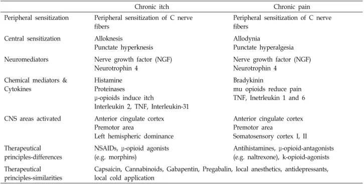
Table 1. Comparison of characteristics of chronic itch and chronic pain
Chronic itch Chronic pain
Peripheral sensitization Peripheral sensitization of C nerve fibers
Peripheral sensitization of C nerve fibers
Central sensitization Alloknesis
Punctate hyperknesis
Allodynia
Punctate hyperalgesia Neuromediators Nerve growth factor (NGF)
Neurotrophin 4
Nerve growth factor (NGF) Neurotrophin 4
Chemical mediators &
Cytokines
Histamine Proteinases
μ-opioids induce itch
Interleukin 2, TNF, Interleukin-31
Bradykinin
mu opioids reduce pain TNF, Inetrleukin 1 and 6
CNS areas activated Anterior cingulate cortex Premotor area
Left hemispheric dominance
Anterior cingulate cortex Premotor area
Somatosensory cortex I, II Therapeutical
principles-differences
NSAIDs, μ-opioid agonists (e.g. morphins)
Antihistamines, μ-opioid-antagonists (e.g. naltrexone), k-opioid-agonists Therapeutical
principles-similarities
Capsaicin, Cannabinoids, Gabapentin, Pregabalin, local anesthetics, antidepressants, local cold application
하여 긁기 행동을 유발한다(Fig. 2) [4]. 척수시상경로에서도 히스타민-의존적, 히스타민-비의존적 경로가 여전히 구분되어 전달됨을 여러 연구를 통해서 확인할 수 있다[14]. 척수시상경 로 뉴런의 cowhage와 히스타민에 대한 전기생리학적 연구 (electrophysiological itch studies)에서 이 둘은 각자 서로 다 른 뉴런의 반응을 이끌어냄을 알 수 있다[11-13]. 척수시상경 로뿐 아니라 대뇌의 영상학적 연구에서도 cowhage와 히스타 민이 대뇌의 서로 다른 부위를 활성화시킨다는 것을 확인할 수 있다[34].
가려움증과 통증
대표적인 유해자극(nociceptive stimuli)으로서의 가려움증 과 통증은 그 유사성과 차이점에 관련하여 관심의 대상이었다 (Table 1) [48, 61]. 과거 한 때에는 가려움증과 통증은 하나의 감각으로 유해자극수용체(nociceptive)를 통한 자극의 강도가 강할 때는 통증으로, 약할 때는 가려움증으로 인식한다는 주 장도 있었다. 그러나 밝혀진 바에 따르면 가려움증과 통증은 독립적인 신경전달체계를 가지고 있고 두 신경체계는 서로 억제작용을 한다[4].
가려움증과 통증이 독립적인 신경전달체계를 가지고 있으 며 피부의 감각신경은 이 두 자극을 구분하여 인지한다는 측 면에서 보면, 피부의 감각 신경에 존재하는 가려움증 수용체 는 다른 종류의 자극에 비해 가려움증 매개체에 의한 자극에 만 상당히 낮은 역치를 갖는다. 가려움증의 선택적 민감성 이 론은 최근 특정 자극이 어떻게 통증이 아닌 가려움증을 유발 할 수 있는지에 대해 설명할 수 있는 이론으로 널리 받아들여 지고 있다[35]. 최근 연구결과에서는 가려움증에 대한 뉴런의
선택적 소집단(selective subpopulation)의 존재를 제시한다.
GRPR과 NPRA (Natriuretic polypeptide receptor subtype A) 를 발현하는 척수 뉴런이 가려움증 행동에 영향을 주며 통증 행동에는 영향이 없음이 밝혀졌고[29, 53, 54], Bhlhb5 (Basic helix-loop-helix B5) 전사인자를 발현하는 척수 후각의 억제성 뉴런은 가려움증 신호는 억제하지만 통증 신호는 억제하지 못한다는 것이 밝혀진 바 있다[41]. MrgprA3 양성 뉴런이 없 는 동물모델에서 가려움증 행동은 감소되었으나 통증에는 정 상 민감도를 보였으며[18] 가려움증-민감 뉴런에 대한 약리학 적 억제(pharmacological silencing)가 통증행동에는 효과가 없었다는 보고도 있었다[40]. 이러한 연구결과는 말초에서 가 려움증과 통증 자극의 구분은 서로 다른 뉴런에 의해 발생하 고 가려움증 경로와 통증 경로는 구분되어 있다는 것을 의미 한다[15].
한편 가려움증과 통증의 신경체계가 서로 억제작용을 한다
는 것에 대한 근거로 통증조절제 기전에 대한 연구결과가 있
다. 통증조절을 위해 사용하는 아편계 약물이 전신 가려움증
을 일으킨다는 사실로부터 가려움증과 통증은 상호 길항작용
하며, 이를 근거로 가려움증 치료에 μ-아편유사체 수용체 작용
제(μ-opioid agonist)를 사용한다[22, 23]. 가려움증과 통증의
신경체계에 대하여 가려움증이 결국은 긁기를 유발하는 것을
고려한다면, 긁기라는 유해자극이 C와 Aδ 신경섬유를 자극함
으로써 피부에는 통증이 유발되고 동시에 가려움증 신호를
억제하여 더 이상 가려움증을 느끼지 않게 되는 것이 설명된
다. 이를 설명하는 기전은 다양하게 제시되고 있다[11]. 중뇌로
부터 내려오는 하행 억제 신호(descending inhibition)가 척수
레벨의 가려움증 신호를 억제한다는 기전[30], 가려움증을 인
Table 2. Itch mediator
Mediator Receptor Expression, sources Associated mechanism
Acetylcholine Nicotinic(nAchR), muscarinic(mAchR)
Cholinergic nerves, keratinocytes, phocytes, fibroblasts
Itch in atopic dermatitis mAchR3
Cspsaicin TRPV1 receptor TRPV afferent neurons,
keratinocytes, Dendritic cells, mast cells
Effective in some choronic itch condition
CGRP CGRP receptor Sensory nerve fibers,
central terminals
Transmission of itch in spinal cord
CRH, POMC CRH-R1, R2 Keratinocytes, mast cells Releases histamine from mast cells
Cytokines IL31 T cells, macrophages IL-2l IL031 increased in atopic
dermatitis, pruritic in the skin Endocannabinoids CBs(CB1, CB2) Nerve, immune cells, keratinocytes,
hair follicles
Reduces itch peripherallyl; CB1 suppresses histamine-induced pruritus
ETs ET-A, ET-B Endothelium, mast cells Direct pruritic(burning itch)
Histamine H1, H4 Sensory nerve fibers Typical pruritogen
Kallikrein Proteases
PARs Leukocyte, keratinocytes,
endothelial cells, mast cells, platelets
PAR-2 increases itch, upregulated in itchy dermatoses
Kinins Bradykinin receptors
(B1R and B2R)
Endothelial cells, immunocytes B2R antagonist reduces itch
Leukotrien B4 Leukotrien receptor
Sensory nerve fibers, keratinocytes Induced itch; mediates SP Induction of itch
NKA, SP Tachykinin receptor Sensory nerve fibers SP releases TNFα, histamine, LB4, and PGs from mast cells
NGF, BDNF, NT
TrkA(NGF), TrkB(NT-4, BNDF), TrkC(NT-3)
Keratinocytes, mast cells, fibroblasts, eosinophils
Involved in peripheral sensitization and inflammation
Opioids Ρ, κ, δ Neurons, keratinocytes μ Antagonist and κ agonist reduce
itch; μ agonist are pruritic PACAP and VIP VPAC receptor Autonomic and sensory nerve
fibers, lymphocytes, dermal endothelial cells, Merkel cells
Induce histamine release from mast cells
PGs Prostanoid receptor Sensory nerve fibers, keratinocytes PGE2 induce itch in humans(not mice); PG2 reduces
immunoglobulin E-mediated scratching in mice
Purines and Adenosine Triphosphate
P2X, P2Y Sensory nerve fibers, Schwann cells, immune cells
Serotonin SHT receptors Dorsal root ganglion Induce itch when injected
intradermally, enhanced by PGE2 식하는 척수 신경절 뉴런(pruriceptive dorsal root ganglia
neuron)의 중심 종말(central terminal)이 반대 신호(counter- stimuli)에 의해 활성화되는 역행 신호전달 경로(retrograde signaling)에 의해 조절된다는 기전, 척수에 존재하는 억제성 개재뉴런(inhibitory interneuron)이 유해자극의 인식을 활성
화하고 가려움증 신호를 억제한다는 기전[41] 등이 보고되고 있다.
피부 신경펩티드와 수용체
가려움증 발생에 중요한 역할을 하는 말초신경성 및 중추신
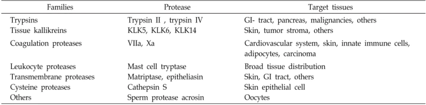
Table 3. Proteases that activate PAR2
Families Protease Target tissues
Trypsins Tissue kallikreins
Trypsin II , trypsin IV KLK5, KLK6, KLK14
GI- tract, pancreas, malignancies, others Skin, tumor stroma, others
Coagulation proteases VIIa, Xa Cardiovascular system, skin, innate immune cells, adipocytes, carcinoma
Leukocyte proteases Transmembrane proteases Cysteine proteases Others
Mast cell tryptase Matriptase, epitheliasin Cathepsin S
Sperm protease acrosin
Broad tissue distribution Skin, GI tract, others Skin epithelial cell Oocytes
경성 매개체들은 다양하게 밝혀져 있다. 대표적인 매개체는 히스타민, 단백분해효소(protease), 아편유사제(opioid), sub- stance P, Mrgpr family, CGRP (Calcitonin gene-related pep- tide) 등이다(Table 2) [28].
히스타민
히스타민은 생리활성 아민으로서 조직에 존재하는 히스타 민은 대부분 비만세포(mast cell)나 각질세포(keratinocyte) 내 에 과립으로 존재하다가 자극들에 의해 세포 밖으로 유리된다 [37]. 히스타민에 의해 각질세포(keratinocyte), 수지상세포 (dendritic cell), 비만세포의 일차성 구심성 뉴런(primary af- ferent neuron)에 발현되어 있는 TRPV1 (Transient receptor potential vanilloid 1) 이온 채널이 활성화되어 glutamate, GRP 같은 신경펩티드가 유리되어 가려움증 신호가 전달된다 [46]. 히스타민이 유두 진피(papillary dermis)에 존재하는 비 만세포에서 분비되면 두드러기(urticaria)가 발생하여 팽진 (wheal), 발적(flare), 가려움증이 유발되며, 반면 망상 진피 (reticular dermis) 또는 피하 조직(subcutaneous tissue)에서 분비되면 혈관부종(angioedema)이 발생하여 가려움증보다 오히려 통증이 유발된다[19]. 히스타민과 히스타민 수용체의 작용은 대체로 빠른 상호작용을 보이기 때문에 만성적인 가려 움증보다는 일시적 가려움증에 그 역할을 하는 것으로 알려져 있다. 이러한 측면에서 만성 가려움증의 대표 질환인 아토피 피부염에서 항히스타민제의 치료적 역할은 제한적으로 나타 나게 되는 것이다[60].
최근 H4 수용체(Histamine 4 receptor)에 대한 연구가 활발 하게 진행되고 있는데, H4 수용체는 비만세포뿐 아니라 염증 반응을 매개하는 T 림프구 표면에도 존재하며 염증반응에 관 여하는 것으로 알려져 있다[9]. 실제 Gutzmer 등의 연구에 따 르면 아토피환자의 도움 T세포(Th2 helper T cell)에 H4 수용 체가 발현되고 있으며 H4 수용체를 상향조절(up-regulation) 시켰을 때 가려움증 매개체인 IL-31 (Interleukin-31)의 분비가 증가되어 가려움증을 발현하게 된다[17]. 기존의 항히스타민 제는 H1 또는 H2 수용체 차단제로서 말초의 다양한 매개체 중 하나만을 차단하며 이러한 작용만으로는 다양한 매개체의 작용에 의해 발생하는 가려움증에 대한 효과가 제한적일 수
밖에 없었다. 그러나 H4 수용체를 차단하는 항히스타민제는 기전에 있어서 단순히 가려움증의 매개체가 그 수용체에 결합 하여 가려움증 신호를 만들어내는 과정을 차단하는 것이 아니 라, 가려움증의 만성화에 중요한 염증반응 자체를 억제할 수 있다는 점에서 그 유용성이 크다. 실제 생쥐 피부에서 아토피 피부염과 유사한 양상의 도움 T세포 매개성 염증에 H4 수용체 억제제를 투약한 후 가려움증 및 염증 반응의 현저한 억제가 확인되었다[9]. 따라서 히스타민이 가려움증의 주매개체이며 그와 관련된 수용체로 기존에 알려진 H1, H2 수용체 외에 H4 가 주요한 작용을 하며 가려움증이 염증세포의 작용과 관련됨 을 알 수 있다.
단백분해효소(Protease)
백혈구(leukocytes), 각질세포, 비만세포, 혈관내피세포 (endothelial cell)에서 분비되며 C 신경섬유에 발현되어 있는 PAR-2 (Protease activated receptor-2)를 활성화시켜 sub- stance P, CGRP를 분비하고 이로 인해 염증반응 및 가려움증 이 유발된다[28]. 내인성 단백분해효소(endogenous protease) 로는 trypsine, tryptase, cathepsin S, kallikrein-related pepti- dases (KLKs)가 있고 특히 KLK-5, KLK-14가 아토피피부염 환자에서 상향조절 되어있다(Table 3) [3, 5, 39, 50]. 외인성 단백분해효소(exogenous protease)로는 cowage, dust mites, cockroaches 등이 있다[21, 33, 38].
아편유사제(Opioid)
중추신경계 및 말초신경성 가려움증 발생에 모두 관여하는
강력한 신경전달물질로 척수레벨에서 μ-아편유사제 수용체
작용제(μ-opioid agonist)들은 가려움증을 발생시키며 κ-아편
유사제 수용체 작용제(κ-opioid agonist)들은 이를 억제한다
[57]. Morphine은 대표적인 μ-아편유사제 수용체 작용제로 히
스타민에 비의존적이며 항히스타민제에 내성을 가진다. 가려
움증을 유발하는 질환군에서 투석 관련 가려움증이나 담즙정
체성 가려움증은 일반적인 가려움 조절제에 반응하지 않으며
항히스타민제 치료효과가 적은 반면, μ-아편유사제 수용체의
길항제인 naloxone과 naltrexone 등에 치료적 효과가 나타나
는데 이는 가려움증의 또다른 매개체의 존재를 시사하는 소견
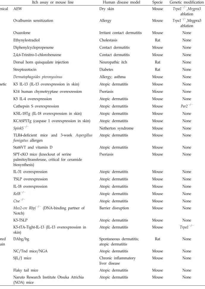
Table 4. Chronic itch models in rodents
Itch assay or mouse line Human disease model Specie Genetic modification
Chemical AEW Dry skin Mouse Trpa1
−/−,Mrgpra3
ablation
Ovalbumin sensitization Allergy Mouse Trpa1
−/−,Mrgpra3
ablation
Oxazolone Irritant contact dermatitis Mouse None
Ethynylestradiol Cholestasis Rat None
Diphenylcyclopropenone Contact dermatitis Mouse None
2,4,6-Trinitro-1-chlorobenzene Contact dermatitis Mouse None
Dorsal horn quisqualate injection Neuropathic itch Rat None
Streptozotocin Diabetes Rat None
Dermatophagoides pteronyssinus Allergy; asthma Mouse None
Genetic K5 IL-13 (IL-13 overexpression in skin) Atopic dermatitis Mouse None
K14 human chymotryptase overexression Psoriasis Mouse None
K5 IL-4 overexpression Atopic dermatitis Mouse None
Cathepsin S overexpression Atopic dermatitis Mouse Par2
−/−K5IL-18Tg (IL-18 overexpression in skin) Atopic dermatitis Mouse None
KCASP1Tg (caspase 1 overexpression in skin) Atopic dermatitis Mouse None
Spink5
−/−Netherton syndrome Mouse None
TLR4-deficient mice and 3-week Aspergillus fumigatus allergen
Atopic dermatitis Mouse None
Stat6VT and vitamin D Atopic dermatitis Mouse None
SPT-cKO mice (knockout of serine palmitoyltransferase, critical for ceramide biosynthesis)
Psoriasis Mouse None
IL-31 overexpression Atopic dermatitis Mouse None
TSLP overexpression Atopic dermatitis Mouse None
IL-18 overexpression Atopic dermatitis Mouse None
RelB
−/−Atopic dermatitis Mouse None
Ctse
−/−Atopic dermatitis Mouse None
Msx2-cre Rbpj
−/−(DNA-binding partner of Notch)
Barrier disruption Mouse None
K5-TSLP Atopic dermatitis Mouse None
K5-tTA-Tight-IL-13 (IL-13 overexpression in skin)
Atopic dermatitis Mouse Trpa1
−/−Inbred strain
DAbg/bg Spontaneous dermatitis;
atopic dermatitis
Rat None
NC/Tnd mice/NGA Atopic dermatitis Mouse None
SJL/J mice Chronic inflammatory
liver disease
Mouse None
Flaky tail mice Atopic dermatitis Mouse None
Naruto Research Institute Otsuka Atrichia (NOA) mice
Atopic dermatitis Mouse None
이다[6, 36, 42].
Substance P
각질세포, 혈관내피세포, 면역세포 등의 표면에 있는 NK1R (Neurokinin-1 receptor)에 작용하여 히스타민 같은 다른 가려 움증 매개체의 분비를 자극하는 신경펩티드이다[28]. NK1R 길항제인 aprepitant가 림프종의 일부인 Sezary syndrome에 서 가려움증을 조절한다는 보고가 있다[16, 49].
Mrgpr (Mas-related G protein-coupled receptor) family에 는 chloroquine 수용체인 MrgprA3, mast- cell amide 2,6-di- chlorobenzamide 수용체인 MrgprC11, B- alanine 수용체인 MrgprD 등의 종류가 있다[25]. 이들은 히스타민과 유사하게 TRPA1 이온 채널과 반응하여 가려움증을 조절하며, 최근 chloroquine 약제에 의한 가려움증 발생이 이와 관련되었다는 보고가 있다[26].
CGRP (Calcitonin gene-related peptide)
신경펩티드 중 피부에 존재하는 종류로 가장 많으며 대개 substance P와 함께 존재한다. 히스타민 분비를 유도하지 않고 혈관에 직접적으로 작용하며 국소 발적을 유발할 수 있다[2, 8, 56].
만성 가려움증과 동물 모델
만성 가려움증의 치료의 대상이 되는 매개체를 확인하기 위해 최근 많은 수의 만성 가려움증 동물모델이 개발되고 있 다(Table 4)[15]. 대부분이 대상 매개체의 유전자를 제거한 유 전자 조작 생쥐로 아토피 피부염, 건선 등 피부 질환뿐 아니라 당뇨와 같은 내과적 질환의 연구에도 이용되고 있다[15]. 앞서 언급된 MrgprA3은 히스타민 비의존성 만성 가려움증의 중요 한 매개체로 MrgprA3 양성 뉴런은 아세톤 에테르 건성피부 유발 모델뿐 아니라 난백알부민 유발 알레르기접촉피부염의 모델로 중요한 역할을 하고 있다. MrgprA3 양성 뉴런이 소실 된 동물모델에서 가려움증 행동이 50% 이상 줄었는데 이것은 MrgprA3 뉴런이 알레르기성 가려움증 기전에 기여한다는 것 을 시사한다[18]. TRPA1을 제거한 생쥐모델에서 피부 부종, 면역세포의 침착, 염증화학매개체 수치의 감소를 보였는데 이 는 TRPA1이 가려움증 전달 기전에 중요한 역할을 함을 의미 한다[24, 58].
결 론
가려움증은 누구나 경험하게 되는 흔한 증상이며 건강에 심각한 위해는 아니지만 개인의 삶의 질을 저하시킬 수 있기 때문에 적절한 치료가 중요하다. 최근 가려움증의 신경전달 경로에 대한 연구가 활발한 연구가 진행되고 있고, 기존에 알 려지지 않았던 새로운 가려움증 매개체의 발견으로 새로운
치료 약제 개발에 근거가 되고 있다. 본 종설에서는 먼저 가려 움증의 분류에 대해 살펴보았다. 기존에 통용되었던 수용체성 가려움증, 신경병증 가려움증, 신경성 가려움증, 심인성 가려 움등의 카테고리에 더불어 최근 가려움증에 대한 다양한 연구 결과에 따라 새로운 분류기준의 변화가 시도되고 있다. 신경 생리학적 신경전달 경로는 크게 히스타민-의존 경로와 히스타 민-비의존 경로로 나뉘며 각 경로에 연관된 다양한 매개체들 이 각각 다양한 연구의 대상이 되고 있다. 가려움증 경로와 통증 경로가 서로 구분되어 있다는 것은 말초에서 가려움증과 통증 자극의 구분이 서로 다른 뉴런에 의해 발생한다는 여러 연구에서 밝혀지고 있다. 특히 가려움증과 통증의 신경체계가 서로 억제작용을 한다는 것에 대한 근거로 통증조절제 기전에 대한 연구결과가 있다. 이러한 신경전달경로에는 다양한 신경 펩티드와 수용체가 존재하며 다양한 연구를 통해 역할이 밝혀 지고 있다.
가려움증은 가장 흔한 증상이고 동시에 통증을 비롯한 여러 다른 증상들에 연관된 인자들이 존재한다는 점에서 임상적인 것뿐 아니라 기초연구자료로 활용할 가치가 있다. 가려움증의 기본적인 발생 기전에 대한 이해는 효과적인 치료법의 개발로 이어질 것이며 기존에 알려지지 않았던 새로운 가려움증 매개 체의 발견과 그에 대한 연구는 현재까지 잘 치료되지 않던 만성 가려움증이나 다른 기전에 의한 가려움증의 치료에 획기 적인 돌파구를 마련할 것으로 생각한다. 특히 통증 등 다른 증상과 구별되는 가려움증의 신경전달경로의 특징을 잘 파악 하고 그에 특이적인 치료법을 개발하는 것이 가려움증 연구가 나아갈 바로 생각한다.
References