279
책임저자: 안창혁, 경기도 의정부시 금오동 65번지
480-130, 의정부성모병원 외과 Tel: 031-820-3998, Fax: 031-847-2717 E-mail: achcolo@catholic.ac.kr
접수일:2009년 2월 14일, 게재승인일:2009년 3월 5일
RKO 대장암 세포주와 MCF-7 유방암 세포주에서 Sodium Butyrate와 5-aza-2’-deoxycytidine에 의한 종양세포 성장억제 상승작용
가톨릭대학교 의과대학 외과학교실
조항주ㆍ박순철ㆍ김기환ㆍ강원경ㆍ조현민ㆍ김정수ㆍ김영애ㆍ안창혁
The Combination Effect of Sodium Butyrate, 5˗aza˗2’˗deoxycytidine on the Tumor Suppressive Activity in RKO Colorectal Cancer and MCF˗7 Breast Cancer Cell Lines
Hang Joo Cho, M.D., Sun Cheol Park, M.D., Kee Whan Kim, M.D., Won Kyung Kang, M.D., Hyun Min Cho, M.D., Jeong Soo Kim, M.D., Young Ae Kim, M.D., Chang Hyeok An, M.D.
Department of Surgery, The Catholic University of Korea College of Medicine, Uijeongbu, Korea
Purpose: It is known that DNA methylation is associated with histone acetylation status in regulation of gene expression. In this study, we investigate the effect of demethylating agents and histone deacetylase (HDAC) in- hibitor on the tumor suppression and the combined effect of two agents according to methylation status in human colon and breast cancer cell lines.
Methods: In this study, the RKO colorectal cancer cell line, MCF˗7 breast cancer cell lines were considered. For each cell line, we used HDAC inhibitor sodium butyrate (SB), demethylating agent 5˗aza˗2’˗deoxycytidine (5˗aza˗
DC) and a combination of both agents. We estimated the percentage of cell survival using the XTT method and experimented with the augmentative effects of both agents.
Results: In RKO cell line in which most of the genes are methylated, 74% of cell survival was shown for 5˗aza˗DC treatment and 83% of cell survival for SB treatment. In MCF˗7 cell line that approximately half of the genes are methylated, 82% cell survival was shown for 5˗aza˗DC treatment and 63% cell survival for SB treatment.
We observed that the survival fraction is lower after the combined treatment of 5˗aza˗DC and SB than that of 5˗aza˗DC or SB alone in both RKO (53%) and MCF˗7 (49%) cell lines (P<0.001).
Conclusion: For highly methylated genes, 5˗aza˗DC is more effective on the tumor suppression than SB. On the other hand, if the methylation of the promoter region is at low density, SB is noted to be more effective than 5˗aza˗DC. Furthermore, the combined treatment of 5˗aza˗DC and SB is more effective than using each agent alone. (J Korean Surg Soc 2009;76:279-284)
Key Words: Epigenetics, Histone deacetylase inhibitor, Demethylating agent 중심 단어: 후성학, 히스톤 탈아세틸화 억제제, 탈메틸화 제제
서 론
Epigenetic change는 DNA의 염기 서열 변화 없이 세포 분 열시에 유전되는 세포의 유전정보를 변화시킬 수 있는 중 요한 세포내 과정이며, 그 중 암 발생과 관련이 있는 epi- genetic 기전에는 DNA 메틸화와 히스톤 변형이 있다.
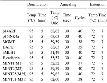
Table 1. Condition of MS-PCR
Denaturation Annealing Extension
Temp (°C)
Time (min)
Temp (°C) U/M
Time
(sec) Cycles Temp (°C)
Time (min)
p14ARF p16INK4a MGMT DAPK hMLH1 E-cadherin MINT1(M1) MINT2(M2) MINT25(M25) MINT31(M31)
95 95 95 95 95 95 95 95 95 95
5 5 5 5 5 5 5 5 5 5
62/62 63/63 59/59 63/63 51/49 55/57 52/52 59/59 59/62 62/60
30 30 30 30 30 30 30 30 30 30
40 40 38 35 40 40 37 40 40 38
72 72 72 72 72 72 72 72 72 72
7 7 7 7 7 7 7 7 7 7 DNA 메틸화는 DNMT (DNA methyltransferase)에 의해 일
어나며 주로 CpG의 염기반복구역인 CpG island의 사이토신 잔기(cytosine residue)에 메틸기가 첨가됨으로서 일어난다.
DNA의 이상(aberrant) 메틸화는 유전자의 조절과 상피의 종 양화에 중요한 기전으로 알려져 있다.(1) 유전자의 기시부 에 있는 CpG island에서 메틸화가 일어나면 유전자의 발현 이 억제되게 되며, 이러한 과메틸화가 종양억제유전자의 기시부(promoter gene)에 일어나면, 전사의 억제를 통한 유 전자의 기능의 소실로 세포증식을 억제하지 못하여 암 발 생에 관여하게 된다.(1-3)
히스톤 변형은 전사를 조절하는 또 하나의 epigentic 기전 이다. 히스톤 옥타머(octamer)가 중심(core)을 형성하며, 주 위로 이중나선구조의 DNA가 감겨져 뉴클레오솜(nucleo- some)을 형성한다. 두 효소가 히스톤의 아세틸화에 관여하 는데 히스톤 acetyltransferase와 히스톤 deacetylase (HDAC) 이다.(4) HDAC는 세포주기의 진행과 분열, 아포토시스 (apoptosis)에 관여하는 유전자의 전사인자 조절로 암 발생 에 관여한다. Epigenetic 기전과 유전적(genetic) 기전의 가장 큰 차이는 epigenetic한 변화는 화학적 물질에 의해서 반대 로의 변화가 가능하다는 것이며, 암의 치료에 있어서 HDAC의 억제를 목표로 연구가 이루어지고 있다.(5,6) 최근에 종양억제유전자에서 히스톤 변형이 DNA 메틸화 와 같이 작용하여 유전자의 기능을 억제한다는 보고가 있 다.(7-11) 저자들은 이상적인 epigenetic 치료 전략으로 메틸 화의 정도가 다른 RKO 대장암 세포주와 MCF-7 유방암 세 포주에서 demethylating 인자인 5-aza-2’-deoxycytidine (5-aza- DC)와 HDAC 억제인자인 sodium butyrate (SB)의 종양억제 효과 및 두 인자의 결합효과에 대해서 연구하였다.
방 법
1) 세포주 배양 및 시약
인간 대장암 세포주 RKO (ATCC, USA), 유방암 세포주 MCF-7 (KCLB, Korea) 그리고 정상 대장 세포주 DDC-112 CoN (ATCC)을 사용하였다. 대장암 세포주 RKO와 유방암 세포주 MCF-7은 10% fatal bovine serum과 1% penicillin/
streptomycin이 첨가된 Dulbecco’s modified Eagle’s medium (DMEM)/F12 (Gibco, Invitrogen Corp., San Diego, CA, USA) 를 사용하여 37°C, 5% CO2가 유지되는 습윤 배양기를 이용 하여 배양하였고 정상 대장 세포주는 10% fatal bovine se- rum 첨가된 Dulbecco's modified Eagle's medium (DMEM)을
사용하여 37°C, 5% CO2가 유지되는 습윤 배양기를 이용하 여 배양하였다.
5-aza-2'-deoxycytidine (Fluka, sigma-aldrich chemic GmbH, Riedstr.)는 phosphate-buffered saline에 녹이고 Sodium buty- rate (Fluka)는 멸균된 증류수에 녹여서 −20°C에 보관하였 다가 필요할 때 꺼내 사용하였다.
2) Bisulfite modification과 methylation-specific PCR
RKO cell에 5-aza-2’-deoxycytidin과 sodium butyrate를 각 각 농도별과 시간별로 처리한 후 QIAamp DNA Mini Kit (Qiagen, Gmbh, Hilden, Germany)를 이용하여 DNA를 추출 하였다. Genomic DNA의 Bisulfite modification 과정은 다음 과 같이 실행하였다.
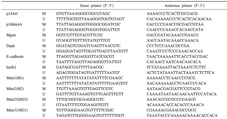
2μg의 DNA를 2 M NAOH로 변성시킨 후 10 mM hydro- quinone (sigma-aldrich Inc., St. Louis, MO, USA) 30μl와 3 M sodium bisulfite (sigma) 520μl를 넣어 50°C에서 16시간 배양하였다. Modified 된 DNA를 Wizard DNA clean-up sys- tem (promega, Madison, WI, USA)으로 정제한 후 3 M NAOH로 다시 변성시킨 다음 100% ethanol과 2.5 M ammo- nium acetate로 침전시킨 후 20μl의 증류수로 녹여 사용하였 다. PCR은 AccuPrime SuperMix I (invitrogen, Life Technolo- gies)을 사용하였으며 Modified genomic DNA 1μl를 넣어 증 폭시켰다. Product는 2.5% agarose gel로 확인하였다. PCR condition은 Table 1, 2에 기록한 것과 같다.
3) Cell proliferation assay
DDC-112 CoN, RKO 그리고 MCF7 cell을 각 3×10³개를
Table 2. MS-PCR primers of specific genes analyzed in this study
Sense primer (5'-3') Antisense primer (5'-3') p14Arf
p16Ink4A
Mgmt
Dapk
E-cadherin
hmlh1
Mint1(M1)
Mint2(M2)
Mint25(M25)
Mint31(M31)
M U M U M U M U M U M U M U M U M U M U
GTGTTAAAGGGCGGCGTAGC TTTTTGGTGTTAAAGGGTGGTGTAGT TTATTAGAGGGTGGGGCGGATCGC TTATTAGAGGGTGGGGTGGATTGT GGTCGTTTGTACGTTCGC
GTAGGTTGTTTGTATGTTTGT GGATAGTCGGATCGAGTTAACGTC GGAGGATAGTTGGATTGAGTTAATGTT TTAGGTTAGAGGGTTATCGCGT TAATTTTAGGTTAGAGGGTTATTGT GATAGCGATTTTTAACGC
AGAGTGGATAGTGATTTTTAATGT AATTTTTTTATATATATTTTCGAAGC AATTTTTTTATATATATTTTTGAAGTGT TTGTTAAAGTGTTGAGTTCGTC GATTTTGTTAAAGTGTTGAGTTTGTT TTTGCGGCGGAAGGCGTC
GTAATTTTGTGGAAGGTGTT TGTTGGGGAAGTGTTTTTCGGC TAGATGTTGGGGAAGTGTTTTTTGGT
AAAACCCTCACTCGCGACG CACAAAAACCCTCACTCACAACAA GACCCCGAACCGCGACCGTAA CAACCCCAAACCACAACCATA GACCGATACAAACCGAACG AACCAATACAAACCAAACA CCCTCCCAAACGCCGA CAAATCCCTCCCAAACACCAA TAACTAAAAATTCACCTACCGAC CACAACCAATCAACAACACA TCTATAAATTACTAAATCTCTTC ACTCTATAAATTACTAAATCTCTTCA AAAAACCTCAACCCCGCG
AACAAAAAACCTCAACCCCACA AATAACGACGATTCCGTACG
CAAAATAATAACAACAATTCCATACA AAACACCGCGCCCGAACG
ACAAAACACCACACCCAAACA CGAAAACGAAACGCCGCG
TAAATACCCAAAAACAAAACACCACA
Table 3. The methylation status of each cell lines, CCD-112, MCF-7, RKO
CCD-112 MCF-7 RKO
MINT1 MINT2 MINT25 MINT31 MGMT P16 P14 E-cadherin DAPK hMLH1 TWIST RARb HIN-1 RASSF1a
U*
U U 50%U/50%M
U U U U U U 80%U/20%M 80%U/20%M
U
M† U U M 50%U/50%M
U U M U 50%U/50%M 30%U/70%M
M M
M M U M U M M M M 50%U/50%M 50%U/50%M 40%U/60%M
M
*U = unmethylated; †M = methylated.
96-well plate에 seeding하고 24시간 후에 5-aza-2’-deoxycyti- dine는 4 uM과 sodium butyrate 1 mM를 각각 그리고 동시에 2가지를 같이 처리한 후 48시간 배양한 다음에 cell pro- liferation kit II (XTT) (Roche Diagnostics GmbH, Mannheim, Germany)를 사용하여 측정하였다.
4) 통계학적 분석
HDAC억제제와 demethylating인자의 효과를 비교하기 위 해 SPSS version 13.0을 이용하여 ANOVA (Analysis of Variance) 검사를 이용하여 결과를 비교하였다. P-value 0.05 미만을 통계적으로 의미 있다고 보았다.
결 과
1) CCD-112 CoN, MCF-7, RKO 세포주의 메틸화 상태
RKO 세포주에서는 대부분의 종양억제유전자가 메틸화 되어 있었으며 MCF-7 세포주에서는 약 반 정도의 종양억 제유전자가 메틸화되어 있었고, CCD-112 CoN 세포주에서 는 메틸화된 종양억제유전자는 없었다(Table 3).
2) RKO 세포주에서 5-aza-2’-deoxycytidine 처리 후 MS-PCR 결과
대조군에서는 거의 모든 유전자가 메틸화되어 있었으나,
5-aza-DC 처리 후에는 세포주에서 비메틸화 밴드가 보이기 시작하였으며 메틸화 밴드가 약간 감소하였다(Fig. 1).
3) RKO 세포주에서 sodium butyrate 처리 후 MS-PCR 결과
대조군과 비교하여 sodium butyrate 처리 후 메틸화 상태 에 거의 변화가 없었다(Fig. 2, Table 4).
Fig. 1. MS-PCR after 5-aza-2’-deoxycytidine (5-aza-DC) treatment.
In the control group, most of the genes were methylated, but cell lines treated with aza-DC showed that demethy- lated bands and methylated bands were slightly decreased.
Fig. 2. MS-PCR after sodium butyrate treatment. Compared to the control group, there were almost no changes in methylation status with the addition of sodium butyrate.
Fig. 3. The effect of 5-azaDC and SB on cell survival.
Table 4. The effects of 5-aza-2’-deoxycytidine and sodium buty- rate on cell survival
Cell survival (%)
P-value
MCF-7 CCD-112 RKO
Control 5-aza-DC*
SB†
SB+5-aza-DC P-value
100 82.6±13.4
63.6±3.9 49.4±6.9
<0.001
100 107.2±3.5 99.9±8.83 97.8±6.83
0.870
100 74.2±6.8 83.1±2.0 52.6±4.6
<0.001
<0.001
<0.001
<0.001
*5-aza-DC = 5-aza-2’-deoxycytidine; †SB = sodium butyrate.
4) XTT result with 5-aza-2’-deoxycytidine and sodium butyrate
MCF-7 세포주에서는 대조군의 100%에 비해서 5-aza-DC 처리시 82.6%, SB 처리시 63.6%의 생존을 보여 SB의 효과 가 더 좋았다. SB와 5-aza-DC를 병합 처리시에는 49.4%로 상승효과를 볼 수 있었다(P<0.001).
RKO 세포주에서는 대조군의 100%에 비해서 5-aza-DC 처리시 74%, SB 처리시 83%의 생존을 보여 5-aza-DC의 효 과가 더 좋았다. SB와 5-aza-DC를 병합 처리시에는 52.6%
로 상승효과를 볼 수 있었다(P<0.001).
CCD-112 세포주에서는 두 인자 모두 생존에 영향이 없었
다(Fig. 3).
고 찰
Epigenetic 분야에서의 암 연구에 관해 최근에는 히스톤 변 형과 DNA 메틸화에 대해서 많은 연구가 이루어지고 있다.
히스톤은 acetyltransferase와 HDAC의 작용에 의한 전사 후 변형에서 중요한 역할을 한다. 종양형성은 히스톤의 acetyl- transferase의 비활성화와 관계되어 있으며, HDAC의 이상 활성이 종양억제유전자의 전사를 억제하여 종양의 형성에 중요한 역할을 하는 것으로 생각되어지고 있다.(12) 히스톤 의 hypoacetylation은 응축된 염색질(chromatin) 구조와 관련 이 있으며, 이 상태에서는 전사가 억제되게 되며, 반대로 hyperacetylation은 개방 염색질 구조를 이루게 되어 전사가 활성화되게 된다.(9) 히스톤의 아세틸화는 가역적인 반응으 로 HDAC 억제제를 통해 응축된 염색질 구조에서 개방 염
색질 구조로 바뀔 수 있다.
DNA의 이상 메틸화는 종양세포에서 흔하며, 종양억제유 전자나 다른 세포주기조절, 아포토시스 또는 DNA 복구에 관여하는 유전자들의 기능을 억제한다.(1-3)
종양세포의 아포토시스에의 저항은 proapoptotic 인자가 메틸화에 의해서 또는 탈아세틸화로 인해서 기능이 소실되 어 발생할 수 있으며 염색질의 응축이 신호전달체계의 신 호를 바꾸어서 prosurvival 경로를 과표현하여서도 발생할 수 있다.(9)
많은 종류의 demethylating agent와 HDAC 억제자가 있지 만, 저자들은 demethylating 인자로서 5-aza-DC를 사용하였 고 HDAC 억제자로서 SB를 사용하였다. 5-aza-DC는 시티딘 (cytidine)과 유사한 물질로 작용기전은 주로 DNMT에 비가 역적인 공유결합을 통하여 DNMT의 활성을 방해하며 메틸 화 수위를 낮춰 유전자의 표현을 조절하는 기능을 한다. 이 런 메틸화는 가역적이므로 demethylating 인자를 통하여 메 틸화에 의해서 비활성화된 유전자를 활성화시킬 수 있다.
SB는 HDAC의 아연의 활성부위를 목표로 하는 단쇄지방산 (short-chain fatty acid)으로서 HDAC의 활성을 억제하여 히 스톤의 아세틸화를 촉진하며, 매우 짧은 반감기를 가지고 있다.(1,3) SB는 in vivo 또는 in vitro에서 종양세포의 아포토 시스를 촉진하고 세포의 분화를 촉진시키며 세포주기를 억 제시킨다.(13) 본 연구에서도 대부분 메틸화되어 있는 RKO 세포주에 5-aza-DC를 처리하였을 때 메틸화 밴드가 줄어들 며, 비메틸화 밴드가 보이기 시작하였다. HDAC 억제제인 SB는 메틸화에 영향이 없었다.
본 연구에서 5-aza-DC의 효과를 보면 MCF-7 세포주에서 는 대조군에 비해서 82%의 세포생존을 보였지만 RKO cell line에서는 대조군에 비해서 74%의 세포생존을 보였다.
RKO 세포주에서는 거의 모든 유전자의 기시부가 메틸화되 어 있었으며, MCF-7 세포주에서는 약 반 정도의 유전자 기 시부가 메틸화되어 있었다. 이런 이유로 MCF-7 세포주 보 다 메틸화가 많이 되어 있는 RKO 세포주에서 demethylating 인자의 효과가 우수하였으며, demethylating 인자는 과메틸 화가 되어 있는 상태에서 효과가 더 우수한 것으로 생각된 다. SB의 효과를 보면 MCF-7 세포주에서는 대조군에 비해 서 64%의 세포생존을 보였지만 RKO 세포주에서는 대조군 에 비해서 83%의 세포생존을 보였다. HDAC의 효과는 메 틸화가 적게 된 MCF-7 유방암 세포주에서 더 우수하였다.
Cameron 등(14)은 HDAC 억제제인 TSA가 과메틸화된 유전 자인 MLH1, TIMP3, CDKN2A에서는 유전자의 발현에 영향
을 미치지 못하였지만 메틸화 되어있지 않은 CDKN2B에는 유전자의 발현을 상향조정(upregulated)시켜, HDAC은 DNA 의 메틸화가 감소할 때 역할이 증가한다고 하였으며, Shen 등(11)도 유전자의 기시부 영역에 메틸화가 낮으면 히스톤 탈아세틸화가 유전자의 전사에서 중요한 역할을 하지만 메 틸화가 상당히 높은 경우에는 히스톤 탈아세틸화는 보조적 역할을 한다고 하여 본 연구의 결과와 일치하였다.
히스톤 탈아세틸화와 DNA 메틸화는 서로 독립적인 epi- genetic 기전이 아니라 이 두 기전은 서로 밀접히 연관되어 있어 서로에게 영향을 준다. 본 연구에서는 MCF-7 세포주 의 경우 세포생존이 5-aza-DC 단독처리시 82%, SB 단독처 리시 63%에 비해서 두 인자를 병합처리하였을 때 49%로 상승효과가 나타났으며, RKO 세포주에서 또한 5-aza-DC 단독처리시 74%, SB 단독처리시 83%, 병합처리시 52%로 상승효과가 나타났다. Cameron 등(14)은 5-aza-DC 단독처리 시 보다 TSA를 병합하였을 때 발현억제된 유전자의 발현이 4배 증가하였다고 하였다. Zhu 등(15)은 폐암 세포주에서 TSA의 아포토시스를 일으키는 역할이 5-aza-DC에 의해서 크게 증가된다고 보고하였다. 병합효과의 우수성에 대한 기전은 아직 명확하게 밝혀지지는 않았으나 다음과 같은 것으로 생각한다. DNA 메틸화는 DNMT 또는 메틸화된 DNA 결합 단백질(methyl DNA binding proteins)을 통하여 HDAC를 증가시켜, 히스톤의 탈아세틸화가 촉진된다.(16,17) 그리고 HDAC는 히스톤 H3 lys9 methyltransferase를 통하여 DNA 메틸화를 촉진한다. 즉, HDAC와 DNA 메틸화는 루프 를 형성하여 서로에게 영향을 주며 서로를 강화하는 역할 을 한다.(18) 그러므로 HDAC 억제제와 demethylating 인자 를 통해서 DNA메틸화와 히스톤의 아세틸화 모두를 차단함 으로서 상승작용이 일어나는 것으로 생각한다. 또한 히스 톤 아세틸화는 응축된 염색질을 풀어서 DNA의 apoptotic endonuclease로의 접근을 증가시킬 수 있다.(15) 다른 기전 으로는 5-aza-DC는 뉴클레오시드 유사형(nucleoside analog) 으로서 DNA backbone에 결합되어 직접 DNA에 손상을 주 게 된다.(19,20) Chai 등(21)은 폐암세포주 A549와 H719에서 5-aza-DC가 [3H]-5-aza-DC가 되어 뉴클레오시드 유사형으 로서 DNA 손상을 줄 때 이를 제거하는 복구과정이 작동하 는데, HDAC 억제제가 이런 복구과정을 방해하여 세포증식 을 억제한다고 하여 두 인자의 종양억제에서의 상승효과를 설명하였다.
결론적으로 5-aza-DC와 SB는 상승작용이 있었다. 단독으 로 사용시에는 고도로 메틸화가 된 상태에서 demethylating
인자의 역할이 상대적으로 크며, 메틸화가 높지 않은 경우 에는 HDAC 억제제의 역할이 상대적으로 높았다. 본 연구 는 세포주를 이용한 실험실연구이며 좀더 명확한 병합 효 과 및 기전을 알기 위해서는 향후 동물생체 내 실험이 필요 할 것으로 생각한다.
REFERENCES
1) Jones PA, Baylin SB. The fundamental role of epigenetic events in cancer. Nat Rev Genet 2002;3:415-28.
2) Szyf M, Pakneshan P, Rabbani SA. DNA methylation and breast cancer. Biochem Pharmacol 2004;68:1187-97.
3) Garinis GA, Patrinos GP, Spanakis NE, Menounos PG. DNA hypermethylation: when tumour suppressor genes go silent.
Hum Genet 2002;111:115-27.
4) Narlikar GJ, Fan HY, Kingston RE. Cooperation between complexes that regulate chromatin structure and transcription.
Cell 2002;108:475-87.
5) Marks P, Rifkind RA, Richon VM, Breslow R, Miller T, Kelly WK. Histone deacetylases and cancer: causes and therapies.
Nat Rev Cancer 2001;1:194-202.
6) Verma M, Srivastava S. Epigenetics in cancer: implications for early detection and prevention. Lancet Oncol 2002;3:755-63.
7) Walton TJ, Li G, Seth R, McArdle SE, Bishop MC, Rees RC.
DNA demethylation and histone deacetylation inhibition co- operate to re-express estrogen receptor beta and induce apopto- sis in prostate cancer cell-lines. Prostate 2008;68:210-22.
8) Murakami J, Asaumi J, Maki Y, Tsujigiwa H, Kuroda M, Nagai N, et al. Effects of demethylating agent 5-aza-2(')-deox- ycytidine and histone deacetylase inhibitor FR901228 on mas- pin gene expression in oral cancer cell lines. Oral Oncol 2004;40:597-603.
9) Bar-Sela G, Jacobs KM, Gius D. Histone deacetylase inhibitor and demethylating agent chromatin compaction and the radia- tion response by cancer cells. Cancer J 2007;13:65-9.
10) Zhu WG, Otterson GA. The interaction of histone deacetylase inhibitors and DNA methyltransferase inhibitors in the treat- ment of human cancer cells. Curr Med Chem Anticancer
Agents 2003;3:187-99.
11) Shen WJ, Dai DQ, Teng Y, Liu HB. Regulation of demethyla- tion and re-expression of RASSF1A gene in gastric cancer cell lines by combined treatment of 5-Aza-CdR and NaB. World J Gastroenterol 2008;14:595-600.
12) Johnstone RW. Histone-deacetylase inhibitors: novel drugs for the treatment of cancer. Nat Rev Drug Discov 2002;1:287-99.
13) Marks PA, Richon VM, Rifkind RA. Histone deacetylase in- hibitors: inducers of differentiation or apoptosis of transformed cells. J Natl Cancer Inst 2000;92:1210-6.
14) Cameron EE, Bachman KE, Myohanen S, Herman JG, Baylin SB. Synergy of demethylation and histone deacetylase inhi- bition in the re-expression of genes silenced in cancer. Nat Genet 1999;21:103-7.
15) Zhu WG, Lakshmanan RR, Beal MD, Otterson GA. DNA methyltransferase inhibition enhances apoptosis induced by histone deacetylase inhibitors. Cancer Res 2001;61:1327-33.
16) Fuks F, Burgers WA, Godin N, Kasai M, Kouzarides T.
Dnmt3a binds deacetylases and is recruited by a sequence- specific repressor to silence transcription. EMBO J 2001;20:
2536-44.
17) Nan X, Ng HH, Johnson CA, Laherty CD, Turner BM, Eisen- man RN, et al. Transcriptional repression by the methyl-CpG- binding protein MeCP2 involves a histone deacetylase com- plex. Nature 1998;393:386-9.
18) Kondo Y, Issa JP. Epigenetic changes in colorectal cancer.
Cancer Metastasis Rev 2004;23:29-39.
19) Zhu WG, Hileman T, Ke Y, Wang P, Lu S, Duan W, et al.
5-aza-2'-deoxycytidine activates the p53/p21Waf1/Cip1 pathway to inhibit cell proliferation. J Biol Chem 2004;279:15161-6.
20) Wang H, Zhao Y, Li L, McNutt MA, Wu L, Lu S, et al. An ATM- and Rad3-related (ATR) signaling pathway and a phos- phorylation-acetylation cascade are involved in activation of p53/p21Waf1/Cip1 in response to 5-aza-2'-deoxycytidine treat- ment. J Biol Chem 2008;283:2564-74.
21) Chai G, Li L, Zhou W, Wu L, Zhao Y, Wang D, et al. HDAC inhibitors act with 5-aza-2'-deoxycytidine to inhibit cell pro- liferation by suppressing removal of incorporated abases in lung cancer cells. PLoS ONE 2008;3:e2445.