ISSN 2234-3806 • eISSN 2234-3814
http://dx.doi.org/10.3343/alm.2014.34.2.155 www.annlabmed.org 155
Ann Lab Med 2014;34:155-158
http://dx.doi.org/10.3343/alm.2014.34.2.155
Letter to the Editor
Diagnostic Hematology
Therapy-Related Acute Megakaryoblastic Leukemia in a Lung Cancer Patient
Jung Joo Moon, M.D., Myung-Hyun Nam, M.D., Chae Seung Lim, M.D., Chang Kyu Lee, M.D., Yunjung Cho, M.D., and Soo-Young Yoon, M.D.
Department of Laboratory Medicine, Korea University College of Medicine, Seoul, Korea
Therapy-related AML (t-AML) is one of the newly expanded dis- ease entities in the 2008 WHO classification, accounting for 10- 20% of all cases of AML, and its incidence is increasing world- wide because of improved survival rates following treatment for other primary cancers [1, 2]. Acute megakaryoblastic leukemia (AMKL) (M7) is the least common of the t-AML French-Ameri- can-British (FAB) subtypes, and only two such cases have been reported to date [3, 4], neither of which was in Korea. AMKL ac- counts for about 7-10% of childhood AML cases (frequently as- sociated with Down syndrome), but only about 1% of adult AML cases [5]. Here, we describe a rare case of therapy-related acute megakaryoblastic leukemia (t-AMKL) with chromosome 5 and 7 abnormalities that presented ten years after chemoradio- therapy in an elderly lung cancer patient.
A 72-yr-old man with lung cancer (non-small cell lung cancer [NSCLC], right lower lobe [RLL], squamous cell cancer, stage T2N1M0) was treated with a combination of radiotherapy and a repeated chemotherapy regimen (4 trials, 13 cycles) consisting of docetaxel, cisplatin, gemcitabine, vinorelbine, gefitinib, irino- tecan, and carboplatin, between March 2003 and February 2005 (Table 1). Subsequently, in April 2005, he underwent sur- gical resection (RLL lobectomy) of the cancer and thereafter achieved complete remission.
He was asymptomatic until being referred to our hospital for further evaluation of pancytopenia discovered at a local hospital,
to which he had been admitted for fever persisting for three days, upper respiratory infection symptoms, and aggravated dys- pnea. He had signs of anemic conjunctiva, but no lymphade- nopathy or organomegaly on physical examination. Abdominal sonography revealed hepatomegaly but no splenomegaly. A complete blood count (CBC) revealed Hb level of 5.2 g/dL; white blood cell counts of 1.7 ×10⁹/L (absolute neutrophil count 0.78 ×10⁹/L); and platelet counts of 34 ×109/L. A peripheral blood smear showed macrocytic normochromic anemia with teardrop cells, a shift to the left in the neutrophilic series together with dysplastic features such as hypogranulation and the Pelger- Huët anomaly in segmented neutrophils, irregularly segmented monocyte nuclei, and giant platelets (Fig. 1A).The plasma Hb (19.5 mg/dL; reference interval [RI], 0.0-5.0 mg/dL) and D-di- mer (4.59 μg/mL; RI, 0-0.5 μg/mL) levels were both elevated.
Most biochemical tests were normal, except for elevated lev- els of serum lactate dehydrogenase (513 IU/L; RI, 0-480 IU/L), beta-2 microglobin (2.9 mg/L; RI, 0-2.4 mg/L), and C-reactive protein (22.24 mg/L; RI, 0-5.0 mg/L). Vitamin B12 was raised at 19,030 pg/mL (RI, 160-970 pg/mL), but the folate level was within the RI. Bone marrow (BM) aspirate smears and a touch print preparation of the BM biopsy revealed hypercellular mar- row, dysplastic changes in all three hematopoietic cell lineages, and 23.5% blasts (Fig. 1B). The blasts were medium to large sized and had round, slightly irregular or indented nuclei, fine
Received: May 29, 2013
Revision received: August 20, 2013 Accepted: October 8, 2013
Corresponding author: Soo-Young Yoon
Department of Laboratory Medicine, Korea University Guro Hospital, 148 Gurodong-ro, Guro-gu, Seoul 152-703, Korea
Tel: +82-2-2626-3246, Fax: +82-2-2626-1465, E-mail: labmd@korea.ac.kr
© The Korean Society for Laboratory Medicine.
This is an Open Access article distributed under the terms of the Creative Commons Attribution Non-Commercial License (http://creativecommons.org/licenses/by-nc/3.0) which permits unrestricted non-commercial use, distribution, and reproduction in any medium, provided the original work is properly cited.
Moon JJ, et al.
Therapy-related AMKL
156 www.annlabmed.org http://dx.doi.org/10.3343/alm.2014.34.2.155 reticular chromatin, one to three inconspicuous nucleoli, baso-
philic agranular cytoplasm, and frequently formed pseudopod.
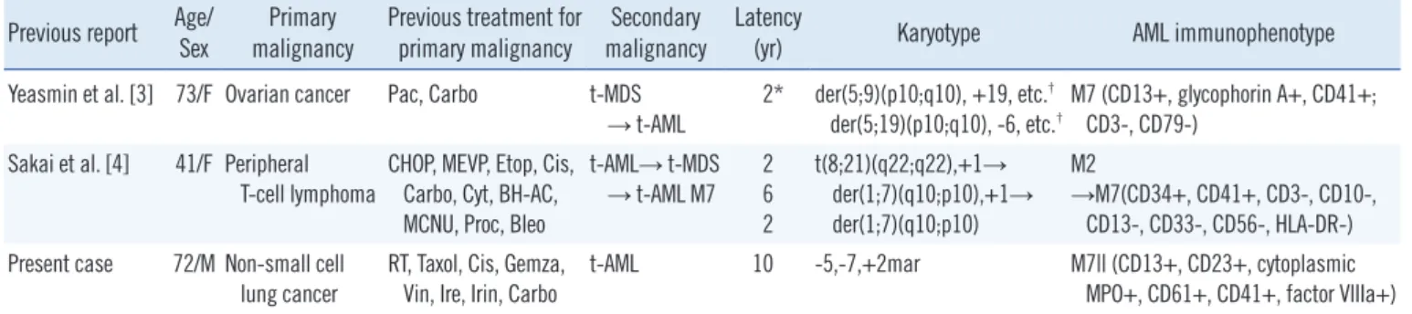
There were relatively fewer erythroid cells, and these had dys- plastic features such as nuclear-cytoplasmic asynchrony. Con- Table 1. Characteristics of previously reported therapy-related acute megakaryoblastic leukemia cases
Previous report Age/
Sex Primary
malignancy Previous treatment for
primary malignancy Secondary
malignancy Latency
(yr) Karyotype AML immunophenotype Yeasmin et al. [3] 73/F Ovarian cancer Pac, Carbo t-MDS
→ t-AML 2* der(5;9)(p10;q10), +19, etc.† der(5;19)(p10;q10), -6, etc.†
M7 (CD13+, glycophorin A+, CD41+;
CD3-, CD79-) Sakai et al. [4] 41/F Peripheral
T-cell lymphoma
CHOP, MEVP, Etop, Cis, Carbo, Cyt, BH-AC, MCNU, Proc, Bleo
t-AML→ t-MDS → t-AML M7 2
6 2
t(8;21)(q22;q22),+1→ der(1;7)(q10;p10),+1→
der(1;7)(q10;p10)
M2
→M7(CD34+, CD41+, CD3-, CD10-, CD13-, CD33-, CD56-, HLA-DR-) Present case 72/M Non-small cell
lung cancer RT, Taxol, Cis, Gemza,
Vin, Ire, Irin, Carbo t-AML 10 -5,-7,+2mar M7|| (CD13+, CD23+, cytoplasmic MPO+, CD61+, CD41+, factor VIIIa+)
*22 months to diagnosis of t-MDS, 3 months to diagnosis of t-AML; †Karyotype of t-MDS: 46,XX,der(5;9)(p10;q10), del(7)(q?), +19, add(3q21), der(12) add(12)(p11.2), t(3;12)(q21;q22), del(12)(p?) [17/20], Karyotype of t-AML: 46,XX,der(5;19)(p10;q10), -6, del(7)(q?), add(12p11.2), -13, -17, +19[20/20].*
Abbreviations: Pac, paclitaxel; Carbo, carboplatin; CHOP, adriamycin, cyclophosphamide, vincristine, prednisolone; MEVP, mitoxantrone, etoposide, vinde- sine, prednisolone; Dox, doxorubicin; Etop, etoposide; Cis, cisplatin; Cyt, cytoxan; BH-AC, behenoylara-C; MCNU, ranimustine; Proc, procainamide; Bleo, bleomycin; RT, radiotherapy; Taxol, docetaxel; Gemza, gemcitabine; Vin, vinorelbine; Ire, iressa; Irin, irinotecan.
A B
C D
E
F
G
22 16 9
X 17 10
Y 18 11 4 3 2
1
12 5
20 14 7
21 15 8
19 mar mar
13 6
Fig. 1. (A) Peripheral blood (PB) smear findings (Wright-Giemsa stain, ×1,000), (B) bone marrow (BM) aspirate smear (Wright-Giemsa stain, ×1,000), and (C) BM biopsy section (H&E stain, ×1,000). Cells in the PB smear (A) had dysplastic features, such as marked aniso- poikilocytosis, and some red blood cells (RBCs) had a teardrop shape. Neutrophils showed the Pelger-Huët anomaly and cytoplasmic hypo- granularity. Leukemic blasts (B), consistent with acute megakaryoblastic leukemia, were of medium to large size and had round, slightly ir- regular or indented nuclei, with fine reticular chromatin, one to three inconspicuous nucleoli, basophilic agranular cytoplasm, and frequent pseudopod formation. The biopsied marrow (C) showed hypercellularity relative to the patient’s age (approximately 70%). (D) The karyotype of the abnormal clone in 2 out of 20 analyzed metaphase cells using G-banding. It was initially interpreted as 46,XY,-5,-7,+2mar. The arrows denote the abnormal chromosomes. (E-G) Interphase FISH for AML1-ETO (E), RELN/TES (F), andTAS2R1/EGR1 (G) at diagnosis. The three green (RUNX1 probe on 21q22) and two orange (RUNX1T1 probe on 8q22) signals (E) were consistent with an additional AML1 signal in 28.2% of the nuclei analyzed. The two green (D5S630 probe on 5p15.31) and one orange (EGR1 probe on 5q31) signal (F) and one green (RELN probe on 7q22) and one orange (TES probe on 7q31) signal (G) were consistent with 5q and 7q deletions, respectively. The two green (D5S630 probe on 5p15.31) signals (F) signified that there were two 5p loci.
Abbreviation: H&E, hematoxylin and eosin.
Moon JJ, et al.
Therapy-related AMKL
http://dx.doi.org/10.3343/alm.2014.34.2.155 www.annlabmed.org 157
versely, there were relatively more granulocytes, but these also showed dysplastic features such as megaloblastoid change, hy- posegmentation, and cytoplasmic hypogranularity. Markedly, more megakaryocytes that varied considerably in size were present, and they were multi-nucleated with hypolobated nuclei.
Micromegakaryocytes, megakaryocytic fragments, and bare megakaryocytic nuclei were frequently observed. It was also noteworthy that basophils accounted for up to 6.7% of the nu- cleated marrow cells.
Cytochemical staining of the BM aspirate revealed that blasts were positive for periodic acid-Schiff, acid phosphatase (punc- tuate), and non-specific esterase, and negative for Sudan black B, myeloperoxidase (MPO), and specific esterase.
The biopsied marrow was hypercellular relative to the patient’s age (approximately 70%) (Fig. 1C), consisting predominantly of dysplastic megakaryocytes of variable size and leukemic blasts.
Immunohistochemical staining of the paraffin-embedded BM bi- opsy specimen showed that the blasts and megakaryocytes were positive for Factor-VIII, CD61, MPO, and c-Kit. There were a re- duced number of glycophorin-positive erythroid cells, and reticu- lin staining showed mild myelofibrosis (grade MF-1) [1].
Immunophenotyping of leukemic blasts in the BM using flow cytometry revealed a weakly positive result for CD45, CD13, CD23, and cytoplasmic MPO and strong positivity for CD61 and CD41, consistent with a megakaryocytic lineage. The same blasts were negative for CD2, CD5, CD7, CD10, CD11a, CD14, CD20, CD22, CD33, CD34, CD64, CD79a, CD34, CD117, HLA- DR, cytoplasmic CD3, cytoplasmic CD19, and terminal deoxy- nucleotidyltransferase (TdT).
G-banded metaphase analysis and FISH were performed on the BM aspirate using standard cytogenetic techniques. The karyotype was 46,XY,-5,-7,+2mar in 2 of the 20 analyzed BM cells (Fig. 1D), and 46,XY in the remaining 18 cells. BCR/ABL, AML1/ETO, PML/RARA, CBFB, MLL, D5S630/EGR1, and RELN/TESFISH probes (Abbott Molecular/Vysis, Des Plaines, IL, USA) and the D5S630/EGR1 probe from Cytocell (Cambridge, UK) showed an additional AML1 signal, a 5q deletion, and a 7q deletion (Fig. 1E-G). The FISH findings suggested that at least part of the marker chromosomes were derived from chromo- some 21 and the short arm of chromosome 5, and not from 5q or 7q. Multiplex reverse transcriptase-PCR (RT-PCR) with the Hemavision kit (DNA Technology, Aarhus, Denmark) did not detect any fusion transcripts.
The patient was diagnosed with t-AMKL and was treated for neutropenic fever and pneumonia. No further therapy was ad- ministered for the leukemia because of his poor general condi-
tion, and he is currently being followed up in our hospital with routine CBC check-ups.
The previous 2008 WHO classification described two subsets of therapy related myeloid neoplasm, consisting of t-AML/ther- apy-related myelodysplasia (t-MDS) and t-AML/t-MDS/therapy- related myeloproliferative neoplasm (t-MPN). The former subset commonly caused by exposure to alkylating agents or radiother- apy has the following characteristics: 1) It typically has a long la- tent period; 2) Its incidence increases with age; 3) It is frequently present with t-MDS, t-AML after a myelodysplastic phase or t- AML with dysplastic features; 4) It has a poorer prognosis; 5) It is associated with an unbalanced loss of genetic material; 6) It commonly affects chromosomes 5 or 7. The other subset is characterized by 1) exposure to topoisomerase II inhibitors; 2) a short latency period; 3) a similar incidence across all ages; 4) the presence of overt t-AML without a preceding myelodysplastic phase or dysplastic features; 5) a more indolent prognosis; and 6) balanced translocations involving 11q23 or 21q22 [1, 2]. The patient in this case was elderly, presented with overt t-AML with prominent dysplastic features after ten years of remission and unbalanced chromosomal abnormalities in chromosomes 5 and 7. Alkylating agents, anti-tubulin agents, topoisomerase I inhibi- tors and ionizing radiation were all used in his treatment, but not topoisomerase II inhibitor. This case therefore showed clinical features typical of the former subset of t-AML/t-MDS. The two previously reported t-AMKL cases also involved dysplastic fea- tures, unbalanced chromosomal rearrangements, and a history of alkylating agent or radiation therapy (Table 1) [3, 4].
AMKL in non-Down syndrome patients is rare, accounting for less than 1% of all adult AML cases [6]. A previous study re- ported t-AMKL in 4 of 37 (10.8%) adult AMKL cases [5], and another study found t-AMKL in 2 of 23 (8.7%) adult AMKL cases [6], and on this basis the incidence of t-AMKL is about 0.1% of all adult AML cases.
Adherence of platelets to leukocytes during flow cytometry and the procedures used for fixation and decalcification in im- munohistochemistry can lead to difficulties in identifying mega- karyocytic lineages [7]. Recent studies have stated that accu- rate diagnosis of AMKL additionally requires that cells are nega- tive for CD11a and HLA-DR, as well as being positive for previ- ously used markers such as CD41, CD42, CD61, or Factor VIII [8]. Likewise, in this case, the patient also showed a CD61+, Factor VIII+, CD11a-, and HLA-DR immunophenotype.
In summary, to the best of our knowledge, this is the third case report of t-AMKL worldwide and the first report of t-AMKL in Korea. Furthermore, it is the first reported case of overt t-
Moon JJ, et al.
Therapy-related AMKL
158 www.annlabmed.org http://dx.doi.org/10.3343/alm.2014.34.2.155 AMKL without any evidence of preceding myelodysplasia.
Authors’ Disclosures of Potential Conflicts of Interest
No potential conflicts of interest relevant to this article were re- ported.
Acknowledgments
This research was supported by the Nano·Material Technology Development Program through the National Research Founda- tion of Korea (NRF) funded by the Ministry of Science, ICT and Future Planning (grant number 2012M3A7B4035286).
REFERENCES
1. Swerdlow SH, Campo E, Harris NL, et al. eds. WHO classification of tu- mours of haematopoietic and lymphoid tissues. 4th ed. Lyon: IARC Press, 2008: 110.
2. Le Beau MM, Albain KS, Larson RA, Vardiman JW, Davis EM, Blough
RR, et al. Clinical and cytogenetic correlations in 63 patients with thera- py-related myelodysplastic syndromes and acute nonlymphocytic leu- kemia. J ClinOncol 1986;4:325-45.
3. Yeasmin S, Nakayama K, Ishibashi M, Oride A, Katagiri A, Purwana IN, et al. Therapy-related myelodysplasia and acute myeloid leukemia fol- lowing paclitaxel- and carboplatin-based chemotherapy in an ovarian cancer patient. Int J Gynecol Cancer 2008;18:1371-6.
4. Sakai C, Matsubayashi K, Saotome T, Ishii A, Kumagai K. Therapy-relat- ed myelodysplastic syndrome with trisomy 1q due to der(1;7) and megakaryoblastic proliferation developing during complete remission of therapy-related acute myeloid leukemia with t(8;21). Intern Med 2004;
43:582-6.
5. Oki Y, Kantarjian HM, Zhou X, Cortes J, Faderi S, Verstovsek S, et al.
Adult acute megakaryocytic leukemia: an analysis of 37 patients treated at M.D. Anderson Cancer Center. Blood 2006;107:880-4.
6. Duchayne E, Fenneteau O, Pages MP, Sainty D, Arnoulet C, Dastugue N, et al. Acute megakaryoblasticleukaemia: a national clinical and biologi- cal study of 53 adult and childhood cases by the GroupeFrançaisd’Hé matologieCellulaire (GFHC). Leuk Lymphoma 2003;44:49-58.
7. Tomer A, Harker LA, Burstein SA. Flow cytometric analysis of normal human megakaryocytes. Blood 1988;71:1244-52.
8. Boztug H, Schumich A, Pötschger U, Mühlegger N, Kolenova A, Rein- hardt K, et al. Blast cell deficiency of CD11a as a marker of acute mega- karyoblastic leukemia and transient myeloproliferative disease in chil- dren with and without Down syndrome. Cytometry B Clin Cytom 2013 Feb. 28 [Epub ahead of print].