*Corresponding author : Myung-June Chung, R&D Center of Cellbiotech Co. Ltd. 134, Gaegok-ri, Wolgot-myun, Gimpo-si, Gyunggi-do 415-871, Korea. E-mail : [email protected]
Lactobacillus casei의 배양물에서 분리한 물질의 항암 효과
김정화․김동명․백홍․이승훈․정명준*
(주)쎌바이오텍 세포공학연구소
Anti-Cancer Effects of Peptides Purified from Culture Supernatant of Lactobacillus casei
Jung-Hwa Kim, Dong-Myung Kim, Hong Baek, Seung-Hoon Lee and Myung-June Chung*
R&D Center of Cellbiotech Co. Ltd.
---
ABSTRACT
This study was conducted to isolate protein components from culture supernatant of Lactobacillus casei. and measure anti-cancer activity. The protein components were isolated A and B on Ultrafiltration membrane(3, 10, 30, 100 KDa). And the protein components A and B were isolated fractions(number 3~9) on FPLC.
Experimental studies were progressed through the cell cytotoxicity and anti-cancer activities. Cell cytotoxicity test using human kidney normal cell(293) showed cytotoxicity of below 20% by the protein components A and B(100 μg/mL). The anti-cancer activity was increased up to 70% by the protein components A and B(100 μg/mL) in AGS(stomach cancer), A549(lung cancer), MCF-7 (breast cancer), SK-OV-3(ovary cancer) and LoVo(colon cancer). Cell cytotoxicity test was showed cytotoxicity of about 50% by the fractions(number 3, 8, 9) isolated FPLC. The others have not the cytotoxicity about the human normal cell. The anti-cancer activity was increased up to 70% by the fraction number 7 in cancer cell line. Therefore the components isolated from culture supernatant of Lactobacillus casei were showed anti-cancer activity.
(Key words : Lactobacillus casei, cytotoxicity, anti-cancer activity, culture supernatant)
---
서 론
최근 유산균에 대한 관심이 높아짐에 따라 그와 관련된 제품들이 부가가치가 높아지고 있다. 이에 따라 유산균에 대한 기능 및 생리적 성질에 관한 많은 연구 보고가 되고 있 다. 그 기능에는 정장 작용(Raiboud, 1980), 항암 효과(Ayebo et al., 1981; Reddy et al., 1973), 혈청 콜레스테롤 저하(Mann and Spoerry, 1974), 비타민의 합성(Alm, 1982) 등의 효과가 보고되었다. 특히 Shackelford(Shackelford et al., 1983) 등도 Streptococcus thermophilus와 Lactobacillus bulgaricus 발효유 가 모두 dimethylhydrazin hydrochloride(DMH)에도 발암 제 어 효과가 현저하다고 하였다.
유산균 중 Lactobacillus 속은 혐기성 또는 통성 혐기성으 로 증식하는 그람 양성 세균으로써, 사람의 구강과 소화기 관 등에서도 발견되며, 자연계에 널리 분포하고 있는 미생
물이다(Pelletier et al., 1997). 인체에 유익한 미생물로써 유 산균은 다양한 발효 유제품의 starter로 널리 이용되고 있으 며, 이러한 젖산균이 함유된 발효 유제품을 섭취함으로써, 인체의 소화관 내에서 유해 세균의 증식을 억제한다는 사실 이 밝혀짐에 따라, 가축이나 사람의 생균제제 또는 정장제 로써 그 이용 가치가 매우 높아지고 있다(Lilly and Stillwel, 1965 ; Kang et al., 2001). 또한, 최근 연구 결과, 장 내 유해 세균의 억제, 항돌연변이능, 항암효과, 유당 이용성 증진, 혈 중 콜레스테롤 저하, 면역력 증진 등 인체의 건강에 유용한 다양한 생리적 기능을 담당하는 것으로 인식되고 있으며 (Gilliland, 1989), 유산균의 생리활성을 비롯한 기능성에 관 한 연구는 매우 다양하고 포괄적인 분야에서 진행되고 있 다. 최근에 유산균이 동물 실험에서 높은 항암 효과를 나타 낸다는 보고 이후로 유산균의 항암 활성에 관한 연구가 활 발하게 진행되었다. 왜냐하면 유산균은 예로부터 요구르트 와 같은 발효유 제품에 이용되어온 그람 양성의 비병원성 균으로써 사람에게 독성이 없는 것으로 인식되어 왔기 때
문이다(Mugitani and Furue, 1987).
많은 연구에서 유산균은 장 내 미생물 균총의 개선에 의 한 발암 물질의 생성 억제능과 장 내 면역계의 활성화에 의 한 암세포 증식 억제능 이외에도 기초적인 생리활성은 유산 균 세포 구성물질의 활용성 및 생균으로서의 적용 범위를 넓히는데 중요한 요소가 될 뿐만 아니라 가열 살균한 유산 간균이 실험동물에서 육종, 유종암, 백혈병의 성장을 강력하 게 억제하는 것으로 보고되었고(Kato et al., 1981; Friend and Shahani, 1984), Matsuzaki 등은 실험동물에서 Lactobacillus casei를 투여하였을 때 Lewis lung carcinoma의 폐 전이와 line-10 hepatoma의 임파적 전이, 고 전이형 B16-melanoma가 현저하게 억제되었다고 보고하였다(Matsuzaki et al., 1985;
Bae et al., 1993). 그러나 생균 또는 사균에 한정하여 연구가 이루어지고 있을 뿐 그 배양액에 대한 연구는 거의 이루어 지지 않고 있는 실정이다.
본 실험에서는 Lactobacillus casei 배양액으로부터 분리한 물질의 여러 가지 암 세포주에 대한 암세포 생육 억제 활성 에 대해 측정함으로써 항암제로 개발하기 위한 가능성을 밝 히기 위한 기초 자료를 확립하고자 하였다.
재료 및 방법
1. Lactobacillus casei의 배양 및 배양물의 분리 Lactobacillus casei를 배양하기 위하여 배지(Glucose 4.0%, Soy-peptone A3 1.5%, yeast extract 1.0%, K2HPO4 0.1%, sodium acetate 0.1%, diammonium citrate 0.1%, MgSO4 0.01%, MnSO4
0.005%)를 제조하고, 여기에 Lactobacillus casei 균주를 접종 (초기 접종 농도 0.5%)하여 37℃에서 약 13~14시간 동안 배 양하였다. 배양시의 pH는 5.5~6.5로 조절하였다.
위와 같이 배양한 다음 Lactobacillus casei를 제거하고 배 양물만을 얻기 위해, 원심분리기를 이용하여, 8000×g로 20 분간 원심 분리하고 0.45 μm 크기의 공극막(pore membrane, Millipore, Bedford, Mass.)을 통과시켜 균체가 포함되지 않은 배양물만을 얻어 실험에 사용하였다.
2. Lactobacillus casei의 배양물에서 단백질의 분리 및 정제
위에서 제조된 Lactobacillus casei 균주의 배양물을 양이 온 교환수지(SK-104, 강산, Na+형), 음이온 교환수지(PA-412, 강염기, Cl-형) 및 합성이온 교환수지(HP-20)를 이용하여 전 처리하고, 상기 전 처리된 배양물에 황산암모늄(ammonium sulfate)을 최종 농도 70%가 되도록 4℃에서 4시간 동안 투여 하여 교반하였다. 교반 후, 4℃에서 8,000×g로 20분간 원심 분리하여 단백질을 침전시키고, 침전된 단백질을 50 mM Tris-HCl 버퍼(pH 7.0)로 용해시켜 단백질을 분리하였다.
3. 단백질의 농축
분리된 단백질을 농축하기 위하여, 분획분자량이 3, 10, 30, 100 KDa인 한외 여과막(ultrafiltration membrane)을 사용 하였으며, 250 mL Stirred Ultrafiltration Cell(Milipore, model 8200, USA)을 이용하여 농축하였다. 농축 순서는 30, 100 KDa 한외 여과막을 사용하여 통과하는(flow-through) 용액을 받 고, 3, 10 KDa 한외 여과막을 사용하여 상등액을 얻어 최종 3~100 KDa, 10~30 KDa의 두 가지 농축물을 이용하여 실 험에 사용하였다.
4. 단백질의 정제
단백질의 농축물에서 항암 활성을 가지는 단백질을 정제하 기 위해서, FPLC(fast protein liquid chromatography, Sephadex 75, Amersham)을 수행하였다. 구체적으로 사용된 buffer는 0.05 M의 인산염 및 0.15 M 염화나트륨이 혼합된 pH 7.2의 용액을 사용하였고, 유속을 0.5 mL/분으로 유지하였으며 시료량은 1 mL(2~4 mg/mL)로 하고 칼럼은 Superdex 75(분 리능 Mr=3,000~70,000)를 사용하여 수행하여 3~100 KDa, 10~30 KDa의 단백질 농축물의 fraction을 fraction collector 로 각 3 mL/fraction으로 순서대로 회수하여 실험에 사용하였 다.
5. 동물세포 배양
본 실험에 이용한 동물세포는 정상 세포로는 293(Kidney, normal)을 이용하였고, 암세포주로는 AGS(Stomach cancer), A549(Lung cancer), MCF-7(Breast cancer), SK-OV-3(Ovary cancer), LoVo(Colon cancer)을 이용하였고, 모든 세포주는 Korean Cell Line Bank(KCLB)로 부터 구입하여 사용하였다.
각 세포의 배양은 10% fetal bovine serum(FBS)과 100 unit/
mL의 penicillin/streptomycin을 1% 첨가한 MEM 배지 및 RPMI1640 배지를 사용하여 37℃, 5% CO2 incubator에서 배 양하였다.
6. 항암 활성 측정(MTT assay)
Lactobacillus casei에서 분리한 단백질 성분에 대한 항암 효과를 측정하기 위해 Charmichael 등의 방법에 따라 MTT assay(Charmichael et al., 1987)를 실시하였다. 3-(4, 5-dimethyl- 2-thiazoly)-2,5-diphenyl-2H-tetrazolium bromide(MTT) assay는 탈수소효소작용에 의하여 노란색의 수용성 기질인 MTT tetrazolium을 청자색을 띄는 비수용성 MTT formazan으로 환원시키는 미토콘드리아의 능력을 이용하는 검사법으로 MTT formazan의 흡광도는 540~550 nm의 파장에서 최대가 되며, 이 파장에서 측정된 흡광도는 살아있고 대사가 왕성 한 세포의 농도를 반영한다. 즉, 정상 세포 및 암세포주를 24 well plate에 적정 수의 세포(4~5×10⁵ cells/mL)를 900 μL
분주하고 37℃, 5% CO2 incubator에서 24시간 배양한 후 대 상 시료를 100 μL씩 첨가하고 다시 37℃, 5% CO2 incubator 에서 24시간 배양한 후 MTT solution(0.5 mg/mL)을 각 well 에 100 μL씩 투여 formazan을 형성시키기 위해 4시간 동안 37℃, 5% CO2 incubator에서 배양한다. 배양한 후 aspirator로 상등액을 조심스럽게 제거한 후 dimethyl sulfoxide(DMSO) 를 각 well에 300 μL씩 첨가 후 상온, 어두운 곳에서 30분간 반응시켜 formazan을 녹인 후 96 well plate로 200 μL를 옮 겨 ELISA reader(BiotrackⅡ plate reader, Amersham)을 이용 하여 540 nm에서 흡광도를 측정하여 정상 세포 및 암세포주 의 성장 억제 효과를 측정하였다. 세포 성장 억제 효과는 다 음과 같은 계산식으로 구하였다.
Growth inhibition rate(%) =
(
1 - 시료첨가구의무첨가구의 흡광도흡광도)
×100
결과 및 고찰
1. Lactobacillus casei의 배양물에서 분리한 단백질 의 농축 및 정제
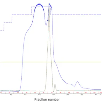
Lactobacillus casei의 배양물을 이용하여 단백질을 분리한 후 3, 10, 30, 100 KDa의 ultrafiltration membrane을 이용하여 최종적으로 3~100 KDa(단백질 성분 A: protein components A)과 10~30 KDa(단백질 성분 B: protein components B)의 범위의 단백질로 농축 제조하여 단백질 정량(Bradford kit)을 통하여 단백질 정량한 후 실험에 사용하였다. 그리고 농축한 단백질을 FPLC(fast protein liquid chromatography, Sephadex 75)을 이용하여 농축된 단백질을 분리 정제하여 fraction collec- tor로 각 3 mL/fraction으로 순서대로 회수하여 항암 활성 측 정에 사용하였다. Fraction별로 나온 peak를 토대로 3번 frac- tion에서 9번 fraction을 이용하여 항암 활성을 측정하여 항암 효과를 지니고 있는 성분을 확인하기 위해 실험에 사용하였 다(Fig. 1, 2).
2. Lactobacillus casei의 배양물에서 분리한 단백질 농축물의 항암 활성 효과
Lactobacillus casei의 배양물에서 분리한 단백질을 3, 10, 30, 100 KDa의 ultrafiltration membrane을 이용 Ultrafiltration 으로 농축한 농축물인 단백질 성분 A(protein components A) 와 단백질 성분 B(protein components B)에 대해 정상 세포의 세포 독성 및 암세포에 대한 생육 억제 활성을 측정하였다.
위의 두 가지 농축물 모두 정상 세포에 대해 농도 의존적으 로 세포 독성이 증가하는 경향을 나타냈지만 최고 농도에서
20% 미만으로 비교적 낮은 세포 독성을 나타내어 정상 세포
Fig. 1. Separation of protein components A(3~100 KDa, Ultra- filtration) from culture supernatant of Lactobacillus casei on FPLC(Sephadex 75) and each fraction(3 mL) was collected(blue line - protein(280 nm); brown line - conductivity).
Fig. 2. Separation of protein components B(10~30 KDa, Ultra- filtration) from culture supernatant of Lactobacillus casei on FPLC(Sephadex 75), and each fraction(3 mL) was collected(blue line - protein(280 nm); brown line - conduc- tivity).
Fig. 3. Cytotoxicity of the protein components A(3~100 KDa, Ultrafiltration) from the culture supernatant of Lacto- bacillus casei on normal cell line, 293(kidney).
Fig. 4. Cytotoxicity of the protein components B(10~30 KDa, Ultrafiltration) from the culture supernatant of Lacto- bacillus casei on normal cell line, 293(kidney).
에 대한 세포 독성은 낮은 것으로 나타났다(Fig. 3, 4).
5가지 암세포주에 대한 암세포 생육 억제 활성을 측정한 결과, 두 가지 농축물 모두 모든 암세포주에 대해서 농도 의 존적으로 암세포의 생육 억제 효과가 증가하는 경향을 나타 내었고, 이 중 최적 농도로는 100 μg/mL의 농도로 두 가지 농축물에서 모두 70% 정도의 암세포 생육 억제 효과를 나타 냄으로서 두 농축물에서 항암 효과를 지니고 있는 단백질 성분을 함유하고 있는 것으로 확인하였다(Fig. 5, 6). 따라서 두 가지 단백질 농축물인 단백질 성분 A(protein components A)와 단백질 성분 B(protein components B)의 항암 효과를 나 타내는 최적 농도는 100 μg/mL로 암세포의 생육 억제 활성 이 70% 정도로 높은 항암 효과를 나타내는 것을 확인할 수 있었다.
3. 단백질 정제 물질에 대한 항암 활성 효과
FPLC를 이용하여 단백질 성분 A(protein components A)와 단백질 성분 B(protein components B)의 농축물을 분리한 분
Fig. 5. Inhibition ratio of the growth at the cancer cell line (stomach, lung, breast, ovary, colon) in adding the protein components A(3~100 KDa, Ultrafiltration) from the culture supernatant of Lactobacillus casei.
Fig. 6. Inhibition ratio of the growth at the cancer cell line (stomach, lung, breast, ovary, colon) in adding the protein components B(10~30 KDa, Ultrafiltration) from the culture supernatant of Lactobacillus casei.
획물을 fraction collector를 이용하여 분획물을 회수하여 실 험에 사용하였는데, 분리된 peak를 중심으로 3번에서 9번 fraction을 이용하여 정상 세포의 세포 독성 및 암세포주에 대한 암세포 생육 억제 활성에 대한 실험을 수행한 결과, 3, 8, 9번 fraction에서는 정상 세포에 대해 50% 정도 높은 세포 독성 효과를 나타내었고, 6, 7번 fraction에서는 세포 독성 효 과를 나타내지 않았다. 반면 암세포에 대한 생육 억제 활성 에서는 다른 분획물들에서는 활성을 나타내지 않았으나, 7 번 fraction에서만 모든 암세포주에 대해 60~70% 정도로 높 은 암세포 생육 억제 활성을 나타내었다(Table 1, 2). 이는 위 의 두 가지 농축물에서 분리한 분획물 모두에서 같은 효과 를 나타내었다. 따라서 Lactobacillus casei의 배양물에서 분 리한 단백질 중 항암 효과를 나타내는 성분을 함유하고 있 는 것으로 나타났다.
Table 1. Inhibition ratio of the growth at the cancer cell line(stomach, lung, breast, ovary, colon) in adding the fractions isolating the protein components A(3~100 KDa, Ultrafiltration) from the culture supernatant of Lactobacillus casei
Fraction number 3 4 5 6 7 8 9
293
(kidney, normal) 53±2 0 0 0 0 40±1 47±2
AGS
(stomach cancer) 0 0 3±1 4 55±4 6±2 0
A549
(lung cancer) 0 0 0 0 63±1 9±2 0
MCF-7
(breast cancer) 2 0 6 8±3 64±2 0 0
SK-OV-3
(ovary cancer) 0 0 4 10±5 63±1 5 5
LoVo
(colon cancer) 0 0 0 0 64±2 5 7
Table 2. Inhibition ratio of the growth at the cancer cell line(stomach, lung, breast, ovary, colon) in adding the fractions isolating the protein components B(10~30 KDa, Ultrafiltration) from the culture supernatant of Lactobacillus casei
Fraction number 3 4 5 6 7 8 9
293
(kidney, normal) 54±2 17±5 42±4 0 0 53±3 52±5
AGS
(stomach cancer) 18±4 16±4 6±3 14±2 65±5 0 0
A549
(lung cancer) 0 6±2 0 0 72±1 0 0
MCF-7
(breast cancer) 15±3 15±5 0 16±3 70±2 3±1 0
SK-OV-3
(ovary cancer) 14±2 10±4 0 0 73±1 7±2 0
LoVo
(colon cancer) 0 0 0 5±1 67±1 0 0
유산균의 한 종류인 Lactobacillus casei는 기존에 많은 효 능을 가지고 있다고 보고되어지고 있고, 계속 연구가 되고 있다. 하지만 이 균주가 배양하면서 분비되어 나오는 물질 이 포함되어 있는 배양물에 대한 연구는 미비한 상태이기 때문에 배양물에서 분리한 물질이 높은 암세포 생육 억제 효과를 나타냈기에 이는 향후 바이오 의약품을 개발하는데 있어서 기초 자료로 활용될 수 있는 가치를 지니고 있을 것 이다.
요 약
최근 유산균에 대한 관심이 많아지고 그와 관련된 수많 은 제품들이 제조되고 유통되고 있다. 이러한 유산균의 기 능으로는 가장 먼저 정장 작용을 들 수 있고, 그 외 항암 효 과, 유당 단백질의 흡수 증진, 혈청 콜레스테롤의 저하 등
많은 기능을 가지고 있다.
이에 따라 유산균의 한 종류인 Lactobacillus casei의 배양 물로부터 단백질을 분리한 것으로 Ultrafiltration membrane (3, 10, 30, 100 KDa)으로 분리 농축한 단백질 물질인 단백질 성분 A(protein components A)와 단백질 성분 B(protein com- ponents B)을 분리하여 실험에 사용하였고, 이 물질을 이용 하여 보다 세밀한 분리를 위해 FPLC(fast protein liquid chro- matography, sephadex 75, Amersham)를 이용하여 분석된 peak 를 이용하여 3번부터 9번까지 분획물을 얻어 실험에 사용하 였다.
위에서 얻은 단백질 물질들을 이용하여 정상 세포와 암 세포주를 이용하여 세포 독성 및 암세포 생육 억제 활성을 측정한 결과, 단백질 성분 A(protein components A)와 단백 질 성분 B(protein components B)의 물질에서 최적 농도인 100 μg/mL의 농도에서 정상 세포에 대한 세포 독성은 20%
정도로 낮은 세포 독성 효과를 나타내 정상 세포에 대한 독 성 효과는 없는 것으로 나타났고, 5가지 암세포주(위암, 폐 암, 유방암, 난소암, 대장암)에 대한 암세포 생육 억제 활성 에서는 70% 정도의 높은 암세포 생육 억제 효과를 나타내 항암 효과를 나타냈다. 그리고 FPLC를 이용하여 분리한 분 획물에서는 3, 8, 9번 분획물에서는 정상 세포에 대한 세포 독성이 50%를 나타내 독성 효과를 나타냈고, 그 외의 분획 물에서는 정상 세포에 대한 독성이 나타나지 않았다. 그 중 7번 분획물에서 암세포에 대한 생육 억제 활성을 나타낸 결 과, 70% 정도의 높은 암세포 생육 억제 활성을 나타내 항암 활성을 지닌 성분으로 확인하였다. 그 외의 분획물에서는 거의 효과를 나타내지 않았다. 따라서 Lactobacillus casei의 배양물에서 분리한 성분이 항암 효과를 나타내고 있는 것을 확인하였다.
감사의 말
본 연구는 지식경제부․한국산업기술 평가원 지원 (주)쎌 바이오텍의 우수제조 기술연구센터(ATC-10026108)의 지원 에 의한 것입니다.
참고문헌
1. Alm, L. 1982. Effect of fermentation on B-vitamin content of milk in Sweden. J. Diary Sci. 65:353-359.
2. Ayebo, A. D., Shahani, K. M. and Dam, R. 1981. Antitumor component(s) of yogurt: Fractionation. J. Diary Sci. 64:
2318-2323.
3. Bae, H. S., Baek, Y. J. and Yoon, Y. H. 1993. Antitumor activity of Lactobacillus casei against Sarcoma 180 and Lewis lung carcinoma in mice. Kor. J. Appl. Microbiol.
Biotechnol. 21:247-255.
4. Charmichael, J., Degraf, W. G., Gazdar, A. F., Minna, J. D.
and Michell, J. B. 1987. Evaluation of a tetrazolium based semiautomated colorimetric assay. assissment of chemosen- sitivity testing. Cancer Res. 47:936-942.
5. Friend, B. A. and Shahani, K. M. 1984. Antitumor pro- perties of Lactobacilli and dairy products fermented by
Lactobacilli. J. Food Prot. 47:717-723.
6. Gilliland, S. E. 1989. Acidophilus milk products: A review of potential benefits to consumers. J. Dairy Sci. 72:2483- 2494.
7. Kang, D. G., Kang, S. P., Chang, D. H., Kim S. H. and Yoon, S. S. 2001. Isolation and characterization of Lactobacillus strains isolated from Korean feces. Korean J. Food Sci.
Technol. 33:567-573.
8. Kato, I., Kobayaahi, S., Yokokura, T. and Mutai, M. 1981.
Antitumor activity of Lactobacillus casei in mice. Gann.
72:517-523.
9. Lilly, D. M. and Stillwel, R. H. 1965. Probiots: Growth promoting factors produced by microorganism. Science 147:747-748.
10. Mann, G. V. and Spoerry, A. 1974. Studies of a surfactant and cholesteremia in the Maasai. Amer. J. Clin. Nutr. 27:
464-469.
11. Matsuzaki, T., Yokokura, T. and Azuma, I. 1985. Anti-tumor activity of Lactobacillus casei on Lewis lung carcinoma and line-10 hepatoma in syngeneic mice and guinea pigs.
Cancer Immunol. Immunother. 20:18-22.
12. Mugitani, H. and Furue, H. 1987. Evaluation on the safety of LC9018-study of single subcataneous administration of LC9018 to healthy men. Biotherapy 1:286-289.
13. Pelletier, C., Bouley, C., Cayuela, C., Bouttier, S., Bourlious, P. and Bellom-Fontaine, M. N. 1997. Cell surface charac- teristics of Lactobacillus casei subsp. casei, Lactobacillus paracasei subsp. paracasei, and Lactobacillus rhamnosus strains. Appl. Environ. Microbiol. 63:1725-1731.
14. Raiboud, P. 1980. The 3rd International Academic Seminar on Lactobacillus and Health, Seoul. p 23.
15. Reddy, G. V., Shahani, K. M. and Banerjee, M. R. 1973.
Inhibitory effect of yoghurt on Ehrlich ascites tumor cell proliferation. J. Natl. Cancer Inst. 50:815-817.
16. Shackelford, L. A., Rao, D. R., Chawan, C. B. and Pulusani, S. R. 1983. Effect of feeding fermented milk on the in- cidence of chemically induced colon tumors in rats. Nutr.
Cancer 5:159-164.