INTRODUCTION
The bone marrow stroma contains primitive and potentially self-renewing cells that can differentiate into various mesen- chymal cells. These mesenchymal stem cells (MSC) are capa- ble of supporting the expansion and differentiation of hema- topoietic stem cells (HSC) in vitro and enhance hematopoi- etic engraftment in vivo (1-5). Furthermore, MSC express class I, but not class II, histocompatibility antigens, and they are not immunogenic in in vitro assays and in preclinical or clinical models.
Recently, it has been reported that there are mesenchymal precursors in cord blood and peripheral blood of normal indi- viduals. The results obtained demonstrate that after systemic infusion of cord blood MSC into nude mice, human DNA was found in the marrow and in ex vivo expanded stromal cells grown from the marrow of transplanted animals. Several groups also have reported bone and fat differentiation capac- ity from cord blood-derived nonhematopoietic cells (6-10).
Despite the low initial frequency in cord blood, MSC have a
tremendous expansion potential in culture. Due to the lim- ited number of HSC in a given cord blood unit, the outcome of HSC transplantation (HSCT) could be improved by coen- graftment of the MSC in addition to the hematopoietic pre- cursors.
Several lines of evidence suggest that MSC produce some essential hematopoietic growth factors (1-5). Based on these reports, we hypothesized that exogenously administered MSC with cytokine gene-transfected would show additive or aug- mented effects on HSC expansion. In this study, mononuclear cells (MNC) were harvested from cord blood and seeded in long-term culture for ex vivo MSC expansion. We transfected culture-expanded MSC with granulocyte macrophage-colony stimulating factor (GM-CSF) and stem cell factor (SCF) cyto- kine genes and then cotransplanted with cord blood MNC in nonobese diabetic/severe combined immunodeficiency (NOD/SCID) mice to further promote HSC engraftment.
Although CD34+ cells can be selected for the preparation of HSC, many cord blood HSCT practically use unselected MNC. Therefore, we chose unselected MNC samples instead
Jin-Yeong Han, Rhee Young Goh, Su Yeong Seo*, Tae Ho Hwang�, Hyuk Chan Kwon�, Sung Hyun Kim�, Jae Seok Kim�, Hyo Jin Kim�, Young Ho Lee�
Departments of Laboratory Medicine, Microbiology*, Pharmacology�, and Internal Medicine�, Dong-A University College of Medicine, Busan; Department of Pediatrics�, Hanyang University College of Medicine, Seoul, Korea
Address for correspondence Jin-Yeong Han, M.D.
Department of Laboratory Medicine, Dong-A University College of Medicine, 1 3-ga, Dongdaesin-dong, Seo-gu, Busan 602-715, Korea
Tel : +82.51-240-5323, Fax : +82.51-255-9366 E-mail : [email protected]
*This study was supported by a grant of the Korea Health 21 R&D Project, Ministry of Health & Welfare, Republic of Korea (03-PJ1-PG3-20500-0065).
242
Cotransplantation of Cord Blood Hematopoietic Stem Cells and Culture-Expanded and GM-CSF-/SCF-Transfected Mesenchymal Stem Cells in SCID Mice
Received : 8 June 2006 Accepted : 18 September 2006 Mesenchymal stem cells (MSC) are multipotent in nature and believed to facilitate
the engraftment of hematopoietic stem cells (HSC) when transplanted simultane- ously in animal studies and even in human trials. In this study, we transfected cul- ture-expanded MSC with granulocyte macrophage-colony stimulating factor (GM- CSF) and stem cell factor (SCF) cytokine genes and then cotransplanted with mono- nuclear cells (MNC) to further promote HSC engraftment. MNC were harvested from cord blood and seeded in long-term culture for ex vivo MSC expansion. A total of 1××107MNC plus MSC/ L were introduced to the tail vein of nonobese diabetic/
severe combined immunodeficiency mice. After 6-8 weeks later, homing and engraft- ment of human cells were determined by flow cytometry and fluorescence in situ hybridization studies. The total nucleated cell count and the engraftment of CD45+/
CD34+ cells and XX or XY positive human cells were significantly increased in cotrans- planted mice and even higher with the cytokine gene-transfected MSC (GM-CSF>
SCF, p<0.05) than in transplantation of MNC alone. These results suggest that MSC transfected with hematopoietic growth factor genes are capable of enhanc- ing the hematopoietic engraftment. Delivering genes involved in homing and cell adhesions, CXCR4 or VLA, would further increase the efficiency of stem cell trans- plantation in the future.
Key Words : Mesenchymal Stem Cell; Hematopoietic Stem Cell Transplantation; GM-CSF; Stem Cell Factor;
Engraftment, Transfection; Cord Blood
of selected samples as the source of stem cells for transplan- tation. Our results show that human cells were significantly increased in cotransplanted mice, and the number was even higher with the cytokine gene-transfected MSC than with transplantation of MNC alone. These data not only provide direct evidence for the presence of MSC in cord blood, but could also add to therapeutic profile of expanded MSC for clinical use.
MATERIALS AND METHODS Ex vivo MSC culture
Cord blood samples were collected from the umbilical vein after full-term vaginal delivery. Informed consent was ob- tained from all mothers. The mononuclear cell fractions from cord blood were suspended at a concentration of 1×10⁶cells/
mL in MesenCult medium (StemCell Technologies, Inc., Vancouver, BC, Canada) and seeded in T-25 flasks. Cultures were maintained at 37℃in humidified air and 5% CO2with a regular change of medium. After removal of non-adherent cells, the adherent layer was further cultured until 80-90%
confluence. At this point, the cell population was expanded by successive passage and resulting adherent cells were used for experiments.
The adherent cells were detached with 0.05% trypsin-ethy- lenediamine tetraacetic acid and washed with phosphate- buffered saline (PBS). We observed morphology of detached cells after Wright-Giemsa staining. The phenotype and puri- ty of MSC were assessed by flow cytometry. The cells were washed with PBS containing 2% bovine serum albumin, and incubated with FITC-conjugated CD45 (BD Biosciences, San Jose, CA, U.S.A.), PE-conjugated CD73 (BD Biosciences), CD105 (DiNonA, Inc., Seoul, Korea), and CD166 (BD Bio- sciences) on ice for 30 min in the dark. Those 3 monoclonal antibodies were chosen because of the popularity and acces- sibility. After fixation with 1% paraformaldehyde, flow cyto- metry was performed on the FACSort instrument (Becton Dickinson, San Jose, CA, U.S.A.).
GM-CSF/SCF gene transfection
For plasmid transfections, pEGFP-N1 DNA (Clontech, Palo Alto, CA, U.S.A.) that encodes an enhanced green fluo- rescent protein (EGFP) was used. Expanded MSC were trans- fected with an EGFP-expressing vectors after cloning with GM-CSF (pEGFP-N1/GM-CSF) or SCF (pEGFP-N1/SCF).
To create pEGFP-N1/GM-CSF or pEGFP-N1/SCF, human peripheral blood monocytes were used for reverse transcrip- tion-polymerase chain reaction. After sequencing of GM- CSF and SCF genes, EcoRI was used for digestion to insert cDNA3.1 and NotI was treated for SCF. Stable MSC trans- fectants that expressed high levels of GFP were cloned by
limiting dilution and selected using 2 mg/mL neomycin (Invitrogen Corp., Carlsbad, CA, U.S.A.). The GFP-express- ing cells were screened by a microplate spectrofluorometer (Shimadzu, Kyoto, Japan) and confirmed by an enzyme-linked immunosorbent assay (ELISA) (R & D Systems, Minneapo- lis, MN, U.S.A.). Stable clones of MSC with GFP and GM- CSF or SCF were maintained in Dulbecco Modified Eagle Medium (Gibco Laboratories, Grand Island, NY, U.S.A.) in the presence of neomycin until transplantation. GFP-positive MSC were visualized using a fluorescence microscope (Olym- pus, Japan) and then used for transplantation to NOD/SCID mice.
Transplantation and analysis of engraftment
NOD/SCID mice were purchased from Korea Research Institute of Bioscience and Biotechnology (KRIBB, Daejeon, Korea) and were maintained in sterile microisolator cages.
All mice received sublethal total body irradiation with a sin- gle dose of 250 cGy. Twelve to 24 hr after irradiation, cells in a final volume of 200 L were injected through the tail vein. We designed 4 experimental groups, and each group received 1×10⁷cells as a total: group 1 (n=5) received MNC only, group 2 (n=7) received a mixture of MNC plus MSC, 0.5×10⁷respectively, group 3 (n=7) received a mixture of MNC plus GM-CSF MSC, 0.5×10⁷respectively, and group 4 (n=6) received a mixture of MNC plus SCF MSC, 0.5×10⁷ respectively, per mouse. MNC were isolated from cord blood using a density gradient centrifugation. All cells were used fresh without cryopreservation.
Six to 8 weeks later, blood samples were collected from the retroorbital venous plexus of the NOD/SCID mice. An aliquot of the blood was used for complete blood cell counts by Sys- mex XE-2100 (Sysmex Corp., Kobe, Japan). Human cell engraftment was detected by flow cytometry, using FITC- conjugated CD45 (BD Biosciences) and PE-conjugated CD34 (BD Biosciences) antibodies. Positive cells were identified by comparison with isotype controls. A part of venous blood was also exposed to a hypotonic solution, and fixed in metha- nol/acetic acid fixative. Nuclei were then stained with CEP X (DXZ1) alpha satellite probe (Vysis, Inc., Downers Grove, IL, U.S.A.) and CEP Y (DYZ1) satellite III probe (Vysis) for fluorescence in situ hybridization (FISH) identification of human cells. A total of at least 500-700 nuclei were exam- ined in each case under a fluorescent microscope.
The results were expressed as mean±standard deviation.
Differences in engraftment percentages between experimen- tal conditions were analyzed by Student-t test. A p value less than 0.05 was considered statistically significant.
RESULTS
Adherent MSC could be expanded from all of the cord
blood analyzed. Primary cultures of MSC mainly consisted of colonies of spindle-shaped fibroblastoid cells (Fig. 1A).
Confluence was usually reached after 2-3 weeks. Wright-Gie- msa staining of detached MSC showed a large cell popula- tion characterized by abundant cytoplasms with occasional vacuoles and projections, fine nuclear chromatins, and one or more nucleoli (Fig. 1B). Flow cytometry analyses demon- strated that MSC expressed CD73, CD105, and CD166 in all cases, but were negative for CD45 (Fig. 2). The purity of expanded MSC was 85-97%.
At 24 hr after transfection, increased GFP expression was identified both by spectrofluorometry and ELISA. The trans- fected MSC showed visible GFP expression as early as 3 hr after transfection. The highest efficiency of the transfection was obtained after 5-7 days of culture both with pEGFP-N1/
GM-CSF and pEGFP-N1/SCF (Fig. 3, 4). On average 50- 70% of transfected MSC expressed GFP. Detached MSC were then ready for transplantation. ELISA detected increased GM- CSF levels from MSC cultures after transfection (273.78± 148.33 pg/mL) compared to those before transfection (34.35
±17.06 pg/mL) (p<0.05, n=4). SCF also increased from 1.30
±1.05 pg/mL to 24.10±20.21 pg/mL post transfection, however, there were no statistical differences (p>0.05, n=4).
In MNC transplantation alone, the absolute numbers of total nucleated cell (TNC) count was 0.08±0.04 (×103/ L) (n=5) comparable to 0.13±0.05 (×103/ L) (n=7) in mice transplanted with MNC plus MSC (Table 1). However, higher levels of human XX or XY cell by FISH (Fig. 5) were found in transplantation of MNC plus MSC, 0.41±0.41 (×103/ L) vs. 1.52±0.81 (×103/ L) (p<0.05). Cotransplantation of MNC and GM-CSF-transfected MSC most enhanced eng- raftment. The TNC count, 4.63±0.96 (×103/ L) (n=7), and the percentage of XX or XY cells, 5.68±0.58 (×103/ L), were significantly higher (p<0.05) than those after transplan- tation of MNC alone. Also, higher levels of TNC count, 2.93
±0.33 (×103/ L) (n=6), and XX or XY cells, 3.43±0.49 (×103/ L), were obtained following cotransplantation of MNC plus SCF-transfected MSC (p<0.05). Engraftment study with immunophenotypic analysis of CD45+/CD34+ cells by flow cytometry showed similar results (Table 1, Fig. 6).
Fig. 1.Inverted phase contrast microscopic findings of cultured cord blood MSC at passage 2, day 14 (A) (×150). Wright-Giem- sa stained smear of the harvested MSC (B) (×1,000).
A B
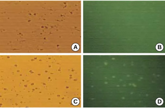
Fig. 3.GM-CSF transfection studies. Microscopic images of phase contrast (A) and fluorescence microscopes (B) before transfection.
Micrographs of phase contrast (C) and fluorescence microscopes (D) post transfection day 5. The panel D shows GFP fluorescence, indicating GFP-expressing MSC after transfection (×200).
A B
C D
Fig. 4.SCF transfection studies. Microscopic images of phase con- trast (A) and fluorescence microscopes (B) before transfection.
Micrographs of phase contrast (C) and fluorescence microscopes (D) post transfection day 7. The panel D shows GFP fluorescence, indicating GFP-expressing MSC after transfection (×200).
A B
C D
SSC-H\SSC-Height
1,000 800 600 400 200 0
0 200 400 600 800 1,000 FSC-H\FSC-Height
Fig. 2.Representative immunophenotypic analysis of MSC ex vivo cultures from the cord blood. CD45-/CD73+ (95.7%), CD45-/CD 105+ (97.4%), and CD45-/CD166+ cells (94.9%) were evident by flow cytometry.
FL2-H\CD73 PE
104 103 102 101 100
100 101 102 103 104 FL1-H\CD45 FITC
FL2-H\CD105 PE
104 103 102 101 100
100 101 102 103 104 FL1-H\CD45 FITC
FL2-H\CD166 PE
104 103 102 101 100
100 101 102 103 104 FL1-H\CD45 FITC
DISCUSSION
Genetic modification of stem cells is an attractive target for gene therapy because of their higher proliferative capaci- ty and long-term survival compared with other somatic cells.
MSC have been demonstrated to be able to express exogenous protein for an extended period of time and to maintain this ability after transplantation in vitro (1, 11-14). Gene modi- fied MSC are useful as therapeutic tools for various tumors.
MSC with forced expression of interferon beta were shown to inhibit the growth of malignant cells in vivo (11, 12). Gene- modification of MSC with therapeutic cytokines also clearly augmented the antitumor effect and prolonged the survival of tumor-bearing animals (13). Recently more efficient non- viral DNA transfection methods have been introduced to ob- tain stably transfected human cells (14, 15).
Ex vivo expanded MSC have been infused in several trans- plantation studies. It is possible that a therapeutic benefit from MSC could be obtained by local production of growth factors and the provision of temporary immunosuppression.
Human MSC have a high expansion potential, genetic stabili- ty, and reproducible characteristics proven in widely dis-
persed laboratories (1-3). No adverse events during or after MSC infusion have been reported, and no ectopic tissue for- mation has been observed.
Based on these results, we hypothesized that exogenously administered MSC with cytokine gene-transfected would show additive or augmented effects on HSC expansion. Among the various cytokines, we have previously demonstrated that the combination of SCF, thrombopoietin, and flt3 ligand is sufficient for ex vivo expansion of cord blood (16). With the addition of granulocyte (G)-CSF, the clonogenic and homing potentials of cord blood were even higher, although statisti- cally significant differences were not observed. Ramirez et al. (17) also have shown that SCF, interleukin (IL)-3, IL-6, and GM-CSF could induce the up-regulation of several adhe- sion molecules involved in the mobilization and homing of HSC. Currently GM-CSF is one of the most widely used cytokines for the purpose of stem cell mobilization and for the treatment of neutropenic patients. Therefore, we trans- fected culture-expanded cord blood MSC with GM-CSF and SCF cytokine genes, and then cotransplanted with cord blood MNC in NOD/SCID mice to further promote HSC engraft- ment. We demonstrated that human cells were significantly increased in cotransplanted mice and even higher with the cytokine gene-transfected MSC than in transplantation of MNC alone. Cotransplantation of MNC and GM-CSF-trans- fected MSC most enhanced engraftment.
Cotransplantation studies in animal models as well as in humans showed that primary or culture-expanded MSC pro- mote the engraftment of HSC (18-22). Cotransplantation of MSC and cord blood (18, 19, 21) or mobilized peripheral
MNC, mononuclear cells; MSC, mesenchymal stem cells; GM-CSF, granulocyte macrophage-colony stimulating factor; SCF, stem cell factor; TNC, total nucleated cells.
TNC (×103/ L)
Mean±SD p value
XX or XY Cells (%)
Mean±SD p value
CD45+/CD34+ (%)
Mean±SD p value
MNC alone 0.08±0.04 0.41±0.41 0.23±0.11
MNC plus MSC 0.13±0.05 >0.05 1.52±0.81 <0.05 0.83±0.65 >0.05
MNC plus GM-CSF MSC 4.63±0.96 <0.05 5.68±0.58 <0.05 5.92±1.95 <0.05
MNC plus SCF MSC 2.93±0.33 <0.05 3.43±0.49 <0.05 2.29±0.46 <0.05
Table 1.Comparison between HSC transplantation alone and cotransplantation of MNC plus MSC alone or MNC plus GM-CSF-/SCF- transfected MSC
Fig. 5.XY human cells identified from the peripheral blood of NOD/
SCID mice 7 weeks after cotransplantation of GM-CSF-transfect- ed cord blood MSC and MNC (×1,000). One orange signal indi- cates one copy of the X chromosome, and one green signal indi- cates one copy of the Y chromosome.
X
X X
Y
Y Y
Fig. 6.Representative CD34+/CD45+ cells (8.7%) detected from venous blood of NOD/SCID mice 7 weeks after cotransplantation of GM-CSF-transfected cord blood MNC and HSC.
SSC-H\SSC-Height
1,000 800 600 400 200 0
0 200 400 600 800 1,000 FSC-H\FSC-Height
FL2-H\CD34 PE
104
103
102
101
100
100 101 102 103 104 FL1-H\CD45 FITC
blood (20) CD34+ cells resulted in a significantly higher level of engraftment than with transplantation of CD34+ cells only. Cotransplantation of human leukocyte antigen (HLA)- identical sibling culture-expanded MSC with an HLA-iden- tical sibling HSC was also feasible and safe without imme- diate infusional or late MSC-associated toxicities (22). The mechanism of the enhancing effect is still unknown. Several lines of evidence suggest that MSC produce several essential hematopoietic growth factors including IL-6, IL-11, SCF, leukemia inhibitory factor, and flt3 ligand and express adhe- sion molecules and extracellular matrix proteins that are known to play an important role in HSC homing such as VCAM1, E-selectin, collagen I, and fibronectin (1-5, 20, 23). Different studies report that MSC have an immunomod- ulatory effect and are able to suppress proliferation of T lym- phocytes both by cell-cell interaction and via soluble factors (22-25).
It has been claimed that there are mesenchymal precur- sors in cord blood and peripheral blood of normal individu- als (6-8, 26, 27). Despite the low initial frequency in cord blood, MSC have a tremendous expansion potential in cul- ture. Compared to bone marrow MSC, cord blood cultures were slower to establish, but once established, the growth was maintained over multiple passages. Using optimized iso- lation and culture conditions, cells showing a characteristic mesenchymal morphology and immune phenotype were iso- lated. The frequency of MSC ranged from 0-2.3 clones per 1
×108MNC. Due to the limited number of HSC in a given cord blood unit, the outcome of HSC transplantation could be improved by coengraftment of the MSC in addition to the hematopoietic precursors.
Erices et al. (8) previously investigated the capability of transplanted cord blood MSC to home and survive in the marrow of nude mice. They have detected human DNA after transplantation in the marrow of recipients as well as in ex vivo-expanded stromal cells prepared from the marrow of transplants. In this study we found that cord blood MSC cotransplantation enhances human cell engraftment. This enhancement was greater after cotransplantation of GM-CSF- and SCF-transfected MSC. According to Wagner et al. (26) several genes involved in mesodermal differentiation were differently expressed on MSC from human bone marrow, adipose tissue, or umbilical cord blood. These results implied that potentials of mesodermal development might be differ- ent depending on MSC preparations. By upregulating genes involved in proliferation and homing of cord blood MSC, this might contribute to establish a reliable genetic modifi- cation system for clinical applications.
MSC are multipotent and believed to facilitate the engraft- ment of HSC when transplanted simultaneously in animal studies, and recently, even in human trials. In this study, we transfected culture-expanded cord blood MSC with GM-CSF and SCF cytokine genes and then cotransplanted with cord blood HSC to further promote HSC engraftment. The TNC
count and the engraftment of CD45+/CD34+ cells and XX or XY positive human cells were significantly increased in cotransplanted mice and even higher with the cytokine gene- transfected MSC (GM-CSF>SCF, p<0.05) than in transplan- tation of HSC alone. Our results indicate that MSC may serve as a platform to deliver hematopoietic growth factors in clin- ical stem cell transplantation. Delivering genes involved in homing and cell adhesions, CXCR4 or VLA, would further increase the efficiency of stem cell transplantation in the future.
REFERENCES
1. Kassem M. Mesenchymal stem cells: biological characteristics and potential clinical applications. Cloning Stem Cells 2004; 6: 369-74.
2. Le Blanc K, Pittenger MF. Mesenchymal stem cells: progress toward promise. Cytotherapy 2005; 7: 36-45.
3. Kan I, Melamed E, Offen D. Integral therapeutic potential of bone marrow mesenchymal stem cells. Curr Drug Targets 2005; 6: 31-41.
4. Jiang Y, Jahagirdar BN, Reinhardt RL, Schwartz RE, Keene CD, Ortiz-Gonzalez XR, Reyes M, Lenvik T, Lund T, Blackstad M, Du J, Aldrich S, Lisberg A, Low WC, Largaespada DA, Verfaillie CM.
Pluripotency of mesenchymal stem cells derived from adult marrow.
Nature 2002; 418: 41-9.
5. Jones EA, Kinsey SE, English A, Jones RA, Straszynski L, Mered- ith DM, Markham AF, Jack A, Emery P, McGonagle D. Isolation and characterization of bone marrow multipotential mesenchymal progenitor cells. Arthritis Rheum 2002; 46: 3349-60.
6. Erices A, Conget P, Minguell JJ. Mesenchymal progenitor cells in human umbilical cord blood. Br J Haematol 2000; 109: 235-42.
7. Goodwin HS, Bicknese AR, Chien SN, Bogucki BD, Oliver DA, Quinn CO, Wall DA. Multilineage differentiation activity by cells isolated from umbilical cord blood: expression of bone, fat, and neu- ral markers. Biol Blood Marrow Transplant 2001; 7: 581-8.
8. Erices AA, Allers CI, Conget PA, Rojas CV, Minguell JJ. Human cord blood-derived mesenchymal stem cells home and survive in the marrow of immunodeficient mice after systemic infusion. Cell Trans- plant 2003; 12: 555-61.
9. Huss R, Lange C, Weissinger EM, Kolb HJ, Thalmeier K. Evidence of peripheral blood-derived, plastic-adherent CD34(-/low) hemato- poietic stem cell clones with mesenchymal stem cell characteristics.
Stem Cells 2000; 18: 252-60.
10. Villaron EM, Almeida J, Lopez-Holgado N, Alcoceba M, Sanchez- Abarca LI, Sanchez-Guizo FM, Alberca M, Perez-Simon JA, San Miguel JF, Del Canizo MC. Mesenchymal stem cells are present in peripheral blood and can engraft after allogeneic hematopoietic stem cell transplantation. Haematologica 2004; 89: 1421-7.
11. Studeny M, Marini FC, Champlin RE, Zompetta C, Fidler IJ, And- reeff M. Bone marrow-derived mesenchymal stem cells as vehicles for interferon- delivery into tumors. Cancer Res 2002; 62: 3603-8.
12. Studeny M, Marini FC, Dembinski JL, Zompetta C, Cabreira-Hansen M, Bekele BN, Champlin RE, Andreeff M. Mesenchymal stem cells:
potential precursors for tumor stroma and targeted-delivery vehicles for anticancer agents. J Natl Cancer Inst 2004; 96: 1593-603.
13. Hamada H, Kobune M, Nakamura K, Kawano Y, Kato K, Honmou O, Houkin K, Matsunaga T, Niitsu Y. Mesenchymal stem cells (MSC) as therapeutic cytoreagents for gene therapy. Cancer Sci 2005; 96:
149-56.
14. Nakashima S, Matsuyama Y, Nitta A, Sakai Y, Ishiguro N. Highly efficient transfection of human marrow stromal cells by nucleofec- tion. Transplant Proc 2005; 37: 2290-2.
15. Martinet W, Schrijvers DM, Kockx MM. Nucleofection as an effi- cient nonviral transfection method for human monocytic cells. Bio- technol Lett 2003; 25: 1025-9.
16. Lee YH, Han JY, Seo SY, Kim KH, Lee YA, Lee YS, Lee HS, Hur WJ, Han H, Kwon HC, Kim JS, Kim HJ. Stem cells expressing hom- ing receptors could be expanded from cryopreserved and unselect- ed cord blood. J Korean Med Sci 2001; 19: 635-9.
17. Ramirez M, Segovia JC, Benet I, Arbona C, Guenechea G, Blaya C, Garcia-Conde J, Bueren JA, Prosper C. Ex vivo expansion of umbil- ical cord blood (UCB) CD34+ cells alters the expression and func- tion of 4 1 and 5 1 integrins. Br J Haematol 2001; 115: 213-21.
18. Noort WA, Kruisselbrink AB, in’t Anker PS, Kruger M, van Bezooi- jen RL, de Paus RA, Heemskerk MH, Lowik CW, Falkenburg JH, Willemze R, Fibbe WE. Mesenchymal stem cells promote engraft- ment of human umbilical cord blood-derived CD34+ cells in NOD/
SCID mice. Exp Hematol 2002; 30: 870-8.
19. in’t Anker PS, Noort WA, Kruisselbrink AB, Scherjon SA, Beek- huizen W, Willemze R, Kanhai HH, Fibbe WE. Nonexpanded pri- mary lung and bone marrow-derived mesenchymal cells promote the engraftment of umbilical cord blood-derived CD34+ cells in NOD/
SCID mice. Exp Hematol 2003; 31: 881-9.
20. Angelopoulou M, Novelli E, Grove JE, Rinder HM, Civin C, Cheng L, Krause DS. Cotransplantation of human mesenchymal stem cells enhances human myelopoiesis and megakaryocytopoiesis in NOD/
SCID mice. Exp Hematol 2003; 31: 413-20.
21. Bensidhoum M, Chapel A, Francois S, Demarquay C, Mazurier C,
Fouillard L, Bouchet S, Bertho JM, Gourmelon P, Aigueperse J, Char- bord P, Gorin NC, Thierry D, Lopez M. Homing of in vitro expand- ed Stro-1- or Stro-1+ human mesenchymal stem cells into the NOD/
SCID mouse and their role in supporting human CD34 cell engraft- ment. Blood 2004; 103: 3313-9.
22. Lazarus HM, Koc ON, Devine SM, Curtin P, Maziarz RT, Holland HK, Shpall EJ, McCarthy P, Atkinson K, Cooper BW, Gerson SL, Laughlin MJ, Loberiza Jr FR, Moseley AB, Bacigalupo A. Cotrans- plantation of HLA-identical sibling culture-expanded mesenchymal stem cells and hematopoietic stem cells in hematologic malignancy patients. Biol Blood Marrow Transplant 2005; 11: 389-98.
23. Majumdar MK, Thiede MA, Haynesworth SE, Bruder SP, Gerson SL. Human marrow-derived mesenchymal stem cells (MSCs) express hematopoietic cytokines and support long-term hematopoiesis when differentiated toward Stromal and osteogenic lineages. J Hematother Stem Cell Res 2000; 9: 841-8.
24. Chung NG, Jeong DC, Park SJ, Choi BO, Cho B, Kim HK, Chun CS, Won JH, Han CW. Cotransplantation of marrow stromal cells may prevent lethal graft-versus-host disease in major histocompati- bility complex mismatched murine hematopoietic stem cell transplan- tation. Int J Hematol 2004; 80: 370-6.
25. Maitra B, Szekely E, Gjini K, Laughlin MJ, Dennis J, Haynesworth SE, Koc ON. Human mesenchymal stem cells support unrelated donor hematopoietic stem cells and suppress T-cell activation. Bone Mar- row Transplant 2004; 33: 597-604.
26. Wagner W, Wein F, Seckinger A, Frankhauser M, Wirkner U, Krause U, Blake J, Schwager C, Eckstein V, Ansorge W, Ho AD. Compar- ative characteristics of mesenchymal stem cells from human bone marrow, adipose tissue, and umbilical cord blood. Exp Hematol 2005;
33: 1402-16.
27. Bieback K, Kern S, Kluter H, Eichler H. Clinical parameters for the isolation of mesenchymal stem cells from umbilical cord blood. Stem Cells 2004; 22: 625-34.