大 훌훌 放 射 線톨홉 學會誌第21 卷第5 號pp.777-782, 1985 Journal of Korean Radiological Society
,
Vo 1. 21,
No.5,
1985題部內 R農傷의 超音波所見
延世大學校 醫科大學 放射線科學敎室
李↑吉雨
·
李鍾太•
씁〕亨植·
全 JII훌愛 - Abstract -Sonographic Findings of the Intra.Abdominal Abscess
K.W. lee
,
M.D.,
J.T. lee,
M.D.,
H.S. Yoo,
M.D.,
S.Y. Jun,
M.D.Department of Radiology, College of Medicine, Yonse; Universitv
With the recel1t development of real time technology, ultrasound is now uniquely well suited fOf the detec.
tion of abdominal abscess. The high accuracy of ultrasound in detecting abdominal abscess has been widely documented, also. The typical ultrasound appearance of an abscess is that of a ffuid collection with diffuse, weak internal echos, representing the internal contents. Authors analyzed echogenicity and regularity of the abscesses in surgically and pathologically confirmed intra-abdominal abscesses, 38 cases except intra-abdominal
。 rgan abscesses
1. 38 cases consist of 30 intra-abdominal abscesses, 4 pelvic abscesses, 4 retroperitoneal abscesses 2. Numbers of the abscesses are 32 single and 6 multiful‘
3. Echgenicity consist of 9 cystic
,
14 semisolid,
6 solid,
9 mixed.4. Mean size of abscess is 6
x
5 cm in single. but, not measureable in multiful5. Abscess wall have relatively irregular inner wall (28 cases) and relat얘Iy regular outer wall (21 cases) in our cases Consequently, echogenicity of the abscess is variable
But, relatively semisolid. Abscess have relatively irregular inner wall and relatively regular outer wall on ultrasonogram.
1. 絡 드ð a빼
題部內 뼈陽의 예후는 治爾하는 경우는 30 %의 mo.
le 超音波機術이 發展되면서 題部內 뼈場의 음?斷에는 획기적인 發展을 이룩하게 되었다.,
비록 超音波的으로 題部內 流體홉積의 籃別이 쉽지는 않지만5l 臨皮的 효狀과 그 解ffU學的 部位에 의해서 題
rtality, 治續안하는 경우는 80-100%의 mortality를 갖 部內 g農場의 超音波에 의한 조기진단 및 治續에 그 有
는 흉愚으로써 1) 조기진단과 治癡는 중요하다고 생 각 用性이 增加함에 따라 6' 著者들은 題部內 觸場으로 外 된다. 1970 年代 初期까지 題部內 觸覆의 談斷과 局所 科的 또는 病理學的으로 確談된 총 38 17IJ를 對象으로 化하는 데는 主로 臨똥뾰狀과 單純 X 線擺影에 依하였 o 農傷의 에코와 慶의 規則性에 대한 超音波所見을 分析 며 2,3) 그외 많은 %究가 있었지만 1974 年 Gray Sca. 比較하여 文敵考察과 함께 報告하는 바이다.
이 논문은 1985 년 9 월 12 일에 접 수하여 1985년 10 월 5 일에 채택되었음.
- 777-
-大韓放射線훌훌學會誌 . 第 21卷 第5號 1985-
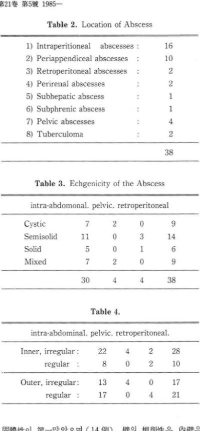
Table 2. Location of Abscess
1) Intraperitioneal
abscesses 2) Periappendiceal abscesses
3)Retroperitoneal abscesses
4)Perirenal abscesses
5)Subhepatic abscess
6)Subphrenic abscess 7)
Pelvicabscesses
8)Tuberculoma
m m
2 2 1 1 4 2 延쁘·大學校 醫科大學 放射線科學敎室에서는 1982 年
1 月부터 1984 年 9 月까지 題部內 體覆이 의심되어 超 音波檢훌를 施行한 愚者中 外科的 또는 病理學的으로 確談된 38 Ø1J를 對象으로 하였다.
단 題部內 魔器의 R農場은 對象에서 除外시켰다.
Real time
Scanner
는Toshiba SAL -
30 A를 썼 고Contact scanner
는SEARLE
PHO/SONIC-SM
ALPHA를 flJ用하였다.
2.
對象 및 方法38
Table 3. Echgenicity of
theAbscess 3.
結 果intra-abdomona
l.pelvic. retroperitoneal
Cystic
Semis이icl
Solicl Mixecl
9 M 6 9 0 3 l 0 2
nU nU
nι
7 l 1L
「D
7I
-
A) 年敵 및 性져IJ 分布
性別分布는 男子가 20 Ø1J 女子가 18 Ø1J로 서로 비슷 하며 年觀別로는 30 代가 11 例로가장 많았다 (Table 1).
38 4 4 30 Table 1.
Age & Sex Distribution
-F K-M
Total
Table 4.
intra-abclomina
l.pelvic. retroperitonea
l.28 10 2
2
4 022
8 Inner
,irregular
regular
76
n
5 6 3 l
2 7 4
”O
A양
AaT
1A
「D
nU
Age
0- 20 21-30 31- 40 41-50 51-60 +61
1
3
1721 0 4 4
0
13 17Outer
,irregular:
regular
固體f똥이 第-많았으며 (14Ø1J), 慶의 規f{IJ샘E 은 內융좋은 不規則하였던 i列가 28 例로 더 많았으며 外율좋은 規則的 인 쪽이 21 例로 더 많았다
(Table
3,
4).各 位置의 g農場에 超音波효例를
Fig.
1-7 에서 보여 주고있다1960 年代 初期부터 1974 年에
Gray Scale
超흉波 機術이 공급되 기 전 까지 여 러 가지 題흩內 R잃陽의 조기 발견과 局所化에 대한 報告가 나왔지만 2,3,4,7,8,9,10>1974 年
Gray Scale
超音波裝備가 나온 뒤 로는 g農覆 찢쫓4.
考 38D)
째얘 轉 般
에코는 半 R農이 題용內 전체에 있는 R흥흩內 R農場이 16 Ø1J로 第 -많았£며 그외 體場의 位置는 Table
2
와 같다.c) H農場의 數 및 2fS均크기 는 單發性인 경 우가 이였고 多發性인 경우가 6 例이었다 2fS均크기는 性인 경우가 6
x
5cm
이었고 多發性인 경우는 이 不加能하였다.18
各 位置에 R農場의 에 코 및 慶의 規則性은 20
B) 빼傷의 位置
超훌波所見
- 李f법雨 外 :JlYi部內 ~횡의 超흡波所見 -
의 超音波所見에 特異{生이 t曾加되 었 다.
1974
年F lemm ing J ense n
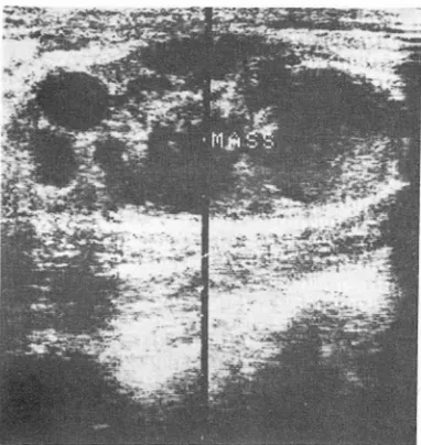
等은 脫홈內 n짧場과 血Fig.
1.Intraperitoneal abscess: abscess is located
anteroinferior site of the Right kidney,has regular
outer wallin shape & se misolid in echogenicity.
Fig. 2. Intrapertioneal abscess: abscess is located superior
睡의 등?斷에 는 超音波檢좁가 가치 있는 方法이 라 하였으 며 거의 묘두 에코 Free 로 나타난다고 하였다.
또한 뼈場파 血睡은 臨tR的 根據에 의해 鍵別이 되야 한다고 했으며 양쪽 오두 內部에 코플 갖는네 그것은 各 各 血塊와 破壞物의 破片째문이라고 했다 "
1974
年Nabil F.
M혀이ad 等은A & B mode
스 캔무로 手術後 題뾰內 뼈場은 超音波談斷에서CD
잘 구 별되는 가장자리를 갖고, (J! 檢훌동안 모양의 變化가 없으며, @ 깊은 구조까지 音波가 전달된다고 하였다:장 (陽) 파 짧別은 φ 불분명 한 8융의 용좋
@
가스가 언어!존재하는 것으로 하였다7>
著者들의 liFf究는 題홈內 뼈場은 覆8탱性에코에서 固體 性에 코까지 다양하였으며 8행場의 률훌은 半에 서 規則的,半 에서 不規則的하였다.
1976
年Morton Schneider
等은 賢魔 및 賢驗周圍에~ftJ9i:愚의 超音波的 所見中에 서 賢周圍觸場을 除外하 고는 談斷的 價f直가 적다고 하였으며 또한 뽑周圍體揚 은 ψ 周圍魔器에 이 동을 주며
@ Echoless
하나So-
nolucent
하지 않고 @Sound beam
이 약해 진 다고 하였site of the uterus and bladder
, has irregular outer Fig. 3. Periappendiceal abscess: abscess has solid in
wallin
shape &mixed in
echogenicity.echogenicity and
irregular outerwall in shape
- 779-
-大햄放射線醫웰會찮 : 第21 卷 第 51lií
1985-
Fig.
4. Tuberculoma:this solid elliptical mass with multiful
highechogenic septum is confirmed intra- abdominal tuberculoma.
Fig. 5. Subphrenic abscess: abscess has
semisolidin echogenicity and regular outer wall in
shape.PE: pleural effusion
으며 賢周圍血睡과 題模後앓 睡錫을 鍵었Ij해 야 된다고 하 였다.
賢魔에 超音波的 利用은 經皮的 接近을 위한 안내 자 로써가 제일 좋다고 하였다 1 1>
著者플의 맑究는 賢周圍體場이 2 例로써 에 코는 固體 的 또는 半固體的이 였고 比較的 잘 形成된 활을 갖고 있었다.
1977
年에 는F aye C. Laing
等은 題模後뾰의 睡塊를 發見하는데 超音波檢훌가 나오기전의 응?斷的 方法은 非 特異的이었으며 @흥體後脫의 R農場에 超흡波所見은CD
題 鷹後置의 非對稱性 @ 가장자리가 잘 그려지는 睡塊 @ 周團廳器의 變位 @ð콩合 또는 훌뼈性에코를 갖고 @ 後 音影의 增加가 보인다고 하였다.8흉模後脫의 體, 血被,1*는 超音波的으로 籃別이 안 되고 臨tf:tlE狀과 관계되야 한다고 했다 12'
著者들은 題體後홈8農場 01 2 例있었으며 外율좋은 모두 規J'!1j的이고 內율좋은 各 1例씩 規則的, 不規則的이었다.
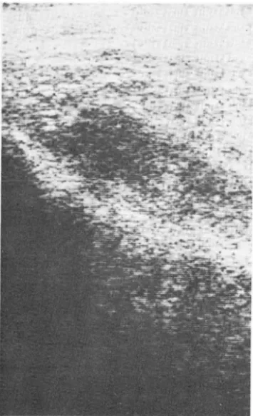
Fig.
6. Pelvic abscess: on oblique pelvic scan,mixed echo
mass is located postrosuperior
siteof the uterus
and showsirregular outer
wall.- 李1농 i펌 外: H동部內 ~傷의 超풍 i皮所見 -
.. ’ ....
;07-:\"“ ‘ ’
.
‘Fig. 7. Retroperitoneal abscess: this abscess shows relatively regular outer wall in shape and semisolid in
echogenicity1977 年에
Bruce D
,Doust
等은 다른 題股內의 流 體의 훔積으로부터 願場의 超音波的 分離맑究에서 超音 波檢훌는 固體的 病짧로부터 流體性病책의 鍵別응?斷에 基本的인 檢훌이며 ;急性血睡(24 시간 以內)은 어떤 內 部에코플 갖지 않으며 慢性血睡은 彈한 內部에코를 가 지며 그 慶이 不規則하다 했다.題水jJE은 @ 周圍廳器에 압박을 안주며 @ 전체가 에 코프리하고 (3) 慶이 잘 그려지나 不規則하다고 하였다.
R農場은 ú) 內部에코가 있고 @ 不規則한 慶을 가지나 慢性血睡보다 덜 不規則한 즉 微細하게 不規則 하며 @ 周圍廳器에 變位 또는 압박감을 주며 @ 散tE性으로 分 布완 약한 에코를 가지며 자주 띠 (帶) 같은 內部에코를 갖으며 @ 다른 流體훌積보다 적은 後흡影增加가 있고
@ 切斷 斷面은 타원형이라고 하며 觸場의 慶,to용{生血睡,
陽은 다른 流體훌積의 뿔보다 털 規則的이라고 했다 5) 著者플의 @다究는 38171]中 2817U가 內룡좋은 不規則했고 外뿔은 21 例가 規則的이였다
1978 年에
Kenneth J. W. Taylor
등은 복강과 骨盤 홈 R農場에Grey Scale
超音波檢훌의 精確度에서 g農場 은 여 러 양의 破片을 포함하는 흥뼈睡塊로 敏感性은93
%, 特異性은
98.6
%, 全體 精確度는96.8
%로 報춤하였다13 ,
著者들의 líff究中 骨盤홈R없場은 混合에 코가 2 例, 훌훌뼈
에코가 ... 2 例 있였으며 모두에서 觸覆의 휠좋은 不規則하였
다.
1982
年에Eric
,C.
Martin
等은 題용內 觸陽의 治 續法£ζ로는 安全하고 셨果的인 超音波플 利用한 經皮없F g農法을 추천하였으며 6' 著者들도 이 方法으로 治覆를 하고 있다1983 年에 는
Howard W.
Raymo떠 등이 體陽은 部位,期間, 機術的 要素에 따라 純햄-하게 훌훌6힘性에 서 固體性 까지 多養한 超音波所見을 보인다고 하였으며 전형적인 超흡波所見은 散tE性으로 分布된 약한 內部에 코플 가지 는 流뽑의 홈積이 며 또한 中隔體 또는 破壞物의 破片을 뜻하는 臨狀 또는 조립한 에코의 지역이 보일 수 있다 고하였다.
後音影은 變化的이 며 慶도 變化하는 規則性을 보여 주고 가스의 훌積도 때때로 發見된다고 하였으며 超音 波的으로는 觸場은 陽, 윷윷8힘, 血雙睡 또는 破壞性 睡陽 과 아주 유사하다고 하였다
…
5.
結 論最近 超훌波裝備의 分解能增加와 더 불어 願部內 R農傷 의 談斷 및 治爾에 상당한 發展이 있었으며 著者들은 題部內 魔器의 腦場을 除外한 題용內 g農覆으로 外科的 또는 病理學的 方法으로 確該된 38 例를 對象으로 觸場 의 에코와 뚫의 規則性을 分析하여 다음과 같은 結論을 얻었다.
A) 體場의 에코
16 例의 g홍홈內 R農場과 10 例의 뾰重周圍購場은 多樣 한 에 코를 보여 주였으며 骨盤內觸場은 混合 또는 훌8包 性에 코 뿐이 였 다. n흥體後홈종8農場, 賢/휩圍 8農場, 結核睡은 各各 2 例였으며 순서 대로 半固體性, 半固體性 및 固體 性, 半固體性 및 混合性에 코이 냐 結核睡은 1 例에 서 中 隔體이 있었다.
B) 뼈場뿔의 規則性
臨홈內 g홉場과 뾰좋흩周圍8農陽은 內설좋은 1:1:較的 不規則 하고 外툴좋은 非特異的이 였다
- 781-
-大짧放射짧醫學會誌 짜 21~ 했5械 1985-
骨盤뾰內 뼈傷은 內• 外탤 모두 不規則的하였으며 題 138: 13-15, }anuary 1982
體後훈H農傷, 뽑周圍8農場, 結核睡은 內현좋은 非特異的이 7. Nabil F. Maklad, Bruce D. Doust, et al.: Ultrasonic 고 外뚫은 포두 規則的이 였 다 diagnosis of postoperative intraabdominal abscesses
Radiology 113:411-422, November 1914
REFERENCES 8. Eugene 0.5., Robert S. Ozeran: Retroperitoneal space abscesses. Surgery, Cynecology & Obstetrics, }une 1969 1. Altemeier W .A., Culbertson W.R. , Fullen W .D., et al.: 1203-1208
intraabominal abscess. Am. }. Surg 125:10-19, 1913. 9. W .A. Altemeier, j. Wesley Alexander: Retroperitoneal 2. Morton A. Meter., joseph. P. Whalen, et al.: Radiologic abscesses. Archives of surgery. Vol. 83. Oct. 1961
feature of extraperitoneal effusions. Radiology 512-524
/04;249-251, August 1912. 10. Mark M. Ravitch, Ormand C. julian, et al.: Current pro-
3. joseph P. Whalen. , Alfred S. Berne. et al.: The ex- blems in surgery subphrenic abscess. january 1912. 1-41
traperit9rJeal perivisceral 떠 t pad. Radiology 92:466-480, 11. Morton Schneider, joshua A. Becker et al.: Sonographic-
March 19.69. radiographic correlation of renal and perirenal infections
4. Flemming jensen, jan Fog Pedersen: The value of A}R 121:1007-1004, 1916
ultrasonic scanning in the diagnosis of intraabdominal 12. Faye C. Laing, Richard P. jacobs.: Value of ultrasonography abscesses & hematoma. Surgery, Cynecology & in the detection of retroperitoneal inflammatory masses Obstetrics. September 1914. Volume. 139. 326-328. Radiology 123:169-112. April 1911
5. Bruce D. Doust, Francisco Quiroz, et al.: Ultrasonic distinc- 13. Kenneth j.W., Jane F. McI et al.: Accuracy of grey scale tion of abscesses from other intraabdominal fluid collec- ultrasound diagnosis of abdominal and pelvic abscesses tions. Radiology 125:213-218, October 1911. in 220 patients. the lancet, january 14, 1918. 83-84 6. Eric C. Martin, Kaern B. Karlson, et al.: Percutaneous 14. Seminars in ultrasound: Abscesses. june 1983. 11-19
drainage of postoperative intraabdominal abscesses. A}R