大韓放射線홉學會誌 Vol. XVIll. No. 4. 1982
-Absσact -
뇌종양에 대한 전산화 단층 촬영
국럽의료원 방사선과
노계동·한상돈 조동일 · 이창준 황인순 · 김한석
Computed Tomographic Evaluation of
Brain
AbscessGae Dong Ro. M.D .• 5ang Don Han, M.D .• Dong 111 Cho, M.D., Chang )oon Lee, M.D., In 500n Whang, M.D., Han 5uk Kim, M.D.
Department of Radíology
,
Natíonal Medlcal CenterThe use of computed tomography is most reliable in diagnosis and management of brain abscess.
Authors analized 17 cases of pathologically and clinically proven brain abscess during the period of 39 monthsfrom )an. 1978 to Mar. 1982 at National Medical Center.
The results were as follows:
1. The sex ratio 9 males to 8 females
,
and no sex difference was seen,
and the greatest number of cases were seen below the age of 30 (65%).2. The otogenic infection was the most frequent p'redisposing factors (8 cases). Other predisposing factors were postoperative infection (2 cases). pulmonary infection (2 cases). and congenital heart disease (2 cases).
The most common site of involvement was posterior fossa (5 cases). Next was temporallobe (4 cases).
and temporoparietallobe (3 cases).
3. Most common presenting symptoms were headache, fever, focal neurological signs, and dizziness.
4. Among the 22 brain abscesses of 17 patients
,
the most frequent CT finding in precontrast scan was a low density surrounded by a faint dense or dense ring (11 cases). Next was purely low density (6 cases). Associated hydrocephalus was found in 4 cases,
and multiple or multiloculated abscess was see n i n 4 cases.5. In post contrast scan, brain abscess usually show complete. oval or round shaped, thin, evening ring enhancement with mild or moderate surrounding edema
,
but there was no specific enhancement.I
서 론망율이 20'" 40 %로 높았마}, 2 , 3 , 4 , 5 , 6, 7)
그러나 전산화 단충 촬영솔이 입상적으로 응용된 이 래, 뇌농양의 유무판옐. 정확한 칭법부위의 측정 및 선 뇌농양의 조기진단과 효과척인 치료는 임상적으로상 별검사로 가치가 있무며 사앙율도 상당한 강소를 가져 당히/어려운 문제로 되어 있다. 과거 뇌농양의 방사선 온 것£로 알려졌다 8.9)
학척 진단방법으로는 혈판 조영솔, 동위원소를 이용한 저자들운 국럽의료원 ’u갱외과에 내원하여 CT 를 시 뇌조사 및 뇌실 촬영술에 의존하여 왔무냐 진단이 늦고 행한 환자중 뱅리조직학쩌 및 임상척으로 확진된 17 예 정확한 뇌농양의 침뱀부위에 대한 평가에 문제점으호 사 를 대상으로 입상소걱 및 CT 소견을 분석 검로하고, 운
*온 논문은 1982 년도 국법의료원 임상연쿠바로 이루어졌음.
-676-
헌고찰과 함께 보고하는 바이 다.
II.
대상 및 방법50 세 이후에서는 5 예가 발생하였마 (Table 1)
2.
유발인자 및 칭범부위뇌농양에 걸리기 쉬운 영인윤 이엽이 (otogenic infe- 1978 년 1월부터 1982 년 3 월까지 국럽의료원 신경 ction)17 에중 8 예로서 가장 않았으며 j 수술후 강영,
외과에 내원하여 전산화 안충촬영을 시행한 환자중, 수 폐질환 l 선천성 심장질환이 각각 2 예의 밴도순드로 보 솔을 시행하여 영리조직학척A로 확진된 13예와 수술 였다.
이 불가능하였으나 임상적으로 확진된 4 에를 포항하여 침엄부위는 후두와가 5 예, 측두엽이 4 예, 측두우정 총 17 예의 22 영변을 대상으로 성옐 및 연령분포,유발 엽이 3 예, 전두엽이 2 예의 순무로 보였다 (Table←. 2).
인자 및 첨범부위, 주증상, 조영증강천 및 초영증강후 소견을 분석하여 보았다
m.
결 과1. 성별 및 연령분포
총 17 에중 낭자가 9 예, 여자가 8 예로 낭여차이가 없었으며 . 30 세 이하에서 11 예 (65%) 로 호발하였고
Table 1. Age and Sex Distribution
Sex Male Female Total
Age 1-9 10-19 20-29 30-39 40-49 50-59 60-69
?‘
1i 껴4
1i 끼j 꺼ι
껴4
?‘
-Q /
1
Total 8 17
3. 증 상
증상윤 발생 부위에 따라 다소 차이 가 있으나 두통이 14 예로 가장 않았으며 , 그 다음으로 발열, 국소적 신 청중상, 현기, 요성 및 구로의 순으로 나타났다 (Tab-
le 3).
4. 증상 발현시기부터 CT 시행까지의 기간과 CT 소견
꺼j j야
A4
증상 말현시기부터 CT 시행까지의 기간윤 17 예 중 15 예가 3 주와 4 주사이였으며, 1 주와 6 주가 각각
1 예 씌 이 었 다 (Table 4).
조영층강전 소견윤 주위 뇌실질보다 저밀도를 보인예 가 22 명변중 18예로 대부분이였으며, 그 중 저밀도를 가지며 주위에 희미한 환상밀도를 가진 예가 11예 (F-
ig. 1), 단순히 저 밀도를 가진 6 예 (Fig. 2), 공기 밀도 를 동반한 저밀도가 1예였다. 그 외 동둥한 밀도 및혼 합밀도가 각각 2 예였다 (Fig. 3). 동반된 수두증윤 4 예 였으며 , 다발성 혹은 다방성이 3 예 (Fig. 4) 에서 보 3
2
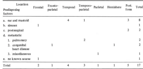
Table 2. Predisposing factors and Causative Factors andLocation of Brain Absceses
Lo ∞tion Frontal Fronto- Temporal Temporo- Parietal Hemishere Post. Total
Predispo síng parietal parietal fossa
factors
a. ear and mastoid 4 3 8
b. sinuses 1
c. postsurgical 2 2
d. metastatic
1. pulmonary 2 2
2. ∞ ngenital 2
h eart d isea se 3. miscellaneous e. no known sourse
Total 2 4 3 5 17
Table 3. Presei1ting Symptoms in 17 cases of Brain Abscess
Symptoms No. of Cases
Headache 14
Fever (380C) 11
Dizziness 7
Nausea and vomiting 4
Seizure 3
Nuchal rigidity 4
Focal neurological
Deficit 10
Table 4. Duration of Symptoms prior to Initial CT
Duration
1 week 2 weeks 3 weeks 4 weeks 6 weeks
Total
A
No. of Cases
8 7
17
였다.
조영증강후 소견윤 22 영연중 19 영연에서 환상조영 증강을 보였 무며 , 그 중 완전 환상이 14 예 (Fig .1,2,4).
불완전 환상이 5 예 (Fig. 3) 였고, 두께 는 앓고 균일한 것이 13 예로 대부분을 차지하였고 앓고 불균일한 것이 4 예 두꺼 운 것 이 2 예 였 으며 , 모양윤 원형 혹윤 난원 형이 16 예호 대부운이며 불규칙한 모양이 3 예였다-그 리고 환상조영층강후 내측벽이 외측벽보다 앓윤 것이 5 예였다 (Fig. 2). 결철성 조영층강운 3 예에서 보였으며 이들운 다발성 뇌농양의 경우에서만 보였다 (Fig. 4).
주위 뇌부종윤 대부분 경 01한 정도 (11 예) 내지 중둥도
(moderate. 7 예)를 보였으며 4 에에서 중둥도
(severe) 를 보였 다.
N. 고 찰
두개강내 염증성 질환올 가진 환자의 치료는 신경진 단학척 방법무로 항상 특이한 영리학척 특성을 냐타내 지 않기 때운에 진단이 어려운 것으로 알려졌고, 또 뇌 파요도솔, 방사선 동위원소를 이용한 뇌조사, 뇌실촬영 숭 및 혈판 조영솔로 국소화농성뇌영과 뇌농양의 강멸 이 항상 가능하지 않았고 사망율과 환한율이 높윤 것 으
B
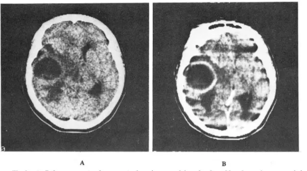
Fig. 1. A. Before contrast enhancement. there is a round low density with a dense ring surrounded by a moderate low density of edema in the left temporal region.
B. After contrast enhancement, there is a complete thin. evening ring enhancement at the site of the dense ring seen in A.
A B
Fig. 2. A. Before contrast enhancement, there is a purely low density in the right temporallobe with severe surrounding edema.
B. After contrast enhancement, there is a complete, thin ring enhancement with slight thinn- ing of medial abscess wall
A B
Fig. 3. A. Before contrast enhancement, there is no abnormality except bony destruction on the left petrous bone.
B. After contrast enhancement, a incomplete, thin ring enhancement in the left cerebellar hemisphere is seen.
679-
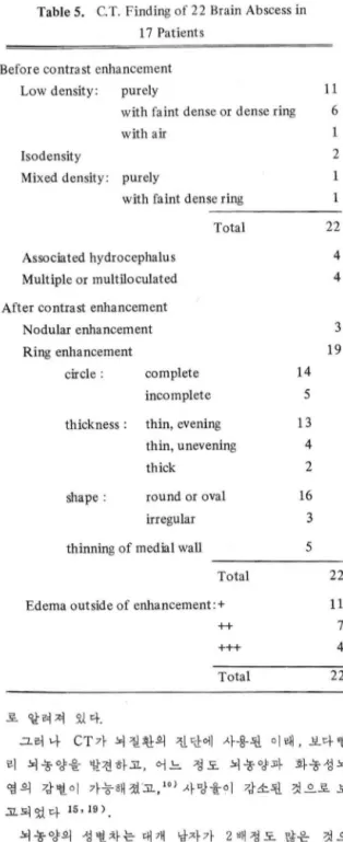
Table 5. C.T. Finding of 22 Brain Abscess in 17 Patients
Before contrast enhancement Low density: purely
with faint dense or dense ring with air
Isodensity
Mixed density: purely
with faint dense ring
Associated hydrocephalus Multiple or multiloculated After contrast enhancement
Nodular enhancement Ring enhancement
circle : complete incomplete
Total
thickness : thin, evening
shape :
thin, unevening thick
round or oval irregular thinning of medial wall
Total Edema outside of enhancement:+
++
+++
Total
로 알려져 있다-
14 5 13 4 2 16 3 5
11 6
2
22 4 4
3 19
22 11 7 4 22
그러 냐 CT 가 뇌질환의 진단에 사용된 이래 , 보다빨 리 뇌놓양을 발견하고, 어느 정도 뇌놓양파 화농성뇌 염의 강멸이 가능해졌고 10) 사망율이 강소된 것으로 보
고되었다 15,19 )
뇌놓양의 성별차는 대개 남자가 2 배정도 않윤 것무 로 알려졌으냐 1 , 2 ,4 ,6 , 8) 저자들의 청우 show10)와 같 이 냥녀차가 없었다. 연령훈포는 15 세 이하에서 Be-
Ilarl 가 43%, Like') 는 3 분의 1 로 가장 않았으며 , 원인윤 어련아이들에서 두부주위의 감염이 장되기 때문 이 아닌 생각했으며, 저자들의 정우 30 세 이하에서 65
%로 호발하였다.
유발언자는 과거에 이영 및 부비강엽, 외상후강엽,
뇌악영 둥의 빈도순 1, 2 ,4 ,6 )이었으냐, 최근에는 이 영 및 부닝|동 강영에 의한 발생율운 적어지고 선천성 심장질환, 획l 질환 1 1, 8 )의 밴도순이었다. 그러고 sh-
OW 6) 의 경우 후두와 뇌농양의 94 %가 이엽에서 발 생한 것이었다. 저자들의 정우 이엽이 8 에, 수술후강 영, 폐질환, 선천성심장질환이 각각 2 예로 과거와 닝l 슷한 말생 빈도순을 보였다.
뇌농양의 칭엄부위는 유말안자가 이영일때는 대개 즉 두엽, 후두와, 선두엽의 빈도순이었고 1 , 3) ,유발언자가 전 천이성일 경우 선두영, 두정엽, 측두엽의 빈도순이었다
.. ,8) 저자들의 경우후두와가 5 예, 측두영 4 예,측두 두정엽 3 예, 전두엽 2 예로 유발언자가 이엽이 않았기 때문에 이와 같윤 반도순 A로 나타난 것£로 생각한다.
입상증상은 두통, 말열, 국소척신청학적소견, 현기,
오심 및 쿠토, 자간동으로 마른 저 자들과 같운 증상을
보였 다 1 , 2 , 3 ,‘, 8)
조영층강천 소견운 whelanl9) 의 정우 총 23 명변중 저밀도가 20 예로 대부분을 차지하였고 그 안에는 희미 한 환상밀도를 동반한 16 에와 단순한 저밀도 4 예를 보 였고, 동등밀도가 3 예를 나타냈다. 저자들의 경우 총
22뱅변중 18예에서 저밀도를 보였고, 그 중희미한환 상밀도 동반이 11예, 단순한 저밀도 6 예와 공기밀도를 동안하여 쉽게 진단된 1예릎 보였다. 그리고 동등밀도 와 혼합밀도가 각각 2 예 호 바슷한 결파를 냐타냈 마.
뇌 놓양의 다발성은 Samson' ) 의 경우 5 ~30 %에서 보 인마고 했고, 저자들의 경우 2 예에서 보였다.
조영증강후소견운 Enzmann 10)의 동물실험에 의하 연 말기뇌염시기에 (4 ~ 9 일)에 두껍게 환상조영층강 이 일어냐고 l 시간후 저밀도 중심지역에 조영증강이 일 어 난다고 했으며 ,수위뇌부종과 덩어리효과 (Mass ef f-
ect) 가 제일 심하다고 했다. 초기피막형성시기 (1 0~
13 일)에 는 환상조영증강되고 시간이 지 낭에 따라 중 싱저밀도 지역에 일부 조영되고 주위뇌부종과 덩어리 효과가 강소된다고 했다. 말기 피막형성시기 ( 14 일 이 후)에 완전히 형성된 앓고 균일한 조영증강이 띈다고 한다 ZimmermanI3, 14) 의 정우 11 일에 초기띄막 형성, 25 일에 완선피악형성이 된다고 했다 whelan9) 의 정우 5 일에서 10 일 사이에는 불춧분한 띄막을 2 주이상에서 완전파악형성을 볼 수 있었무며, 총 20 예중 환상조영풍캉이 20 예고! 그 중 완선환상초영증강이 19 예로 가장 않았으며, 저자들의 경우 그와 비슷하게 완 선하고 앓고 균일한 환상조영증강을 대부분에서 보이
A B
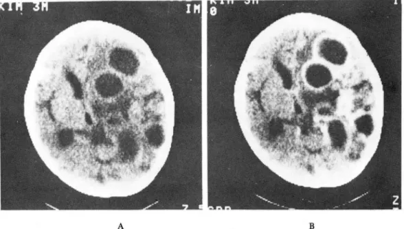
Fig.4. A. Before contrast enhancement, there are multiple low density with faint dense or dense ring surrounded by mild or moderate edema and a isodense nodule in the right hemisphere.
B. After contrast enhancement, multiple, multiloculated, complete or incomplete, thin ring enhancement and a dense nodular enhancement.
고 , 주위 뇌 부종도 정 01 한 것 내 지 중둥도 (moderate)가 17 예로 대부붐을 차지한 것윤 종상 말현후 대부분 3
"'4주에 전산화 단충촬영을 시행하여 말기 띄악형성시 기에 해당되어 그렇게 냐타냐는 것£로 생각된다. 3 예 의 결정성 조영층강운 전부 다딸성무로 생긴 경우에 보 였£며 뇌염시기에 해당되는 것으로 생 각된다.
CT 소견상 감벌을 요하는 질환으로서는 원발성 뇌종 양좋 특히 다형성교아종 (Glioblastoma Multitorme), 전이성 뇌종양, 용해혈종, 뇌정색증 둥이 있마'. whe-
lanl5 ) 과 허승재 16 ) 에 의하면 마형 성교아종과 선이성
뇌 종양윤 환 λ£조영 증강。l 두껍 고 균일하지 않으며 , 환 내 결절조영증강。l 보이 는 것 ξ 로 어느 정도 강멸이 가 능하며 용해혈종과 뇌갱색증윤 강벌이 용이하지 않고 임상증상과 영리검사소견을 참조할 때 보마 협게 강별 된다고 했다.
V. 결 론
1 성옐 및 연령폼포는 납자 9 에, 여자 8 예로 냥 녀차가 없었으며, 30 셰 이하에서 11 예 (65 %)로호발 하였다
2. 유발언자는 17 예중 5 예로 이엽이 가장 않았으 며, 수술후강영, 퍼l 질환, 선천성 심장질환이 각각 2 예 의 빈도순이었다.
칩법부위는 후두와 5 예, 측두영 4 예, 옥두두정영 3 예의 순이었다.
3. 임상증상운 우통, 발열‘ 국소신경학척층상, 현 기 , 오싱 및 우로둥을 냐타내었다
4. 뇌농양의 조영증강전 소견응 저밀도플 보인 것이 대부붐이었으며, 그 중 희미한 환상밀도를 동반한 것이 11예, 단순한 저밀도 6 에, 공기밀도를 통반한 것이 1 예에서 보였 A 며, 동둥밀도 및 혼합일도 각각 2 에였다.
수두증윤 4에에서 보였으며, 다발성 혹윤 다방성은 4 예서 보였다.
5 • 조영증강후 소견윤 22 뱅연중 19 예에서 환상 조 영 증강을 냐타냈으며 , 주로 완전하고, 앓고, 균일한 환 저자는 1978 년 1 월부터 1982 년 3 월까지 영리조 상을 보였다. 동안원 뇌부종은 대부분 경마한 정도내지 칙학적 및 엄상척으로 확진된 17 예의 22 뇌종양 환자 중둥도를 보였다.
의 입상 및 CT 소견을 분석 고찰한 결과 마음과 같운 결론을 얻었다.
-681-
REFERENCES
1. Bellar AJ
,
Sahar A,
Praiss 1 : 8raln abscess. Review of 89 cases over a perlod of 30 years. J Neurol Neurosurg Psychlatry 36:757-768,
7973.2. liske E
,
Weiskers NJ Changlng aspect of braln abscess. Revlew of cases In Wlnscosln 7940 through 7962. Neorology 14:294-300,
1964.3. Morgan H
,
Wood MW,
Murphy F : Experlence with 88 conscutlve cases of braln abscess.J
Neurosurg38:698-704
,
1973.4. Samson DS
,
Clark A current revlew of braln abscess. Am J Med 54:207-270,
1973.5. Shaw MDM
,
Russell JA Cerebellar abscess. j Neurol Neurosurg Psychlatry 38:429-435,
7975.6. Shaw MDM
,
Russell JA Value of computed tomo- graphy in the diagnosls of Intracranlal abscess.J Neurol Neurosurg Psychlatry 40:274-220
,
7977.7. J ouber M J, Stephnov 5 Computed tomography and surglcal treatment in intracranlal suppuration.
Report of 3 conscutive unselected cases of brain abscess and subdural empyeman.
J
Neurosurg 47:73-78
,
1977.8. Rosenblum ML
,
Hoff JT,
Norman D,
et al De- creased mortallty from braln abscess slnce advent of computerized tomorgraphyJ
Neurosurg 49:658-668
,
7978.9. Whelan MA
,
Hilar SK : Computed tomography as a guide In the diagnosls and follow-up of brain abscess.Radlology 135 :663-671
,
1980.10. Enzmann DR
,
Britt RH,
Yeager AS : Experlmental braln abscess evolutlon: computed tomographlc and neuropathologic correlatlon. Raäiology 733:733-722
,
7972.11. New PFT
,
Davis KR,
Ballantine HJ,
et al : Computedtomography in cerebral abscess. Radlology 727 :64 7- 646
,
7976.12. Martin G Non-otogenic cerebral abscess.
J
Neurol Neurosurg Psychiatry 36:607-670,
7973.13. Zimmerman RA
,
Patel 5,
Bilaniuk LT : Demonstra- tion of purulent intracranlal infection by computed tomography. Am J Roentgenol 727:755-765,
1976.
14. Zimmerman RA
,
Bilaniuk L T,
Shipkin PM,
et al Evolutio of cerebral abscess: Correlation of c/lnlcal features wlth computed tomography. Neurology 27:14-79,
7977.15. Weisberg LA Cerebral computed tomography In intracranial inflammatory disorders. A rch Neurol 37:737-742
,
7980.16. 허승제 적용인