I. Introduction
Dental implant therapy is now increasingly recommended for rehabilitation of partially and totally edentulous patients. The frequent application of dental implant as an alternative treatment modality is due to the high clinical success rate attributed to the osseointegration between implants and bone tissues.
Osseointegration is a histologic term meaning
“a direct structural and functional connection between ordered, living bone and the surface of a load-carrying implant at the light microscopic level."1)
For osseointegration to occur, the biocompatibility of implant material, the surface topography of the implant and the state of the host bed are needed to be controlled2).
A number of implant materials-including titanium and hydroxyapatite-are biocompatible.
But, there is less agreement as to what constitutes an ideal design3). The rapidity of
osseointegration may vary with different materials4).
Commercially-pure titanium has a predictable interaction with the environment5,
6).
Good tissue tolerance seems to depend on the adherent oxide layer formed within a millisecond of exposure to air7). This layer, rather than the metal itself, determines the interaction between implants and tissues.8, 9) Also, surface energy properties seem to be important in establishing initial cell adhesion and implant fixation.10~12) A clean, sterile, organic free, highly charged titanium oxide surface was reported to be ideal for bioadhesion10). In alloyed form, titanium is most commonly mixed with 6% aluminum and 4% vanadium(Ti-6Al-4V) for the improvement of the mechanical properties of pure titanium13). Titanium or titanium alloy can be used in form of substructures coated with a thin layer of hydroxyapatite or a plasma-spray technique14, 15). In order to 대한치주과학회지 : Vol. 27, No. 1, 1997
A Histological Study on the Bone Tissues Surrounding Coated and Non-Coated Titanium Implants in Beagle Dogs
Sang-Hoon H. Leem and Sung-Hee. Son
Department of Periodontology, School of Dentistry, Seoul National University
* 이 연구는 1994년도 서울대학교 병원 지정연구비(02-94-250)자원에 의한 결과임
eliminate fatigue phenomenon of dense hydroxyapatite, hydroxyapatite-coating of a biocompatible metal has been proposed16~19). It has been reported that hydroxyapatite-coating of an implant surface significantly enhances bone apposition and attachment strength at the interface.20, 21) But, the interfaces of coated titanium implants is not well documented and coatings still cannot be manufactured to be as dense as sintered hydroxyapatite19). Osteolysis and subsequent implant loosening are potential complications of hydroxyapatite-coating breakdown22), and surfaces with interconnected porosities are prone to implant infection23).
Commercially available dental implants sharply differ in their surface topographies. At the macroscopic level, there are screw-shaped, cylindrical, conical, and blade-shaped dental implants. At the microscopic level, some implants have smooth surfaces, whereas others have machined, textured, or porous surfaces3). Regardless of different surface characteristics, non-coated, coated or plasma- sprayed implants all have the capacity to achieve osseointegration4). Still, surface considerations are clearly important and cell behavior at the implant surface is modified by surface topography3, 24). There may be quantitative differences as to the amount of bone-implant contact with each particular type of surface4).
Despite the extensive literature on dental implants, only very few reports dealt with the healing of implants placed in maxillae. Maxilla differs from mandible in bone histology and anatomic structures, and unfavorable bone conditions of host beds for implantation may be more frequently encountered in the
maxillae.
This study was designed to investigate and compare bone tissues surrounding commercially-pure titanium implants, titanium plasma-sprayed titanium implants and hydroxyapatite-coated titanium implants placed in maxillae and mandibles at different periods of healing.
II. Materials and Methods
1. AnimalsThree inbred-strain, 1 year-old beagle dogs weighing approximately 12kg(certified by B.
K. Co., London, Great Britain) were used.
During the entire experiment, the remaining teeth were cleaned once a week. The animals were fed with a soft food diet.
2. Implants
Implants were received from the manufacturers in aseptic transfer packages with the following identification markings.
1. Commercially-pure titanium implant of thread-screw type : Br nemark implant (Nobelpharma AB, G teborg, Sweden);
3.75mm in diameter, 8.5mm in length 2. Titanium plasma-sprayed titanium
implant of cylinder type : Intramobile cylinder(IMZ) implant (Friedrichsfeld Ltd., Mannheim, Germany) ; 3.3mm in diameter, 8.0mm in length
3. Hydroxyapatite-coated titanium implant of cylinder type : Intramobile cylinder(IMZ) implant (Friedrichsfeld Ltd., Mannheim, Germany) ; 3.3mm in diameter, 8.0mm in length.
3. Surgical Procedures
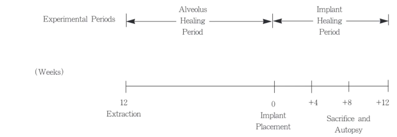
The design of surgical procedures is outlined Fig 1.
Twelve weeks before the implant placement, clinical and radiographic examination confirmed healthy supporting tissues in the maxillary and mandibular premolar regions of 3 dogs. The animals were anesthetized intravenously with sodium pentobarbital(Choong Wae Pharm. Co., Seoul, Korea) of 15mg/kg. The buccal and lingual mucosa were infiltrated with 2% lidocaine anesthetic solution containing 1 : 100,000 epinephrine(Yu Han Co., Seoul, Korea) to a total volume of about 1.7ml per operation site.
Premolars(P1, P2, P3 and P4) were extracted giving unilateral edentulous premolar areas in both jaws. Immediately after the extraction, the buccal and lingual flaps were repositioned and the wounds were closed by sutures with chromic Cat-gut 4-0(Ethicon Ltd., Edinburgh, United Kingdom). Antibiotic regimens of Penicillin G Procaine(Han Dok Remedia Co., Seoul, Korea) of 600,000 unit and Dihydro- streptomycin sulfate(Chong Kun Dang Co.,
Seoul, Korea) of 0.75g equivalent were administered through intramuscular injection for 5 days. The extracted sites were dressed with 10% povidone-iodine ointment (Sam Il Pharm. Co., Seoul, Korea) twice a day for a week. The animals were allowed to heal for 12 weeks.
For the implantation, healthy soft tissues and complete healing of alveoli were confirmed through clinical and radiographic examination. After anesthesia, buccal and lingual mucoperiosteal flaps were raised in pre-extracted premolar regions with modified ridge incision. Implant beds were then prepared and implants were placed using instruments and techniques described by the manufacturers. The implants were positioned approximately 6-7mm apart from each other.
In maxillae, the implants were placed with mesio-distal angulation. Using tension-release suture technique, buccal and lingual flaps were repositioned with chromic Cat-gut 4-0.
Antibiotic coverage and dressing were executed according to the regimens above mentioned. Radiographs were taken immediately after the placement.
Fig 1 Experimental Design of Surgical Procedures 12
Extraction (Weeks)
Experimental Periods
0 Implant Placement
+4 +8 +12
Sacrifice and Autopsy Alveolus
Healing Period
Implant Healing Period
4 weeks, 8 weeks and 12 weeks after implant placement, each dog was sacrificed respectively with an overdose of sodium pentobarbital. Radiographs of the implants in situ were obtained. There had been no direct functional load on the implants because they had been covered with attached gingiva for the entire period of the experiment.
4. Histologic Preparation
After anesthesia, carotid arteries and jugular veins were surgically exposed and tagged.
Through a carotid artery cutdown and cannulation, 3% glutaraldehyde (Sigma Chemical Co., St. Louis, U.S.A.) in 0.1 M phosphate buffer(pH 7.4) was administered in approximately 60 minutes for the fixation.
External jugular veins were severed to allow escape of the perfusate and blood. Jaw sections containing implants were resected using a band saw(PRO-TECH 3202, PRO- TECH POWER Inc., Gardena, U.S.A.) and immersed in fresh fixative. With IsometR low speed diamond wheel saw(B ehler Ltd., Lake Bluff, U.S.A.), each section containing one implant and surrounding tissue was cut longitudinally into three pieces through the implant. Two pieces were prepared separately for decalcified and undecalcified specimens for the examination with light microscope (OLYMPUS BH-2, OLYMPUS Ltd., Tokyo, Japan), and the third one for the examination with scanning electron microscope (JEOL 840A, JEOL Ltd., Tokyo, Japan).
(1) Preparation of decalcified specimens for light microscopic examination
Sections were refixed for an additional 2
days in 3% glutaraldehyde in 0.1 M phosphate buffer(pH 7.4) and processed for demineralization in nitric acid(Sigma Chemical Co., St. Louis, U.S.A.) for a week. The implants were then carefully removed.
Specimens were prepared for routine histologic preparations and embedded in paraffin.
Longitudinal serial sections were cut with sliding microtome(Model 860, American Optical Corp., Buffalo, U.S.A.) set at 4 m, and they were stained with hematoxylin and eosin.
(2) Preparation of undecalcified specimens for light microscopic examination
Sections were cleansed with tap water for 30 minutes for the removal of fixative and were dehydrated in 70% ethanol (Carloerba Reagenti S.R.L., Milano, Italy) for 3 days and 100% ethanol for 2 days. All blocks were then embedded in Osteo-Bed kit(Polyscience Inc., Warrington, U.S.A.) using extended infiltration time. After resin polymerization, each block was cut longitudinally of a thickness of less than 300μm using Isomet low speed diamond wheel saw. After each specimen was sliced of a thickness of less than 20μm with low speed grinding wheel (Dong Yang Science Co., Seoul, Korea ), it was completely polished with 3μm silicon carbide coated lapping and polishing film (South Bay Technology Inc., San Clemente, U.S.A.). Each specimen was stained with silver nitrate using von Kossa's method and toluidine blue.
(3) Preparation of specimens for scanning electron microscopic examination
Implants were detached manually from the
pieces of sample blocks. Specimens were rinsed in 0.1 M phosphate buffer(pH 7.4) and dehydrated in ascending series of ethanol. All Specimens were then dried with critical point dryer(BIO-RAD E3000, BIO-RAD Ltd., Microscience Division, Watford, England) and mounted on metal stubs. Using ion sputtering coater(BIO-RAD E5400, BIO-RAD Ltd., Microscience Division, Watford, England), each specimen was coated with gold-palladium.
III. Results
1. Clinical ObservationAt sacrifice, all implants were covered with healthy soft tissues and the radiographs did not demonstrate any evidence of peri-implant radiolucency.
2. Histological Observation
Inflammation or foreign body reaction could not be observed in the bone tissues interfacing the implants. Neutrophils, plasma cells or multinucleated giant cells could not be found in the bone tissues interfacing the implants.
Osteoclast was not seen near the interface while it could be found in old bone tissue.
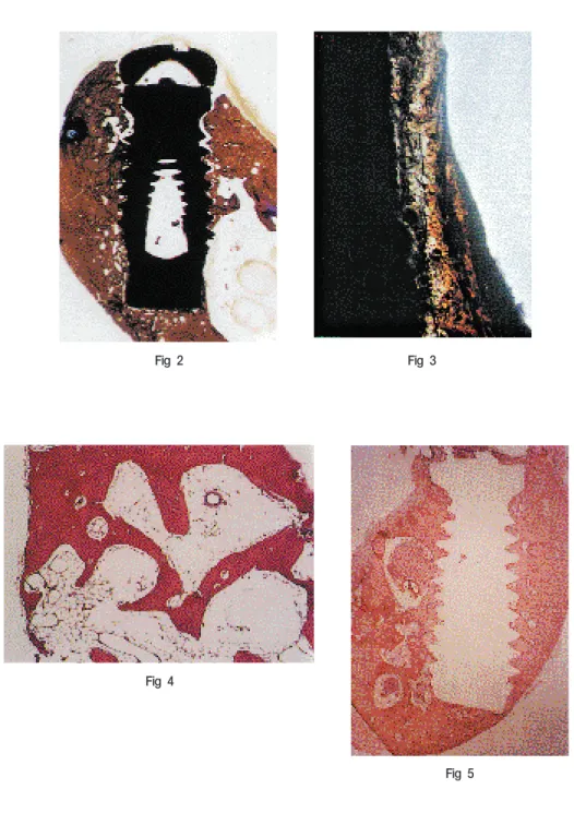
Thick compact bones were observed in mandibles(Fig 2), whereas very thin cortical plates and fine trabeculae were observed in maxillae(Fig 3, 4).
As early as 4 weeks, all implants placed in maxilla and mandible were in direct contact with bone at the interfaces. In cortical bone, direct contact with bone was observed at most surface areas of the implant, while both direct contact with bone and marrow space
interface were observed in spongious bone (Fig 5). The intervening marrow spaces between the mineralized bone and the surface of the implant could be seen throughout the study, though it was observed that the contact with bone gradually increased at the interface of the implant with the lapse of implantation time.
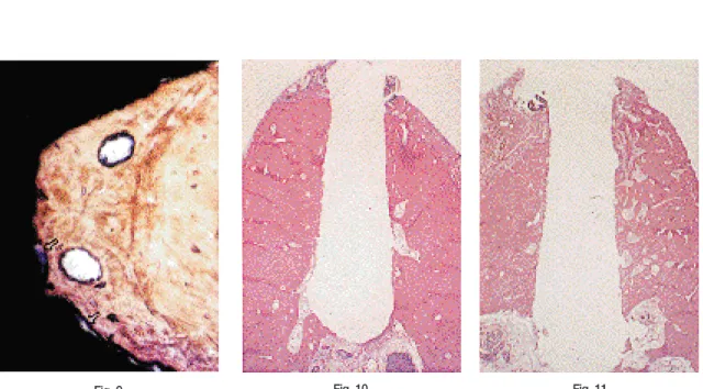
At each period of healing, the amount of contact with bone varied with jaws and implants. An implant placed in mandible showed more contact with bone than the same implant placed in maxilla did, and coated titanium implants showed more contact with bone than commercially-pure titanium implant did. Specimens of commercially-pure titanium implant showed that marrow space contact was prominent in earlier healing period(Fig 6) and marrow space interfaces reduced remarkably with increasing implantation time(Fig 7, 8, 9).
Coastlines of mineralized bone were observed at the interfaces of coated titanium implants(Fig 10, 11). Marrow space interfaces were not prominent even in earlier healing period(Fig 12, 13). There was no distinct difference in the amount of contact with bone between coated titanium implants at each period of healing.
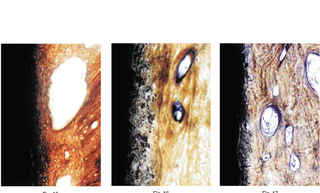
Bumpy implant surfaces were seen in undecalicified specimens of titanium plasma- sprayed implant(Fig 14). In undecalcified specimen of hydroxyapatite-coated implant, relatively even contact with bone was observed at the interface of translucent hydroxyapatite coating(Fig 15, 16, 17). The ruffled border with vacant spaces could be found in decalcified specimen of hydroxyapatite-coated implant. There was an
observation of direct bony contact with titanium surface of hydroxyapatite-coated implant(Fig 18).
In scanning electron microscopic examination, rough and irregular topographies were observed at the surfaces of coated titanium implants, and relatively smooth topography was observed at the machined surface of commercially-pure titanium implant.
Regardless of surface topographies, bone tissues directly attached to the implant surfaces could be found in all specimens investigated. Fibroblast-like mesenchymal cells attached to machined sufaces were observed in 4-week specimens of commercially-pure titanium implants placed in maxilla and mandible(Fig 19). Melted chocolate-like surface characteristics was observed in titanium plasma-sprayed implant(Fig 20).
Manual detachment of the implants from the tissues frequently exposed titanium surfaces of hydroxyapatite-coated titanium implants.
Three different surface topographies were observed in hydroxyapatite-coated implant (Fig 21, 22, 23).
IV. Discussion
Adverse bone reactions could not be found in any of our specimens. Histologic studies have suggested that the presence of implant may have no distinct impact on the normal cellular activity or normal physiologic process25~27) Mineralization phenomena appear similar to those events occurring naturally within bone tissues of jaw. For the calcification to occur at the interface, a connective tissue stroma may first be deposited by osteoblasts. Osteocytes found in
lacunae, close to the implant surfaces, may render services for calcification25).
In our specimens of 4-week healing, all implants were in direct contact with bone at the interfaces, though the amount of contact with bone varied with implants and jaws.
There may be no difference in the rapidity of new bone apposition at the implant surfaces between the same implants placed in maxilla and mandible.
A review of short-term studies suggests that, compared with commercially-pure titanium implant and titanium plasma-sprayed titanium implant, there is a faster integration and a higher percentage of contact with bone in hydroxyapatite-coated titanium implant.
Maximum amount of contact with bone appears to be reached after approximately 12 weeks28~32).
In our specimens, coated titanium implants showed more contact with bone than commercially-pure titanium implant did at each period of healing. There was no distinct difference in the amount of contact with bone between coated titanium implants.
Histomorphometric calculation could not be achieved due to the small number of specimens.
Studies related to biomaterials have suggested that the biocompatibility of implant material and the surface topography of the implant may affect new bone formation at the surface of the implant.
Mechanical tests and histologic investigations demonstrated excellent biocompatibility and tissue tolerance of hydroxyapatite in bone tissues33, 34). The leakage of titanium ions from the implant may result in disturbances in osteogenesis. It has been demonstrated that
titanium leakage from plasma-sprayed surface increases35), whereas metal ionic leakage diminishes in hydroxyapatite-coated titanium implant36).
Compared with commercially-pure titanium implants, coated titanium implants provide greater amount of surface area21). Also, some difference in surface topography between titanium plasma-sprayed titanium implant and hydroxyapatite-coated titanium implant was reported17).
Observations regarding the long-term effects have demonstrated a nonprogressive contact with bone in hydroxyapatite-coated titanium implant and more contact with bone in commercially-pure titanium implant at 1 year20, 37~40). Through mechanical tests, hydroxyapatite-coated titanium implants have showed significantly greater shear strength in short-term studies, and no difference was reported in 6-month specimen35, 41, 42). But, it has not been clearly substantiated why this reversal takes place and whether the phenomenon also occurs in human being43). Bioactive materials, which are thought to be able to generate surface apatite layers, produce strong bond with bone44~46) .However, this process is not restricted only to materials containing calcium and phosphate ions or glass ceramics. Commercially-pure titanium can take up those ions from the surrounding fluids through its oxide layer, and this material may be able to generate a bond with bone47). This process of ion-uptake requires more time, which may explain the slow bone apposition on titanium implants and the increase of bone bonding which is seen from biomechanical tests21).
Considering histologic differences between
jaws, it is natural that an implant placed in mandible should demonstrate more contact with bone than the same implant placed in maxilia do at each period of healing.
Generally, bone trabeculae are delicate and irregularly arranged in the maxilla, and the cortical plates are much thinner in the maxilla than in the mandible, as observed in this study.
Osseointegration in the maxilla may require a longer healing period because the density of available bone has substantial impacts on the initial fixation and the resultant load-bearing capability of the implant. During prolonged healihg period, careful and progressive loadings may improve the density of supporting bone tissue.
It has been suggested that in case of maxilla, a healing period of more than 6 months is required and more than 3 months is required in mandible1, 48~50). But, the healing period should be determined depending on the amount and density of available bone, not merely on the location of edentulous area. A longer duration of edentulousness can severely alter the bone condition of the host bed located anywhere in the mouth. The healing period allotted for clinical functon as well as other elements of treatment plan should be adjusted to each condition of available bone. It has been suggested that hydroxyapatite-coated implant of threaded type is more advantageous in soft bone condition because the coating increases the amount of trabecular bone at the interface, and may even accelerate the bone healing process20, 21). In case that the amount of available bone is not sufficient, graft techniques and guided tissue regeneration
procedures have been recommended, and these combined therapies are considered to be helpful for the increase of bone volume51, 52). The observations of this study indicate that the amounts of contact with bone at the interfaces are dependent upon implants and bone densities of host beds. But, the results obtained from histologic comparison does not indicate the superiority or the inferiority of an implant to others. The long-term usefulness of the implant of a material or a suface topography still remains to be investigated.
V. Conclusion
A histological study was performed to investigate and compare bone tissues surrounding commercially-pure titanium implants, titanium plasma-sprayed titanium implants and hydroxyapatite-coated titanium implants placed in maxillae and mandibles at different periods of healing.
Any adverse reaction could not be found in the bone tissues interfacing the implants. As early as 4 weeks, all implants placed in maxilla and mandible were in direct contact with bone at the interfaces. At each period of healing, an implant placed in mandible demonstrated more contact with bone than the same implant placed in maxilla did, and coated titanium implants showed more contact with bone than commercially-pure titanium implant did.
References
1. Br nemark PI, Zarb G, and Albrektsson T. Tissue Integrated Prosthesis : Osseointegration in Clinical Dentistry.
Chicago : Quintessence Publishing Co., 1985.
2. Albrektsson T, Br nemark PI, Hansson HA, Ivarsson B, Lindstr m J.
Osseointegrated titanium implant.
Requirement for ensuring a long-lasting direct bone anchorage in man. Acta Orthop Scand 1981 ; 52 : 155-170.
3. Brunette DM. The effects of implant surface topography on the behavior of cells. Int J Oral Maxillofac Implants 1988 ; 3 : 231-246.
4. Albrektsson T, Sennerby L. The state of the art in oral implants. J Clin Periodontol 1991 ; 18 : 474-484.
5. Br nemark PI, Albrektsson T. Titanium implants permanently penetrating human skin. S md J Plast Reconstr Surg 1982 ; 16 : 17-21.
6. Steinemann SG. Eulenberger J, Maeusli PA, Schroeder A. Adhesion of bone to implant. Adv Biomater 1986 ; 6 : 409- 414.
7. Kasemo B. Biocompatibility of titanium implants. Surface science aspect. J Prosthet Dent 1983 ; 49 : 832-837.
8. Meffert R. The soft tissue interface in dental implantology. Implantologist 1986
; 5 : 55-58.
9. Meffert R. Implant therapy. In:
Proceedings of the World Workshop in Clinical Periodontics. Chicago : The American Academy of Periodontology 1989 ; 8 : 1-10.
10. Doundoulakis J. Surface analysis of titanium after sterilization. Role in implant-tissue interface and bioadhesion.
J Prosthet Dent 1987 ; 58 : 471-478.
11. Baier R, Meyer A, Natiella J, Natiella R,
Carber J. Surface properties determine bioadhesive outcomes. Methods and results. J Biomed Mater Res 1988 : 337- 355.
12. Hartman L, Meenaghan M, Schaaf N, Hawker P. Effects of pretreatment sterilization and cleaning methods on materials, properties, and osseoinductivity of a threaded implant. J Oral Maxillofac Implants 1989 ; 4 : 11-18.
13. Meffert RM, Langer B, Fritz ME.
Dental Implant. A Review. J Periodontol 1992 ; 63 : 859-870.
14. Putter CD, Lange GLD, Groot KD.
Permucosal dental implants of dense hydroxylapatite. Fixation in alveolar bone. In : Proceedings of the International Congress on Tissue Integration in Oral and Maxillofacial Reconstruction. May 1985, Brussels.
(Excerpta Medica, Current Practice Series #29) 389-394. 15. Denissen HW, Veldhuis AAH, van den Hooff A.
Hydroxylapatite titanium implants. In:
Proceedings of the International Congress on Tissue Integration in Oral and Maxillofacial Reconstruction. May 1985, Brussels.(Excerpta Medica, Current Practice Series #29) 399-405.
16. Putter CD, Groot KD, Sillevis Smitt PAE, Van der Zel JM. In vivo fatigue behavior of permucosal dental implants of calcium hydroxylapatite, comparing non-prestressed with prestressed implants. Tras 9th Ann Meeting Am SocBiomater 1983(Prog & Abst) : 27.
17. Groot KD, Geesink RGT, Klein CPAT, Serekian P. Plasma sprayed coatings of hydroxylapatite. J Biomed Mater Res
1987 ; 21 : 1375-1381.
18. Denissen MW, Kalk W, Nieuport HM, Maltha JC, van den Hooff A.
Mandibular bone response to plasma- sprayed coatings of hydroxyapatite. Int J Prostho 1990 ; 3 : 53-58.
19. Lange GLD, Putter CD. Structure of the bone interface to dental implants in vivo. J Oral Implantol 1993 ; 19 : 123- 135.
20. Block MS, Kent JN, Kay JF. Evaluation of hydroxylapatite-coated titanium dental implants in dogs. J Oral Maxillofac Surg 1987 ; 45 : 601-607.
21. Cook SD, Kay JF, Thomas KA, Jarcho M. Interface mechanics and histology of titanium and hydroxylapatite-coated titanium for dental implant applications.
Int J Oral Maxillofac Implants 1987 ; 2 : 15-22.
22. Bloebaum RD, Dupont JA. Osteolysis from a press-fit hydroxyapatite-coated implant-a case study. J Arthroplasty 1993 ; 8 : 195-202.
23. von Recum AF, Park JB. Permanent percutaneous devices. CRC Crit Rev Bioengineer 1981 : 37-77.
24. Brunette DM. Interactions of epithelial cells with foreign surfaces. CRC Crit Rev Biocompatibil 1986 ; 1 : 323-370.
25. Steflik DE, Hanes PJ, Sisk AL, Parr GR, Song MJ, Lake FT, McKinney RV. Transmission electron microscopic and high voltage electron microscopic observations of the bone and osteocyte activity adjacent to unloaded dental implants placed in dogs. J Periodontol 1992 ; 63 : 443-452.
26. Linder L, Albrektsson T, Br nemark PI,
Hansson HA. Electron microscopic analysis of the bone-titanium interface.
Acta Orthoped Scand 1983 ; 54 : 45-52.
27. Holtrop ME. Light and electron microscopic structure of bone-forming cells. In : Hall BK ed. Bone, Vol. 1.
Caldwell NJ : The Telford Press ; 1990 : 1.
28. Ettinger RL, Spivey JD, Han DH, Koorbusch GF. Measurement of the interface between bone and immediate endosseous implants. A pilot study in dogs. Int J Oral Maxillofac Implants 1993 ; 8 : 420-427.
29. Gottlander M, Albrektsson T.
Histomorphometric studies of hydroxylapatite coated and uncoated CP titanium threaded implants in bone. Int J Oral Maxillofac Implants 1991 ; 6 : 399-404.
30. Weinlaender M, Kenney EB, Lekovic V, Moy PK. Histomorphometry of bone apposition around 3 types of endosseous dental implants. Int J Oral Maxillofac Implants 1992 ; 7 : 491-495.
31. Ducheyne P, Hench LL, Kagen A, Martens M, Bursens A, Mulier JC.
Effect of hydroxyapatite impregnation on skeletal bonding of porous coated implants. J Biomed Mater Res 1980 ; 14 : 225-237. 32.
32. Thomas KA, Kay JF, Cook SD, Jarco M. The effect of surface macrotexture and hydroxylapatite coating on the mechanical strength and histologic profiles of titanium implant materials. J Biomed Mater Res 1987 ; 21 : 1395- 1414.
33. Jarco M. Calcium phosphate ceramics as
hard tissue prosthetics. Clin Orthop 1981
; 157 : 259-278.
34. Groot KD. Bioceramics consisting of calcium phosphate salts. Biomat 1980 ; 1 : 47-50.
35. Osborn JF, Willich P, Meenen N. The release of titanium into human bone from a titanium implant coated with plasma-sprayed titanium. Adv Biomater 1990 ; 9 : 75-80.
36. Ducheyne P, Healy KE. The effect of plasma-sprayed calcium phosphate ceramic coatings on the metal ion release from porous titanium and cobalt- chromium alloys. J Biomed Mater Res 1988 ; 22 : 1137-1l63.
37. Piatelli A, Piatelli M, Romasco N, Trisi P. Histochemical and laser scanning microscopy characterization of the hydroxyapatite-bone interface. An experimental study in rabbits. Int J Oral Maxillofac Implant 1994 ; 9 : 163-167.
38. Gottlander M, Albrektsson T.
Histomorphometric analyses of hydroxyapatite-coated and uncoated titanium implants. The importance of the implant design. Clin Oral Impl Res 1992 ; 3 : 71-76.
39. Gottlander M, Albrektsson T, Carlsson LV. A histomorphometric study of unthreaded hydroxyapatite-coated and titanium-coated implants in rabbit bone.
Int J Oral Maxillofac implants 1992 ; 7 : 485-490.
40. Albrektsson T, Eriksson AR, Friberg B.
Histologic investigations on 33 retrived Nobelpharma implants. Clin Mater 1993
; 12 : 1-9.
41. Oonish H, Yamamoto M, Ishimaru H,
Tsuji E. Kushitani S, Aono M, Ukon Y.
The effect of hydroxyapatite coating on bone growth into titanium alloy implants.
J Bone Joint Surg (Br) 1989 ; 71-B : 213-216.
42. Soballe K, Hansen ES, Brockstedt- Rasmussen H, Pedersen CM, Bunger C.
Hydroxyapatite coating enhances fixation of porous coated implants. Acta Orthop Scand 1990 ; 61 : 299-306.
43. Tarnow DP. Dental implants in periodontal care. Current opinion in periodontology. Philadelphia : Current Science Ltd., 1993 ; 157-162.
44. Groot KD. Degradable ceramics.
Biocompatibility of clinical implant materials. In : Williams DF, editor. Boca Raton(FL), CRC Press 1981 ; 1 : 199- 222.
45. LeGeros R. Daculsi G, Orly I, Gregoire M. Substrate surface dissolution and interfacial biological mineralization. In : Davis JE, editor. The bone-biomaterial interface. Toronto : University of Toronto Press 1991 ; 76-88.
46. Neo M, Nakamura T, Yamamuro T.
Ohtsuki C, Kokubo T. Transmission electron microscopic study of apatite formation on biological ceramics in vivo.
In: Ducheyne P, Kokubo T, Van Blitterswijk CA, editors. Bone bonding biomaterials Leiderdorp(The Netherlands) : Reed Healthcare Communications 1992
; 112-120.
47. Hanawa T. Titanium and its oxide film : A substrate for formation of apatite. In : Davies JE, editor. The bone-biomaterial interface. Toronto : University of Toronto Press 1991 ; 49-61.
48. Br nemark PI, Hansson BO, Adell R, Breine V, Lindstr m J, Hallen O, Ohman A. Osseointegrated implants in the treatment of the edentulous jaw.
Experience from a 10-year period.
Scand J Plastic Reconstru Surg 1977 ; 11 : Suppl. 16.
49. Adell R, Lekholm V, Rockler B, Br nemark PI. A 15-year study of osseointegrated implants in the treatment of the edentulous jaw. Int J Oral Surg 1981 ; 10 : 387-416.
50. Hansson HA, Albrektsson T, Br nemark PI. Structural aspects of the interface between tissue and titanium implants. J Prosth Dent 1983 ; 50 : 108-113.
51. Breine U, Br nemark PI. Reconstruction of alveolar jaw bone. An experimental clinical study of immediate and preformed autologous bone grafts in combination with osseointegrated implants. Scand J Plast Reconstr Surg 1980 ; 14 : 23-48.
52. Dahlin C, Sennerby L, Lekholm U, Lindhe A, Nyman S. Generation of new bone around titanium implants using membrane technique. An experimental study in rabbits. Int J Oral Maxillofac implants. 1989 ; 4 : 19-25.
사진부도 설명
Fig 2 A light micrograph of a ground section showing thick compact bone of mandible. 8- week specimen of commercially-pure titanium implant placed in mandible. × 10
Fig 3 A light micrograph of a ground section showing thin cortical plate of maxilla. 8-week specimen of hydroxyapatite-coated implant. ×100
Fig 4 A light micrograph of a paraffin section showing fine trabeculae and fatty marrow of maxilla. 8-week specimen of hydroxyapatite-coated implant ×100
Fig 5 A light micrograph of a paraffin section showing direct contact with bone and marrow space contact at the interface of the implant. 4-week specimen of commercially-pure titanium implant placed in mandible. ×10
Fig 6 A light micrograph of a paraffin section showing marrow space interface. 4-week specimen of commercially-pure titanium implant placed in mandible. ×100
Fig 7 A light micrograph of a paraffin section showing reduced marrow spaces at the interface. 8-week specimen of commercially-pure titanium implant placed in mandible.
×100
Fig 8 A light micrograph of a ground section showing intimate contact with bone of a lamellar type. Intervening marrow spaces (arrow) are seen at the interface. 12-week specimen of commercially-pure titanium implant placed in mandible. ×200
Fig 9 A light micrograph of a ground section showing intervening marrow spaces (arrow) at the interface. 12-week specimen of commercially-pure titanium implant placed in maxilla. ×400
Fig 10 A light micrograph of a paraffin section showing coastline of mineralized bone at the interface. 8-week specimen of titanium plasma-sprayed implant placed in mandible. ×10 Fig 11 A light micrograph of a paraffin section showing coastline of mineralized bone at the
interface. 8-week specimen of hydroxyapatite-coated implant placed in mandible. ×10 Fig 12 A light micrograph of a paraffin section shows that marrow space interface is not
prominent. 4-week specimen of titanium plasma-sprayed implant placed in mandible. × 100
Fig 13 A light micrograph of a paraffin section shows that marrow space interface is not prominent. Ruffled border with vacant spaces is seen. 4-week specimen of hydroxyapatite-coated implant placed in mandible. ×100
Fig 14 A light micrograph of a ground section showing bone tissues, of a lamella type, directly attached to the bumpy surface of the implant. 4-week specimen of titanium plasma- sprayed implant placed in mandible. ×200
Fig 15 A light micrograph of a ground section showing intimate contact with bone at the interface of translucent hydroxyapatite coating. Lacunae are seen close to the implant surface. 4-week specimen of hydroxyapatite-coated implant placed in mandible. ×200
Fig 16 A light micrograph of a ground section showing intimate contact with bone at the interface of translucent hydroxyapatite coating. 4-week specimen of hydroxyapatite- coated implant placed in maxilla. X400
Fig 17 A light micrograph of a ground section showing intimate contact with bone at the interface of translucent hydroxyapatite coating. 12-week specimen of hydroxyapatite- coated implant placed in mandible. ×200
Fig 18 A light micrograph of a paraffin section showing direct bony contact with titanium surface(arrow). 12-week specimen of hydroxyapatite-coated implant placed in maxilla.
×400
Fig 19 A scanning electron micrograph showing relatively smooth topography of machined surface. MS : machined surface, MC: mesenchymal cell, R : red blood cell, 4-week specimen of commercially-pure titanium implant placed in maxilla. ×800
Fig 20 A scanning electron micrograph showing melted chocolate-like structure of titanium plasma-sprayed surface(TPS). 12-week specimen of titanium plasma-sprayed implant placed in maxilla. ×1000
Fig 21 A scanning electron micrograph showing bone tissues attached to the polished surface(PS). 8-week specimen of hydroxyapatite-coated implant placed in maxilla. × 500
Fig 22 A scanning electron micrograph showing hydroxyapatite-coated surface (HAC). 12- week specimen of hydroxyapatite-coated implant in mandible. ×700
Fig 23 A scanning electron micrograph showing titanium surface(TS) at the detached portion of hydroxyapatite layer. 12-week specimen of hydroxyapatite-coated implant placed in maxilla. ×1000
Fig 2 Fig 3
Fig 4
Fig 5
사진부도 (Ⅰ)
Fig 6
Fig 9 Fig 10 Fig 11
Fig 7 Fig 8
사진부도 (Ⅱ)
Fig 12
Fig 15 Fig 16 Fig 17
Fig 13 Fig 14
사진부도 (Ⅲ)
Fig 18
Fig 19
Fig 20
Fig 21
사진부도 (Ⅳ)
Fig 22 Fig 23
사진부도 (Ⅴ)
-국문초록-
Beagle Dog에 식립된 순수한 타이타늄 임프란트 및 피막처리된 타이타늄 임프란트 주위의 골조직에 관한 조직학적 연구
임상훈·손성희
서울대학교 대학원 치주과학교실
본 연구에서는 악골에 식립된 순수한 타이타늄 임프란트와 타이타늄 플라즈마 처리된 타이타 늄 임프란트 및 수산화인회석으로 피막처리된 타이타늄 임프란트의 주위 골조직을 광학현미경 과 주사전자현미경을 이용하여 비교 관찰하였다.
3마리의 beagle dog에서 상하악 소구치를 편측으로 발거한 후 12주의 치유기를 거쳐 임프란트 를 식립하였으며, 임프란트 식립 후 4주, 8주 및 12주에 실험동물을 각각 희생시켜서 조직 표본 을 제작하였다.
임프란트 주위의 골조직에서 염증이나 이물반응은 관찰되지 않았으며, 정상적인 악골에서와 유사한 골조직 소견이 관찰되었다. 상악골 및 하악골에 식립된 모든 임프란트의 4주 표본에서, 골과 임프란트 면의 직접적인 접촉이 관찰되었다. 동일한 치유기에서는, 상악골에 식립된 임프 란트의 면에 비하여 하악골에 식립된 동종의 임프란트의 면에서 더 많은 양의 골접촉이 관찰되 었으며, 순수한 타이타늄 임프란트의 면에 비하여 피막처리된 타이타늄 임프란트의 면에서 더 많은 양의 골접촉이 관찰되었다.
주요어 : 골조직 / 상악골, 하악골 / 조직학적 연구 ; 치과용 매식체 / 순수한 타이타늄 임프 란트 / 타이타늄 플라즈마 처리된 타이타늄 임프란트 / 수산화인회석으로 피막처리된 타이타늄 임프란트