Received: February 9, 2019 Revised: March 5, 2019 Accepted: April 4, 2019
Address for Correspondence: Soo Ah Kim, Department of Obstetrics and Gynecology, School of Medicine, Chosun University, 309 Pilmun- daero, Dong-gu, Gwangju 61452, Korea
Tel: 82-62-220-3090, E-mail: [email protected], ORCID: https://orcid.org/0000-0002-9049-1741
ORIGINAL ARTICLE
INTRODUCTION
Osteoporosis is regarded as one of the most common diseased entity in modern society. The disease affects more than 33% of female over 50 years old and may result in osteoporotic fractures that may significantly downgrade life quality. And the impact of osteoporosis is most pronounced in elderly women who have the highest risk of fracture [1-3].
The diagnosis is often made by measuring bone mineral density (BMD) using dual energy x-ray ab- sorptiometry (DXA) with the T score less than –2.5
utilized as a cut-off point for diagnosis of osteoporosis [4-7]. Number of treatment modalities have been sug- gested to prevent and to treat osteoporosis. Currently, the most commonly used treatment is using various anti-resorptive (osteoclast) medications including bisphosphonate, selective estrogen receptor modulators (SERMs), and denosumab. While there are number of advantages and disadvantages of each anti-resorptive medication, SERMs is the most commonly used initia- tive medication for postmenopausal osteoporosis.
Recently, nutritional supplements have been sug- gested to be an important adjuvant therapy to maintain
Influence of Supplementary Vitamin D on Bone Mineral
Density When Used in Combination with Selective Estrogen Receptor Modulators
Hyun Ju Liu1, Soo Ah Kim2,3, Da Joung Shim2, Ji Min Jung2, Eun Jeong Lee2
1Department of Obstetrics and Gynecology, Veteran’s Hospital, Gwangju, Korea, 2Department of Obstetrics and Gynecology, Chosun University Hospital, Gwangju, Korea, 3Department of Obstetrics and Gynecology, School of Medicine, Chosun University, Gwangju, Korea
Objectives: Vitamin D is regarded as one of the major nutrients that significantly influence bone metabolism. This study aims to look at the effect of supplementary vitamin D on bone mineral density (BMD) in female osteoporosis patients.
Methods: The retrospective hospital record review was performed on 282 patients who were diagnosed with osteoporosis and treated with selective estrogen receptor modulators (SERMs) between January 2015 and December 2016. Of these patients, 151 were treated with SERMs only while 131 were treated using both SERMs and vitamin D supplements. The BMD and any occurrence of osteoporotic fracture episode were investigated after one year. The result of two groups was compared to find the significance of vitamin D.
Results: Overall, improvement in BMD score was observed in 76% of the patients. The BMD of the SERMs only group improved by 3%
in spine and 1% in the hip while that of the SERMs with vitamin D group improved by 6% and 1% respectively. Statistical significance was noticed in the spine only. One distal radius fracture and one single level vertebral fracture occurred in patients of SERMs group while two distal radius fractures occurred in SERMs with vitamin D group. There was no occurrence of around hip fracture in both groups.
Conclusions: The result of the current study suggests that additional vitamin D may have some additive effect on improving BMD of the spine. Further study with the larger study population and the extended study period is recommended.
Key Words: Bone density, Osteoporosis, Selective estrogen receptor modulators, Vitamin D
bone metabolism. This includes calcium and vitamin D which plays a significant role in bone remodeling [8].
However, while there are number reports on the role of vitamin D on BMD, there are only handful of reports when this is prescribed in combination with SERMs specifically.
Thus in this study, we aim to evaluate the effect of us- ing supplementary vitamin D to the female osteoporo- sis patients who are treated with SERMs.
MATERIALS AND METHODS
Cohort selection
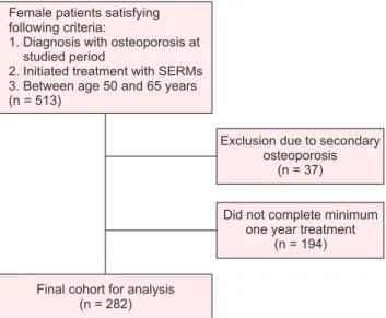
Retrospective chart review was performed on patients who were diagnosed with osteoporosis and initiated os- teoporosis medications between January 2015 and De- cember 2016. The inclusion criteria of the study are the patient 1) between the age of 50 and 65 years, 2) of the female gender, 3) had undergone menopause, 4) used SERMs as their primary osteoporosis medication, and 5) this who have taken the medication continuously for a minimum of 1 year. The exclusion criteria were the patients who had underlying diseases or medication that may potentially develop secondary osteoporosis.
Due to the recent high awareness of the importance of vitamin D supplements on osteoporosis, the prescrip- tion of vitamin D was routinely done by the senior au- thor starting from January 2016. Therefore, the patients who were prescribed with SERMs between January 2015 and December 2015 did not receive vitamin D while the all of the patients who were prescribed after January 2016 received vitamin D supplements. Two- hundred eighty-two patients fulfilled our inclusion and exclusion criteria and this cohort constitute the bases of our study. Of these patients, 151 were treated with SERMs only while 131 were treated with SERMs and vitamin D supplements (Fig. 1).
Study protocol
In order to compare the epidemiologic characteristics of the two groups, the following factors were recorded;
age, height, weight and the time since the menopause occurred.
In all patients, BMD was measured using DXA scan and this was done prior to the medication and was re- peated at 1 year following the index prescription. The measurements at the total spine and total hip were recorded for comparison. Our initial protocol was to exclude the part of the spine that involves compression
fracture. However, none of the patients had compres- sion fracture when initial BMD was taken. Of the pa- tients who developed spinal compression fracture dur- ing the follow-up, the affected vertebra was excluded when BMD was measured. The patients were also checked with the occurrence of osteoporotic fractures of any site including, around the hip, wrist, and ankle.
For the prescription of SERMs, raloxifene (Evista®; Eli Lilly, Indianapolis, IN, USA) was prescribed in all patients. The prescribed SERMs is a daily medication which is taken orally. The patients were asked to take the medication following breakfast each day. For vi- tamin D supplements, daily oral pill of 1,000 IU (D- MAC®; Dalim BioTech, Seoul, Korea) was used. This was again, asked to take at the time of the SERMs administration. In addition to the prescription of the SERMs and vitamin D supplements, all patients had a prescription of daily calcium. Three-month prescrip- tion was given to the patient and was re-prescribed at each 3-month visit to the outpatient office. The patients were asked of any complication that may be related to SERMs, vitamin D and calcium and supplementary medication was given when necessary. The patients who had discontinued taking the medication were con- sidered as the patient who did not take medication for minimal of 1 year and thus excluded from the study.
Statistical analysis
Statistical analysis was performed using SPSS ver. 18 software (IBM Corp., Armonk, NY, USA) and acquired
Fig. 1. Flowchart of cohort selection. SERMs: selective estrogen receptor modulators.
data are expressed as mean ± standard deviation. For the simple comparison of the two groups, Student t test was used. For the comparison of the change in BMD between the two groups, paired t test was utilized. The P value less than 0.05 was considered to be statistically significant.
RESULTS
The mean age of the patients was 58.9 ± 4.1 years with mean body mass index of 23.1 ± 4.1 kg/cm2. The time from the menopause was mean of 6.3 ± 4.0 years. The epidemiologic data of each group are described in Table 1.Initially, the mean BMD of the patient cohort as mea- sured by g/cm2 was 0.812 ± 0.231 in spine and 0.641 ± 0.154 in hip. There was no significant difference of the initial BMD when SERMs only group was compared with SERMs with vitamin D group (P = 0.387 in spine, P = 0.739 in hip). The patient of 68.2% had improved BMD at one year follow up (66.8% in SERMs group and 71.2% in SERMs with vitamin D group). The BMD of the SERMs only group improved by 3% in spine and 1% in hip while that of the SERMs with vitamin D group improved by 6% and 1%, respectively. When two
groups were compared, significance improvement in spine (P = 0.045) was noticed with additional vitamin D yet no significance or trend was found in hip (P = 0.157). The BMD of the two groups are described in Table 2.
Regarding osteoporotic fracture, three patients de- veloped distal radius fracture (1 in SERMs only group and 2 in SERMs with vitamin D group) as a resultant of slip down. One patient in the SERMs group developed single level vertebral fracture at the 4th lumbar spine.
The summary of the occurred fractures and their treat- ments are summarized in Table 3. There was no sig- nificant difference on osteoporotic fracture occurrence between two groups (P = 0.8931).
DISCUSSION
The result of the current study shows that there is a potential tendency that vitamin D may further improve BMD in the spine if it is used in combination with SERMs. However, we were unable to notice any alteration
Table 1. Epidemiologic characteristics of patient cohort Characteristic
Group
P value SERMs only
(n = 151)
SERMs with vitamin D (n = 131)
Age (y) 59.2 ± 4.8 58.6 ± 5.4 0.573
Weight (kg) 56.4 ± 8.2 55.3 ± 9.1 0.463
Height (cm) 156.1 ± 7.8 154.8 ± 9.2 0.398
Body mass index (kg/cm2) 23.1 ± 3.9 23.0 ± 4.1 0.653 Time since menopause (y) 6.1 ± 4.3 6.4 ± 3.9 0.476 Data are presented as mean ± standard deviation.
SERMs: selective estrogen receptor modulator.
Table 2. Bone mineral density (BMD) of patient cohort BMD (g/cm2)
Group
P value SERMs only
(n = 151)
SERMs with vitamin D (n = 131) Spine
Initial 0.810 ± 0.221 0.814 ± 0.242 0.387 After 1 year 0.834 ± 0.212 0.862 ± 0.187 0.461 BMD change 0.024 ± 0.098 0.048 ± 0.062 0.045 Hip
Initial 0.636 ± 0.192 0.641 ± 0.119 0.739 After 1 year 0.642 ± 0.168 0.647 ± 0.198 0.287 BMD change 0.006 ± 0.012 0.006 ± 0.009 0.157 Data are presented as mean ± standard deviation.
SERMs: selective estrogen receptor modulators.
Table 3. Summary of osteoporotic fractures occurred during the follow up
Age (y) Treatment group Time since index prescription (mo) Fracture site Treatment
56 SERMs only 3 Wrist, right Operation
65 SERMs only 9 Lumbar 4 spine Conservative treatment
62 SERMs + vitamin D 11 Wrist, left Cast application
59 SERMs + vitamin D 7 Wrist, right Operation
SERMs: selective estrogen receptor modulators.
in osteoporotic fracture risk with the use of vitamin D.
Vitamin D is produced in the skin, which has main role in bone and mineral metabolism [9]. Nowadays, vitamin D deficiency is correlated with cardiovascular disease by vitamin D receptor signaling [10]. Vitamin D is important for the treatment of osteoporosis as de- ficient vitamin D may result in defective bone mineral- ization which may eventually lead to low BMD [11,12].
The primary function of vitamin D is regulation of cal- cium absorption in the intestine and stimulating bone resorption to maintain serum calcium concentration.
Therefore, theoretically sufficient vitamin D may maxi- mize the effect of the anti-resorptive therapy and con- sequently lower the fracture risk [13,14]. The vitamin D can be derived from diet, sunlight and through other supplements. However, a recent study utilizing Korea National Health and Nutrition Examination Surveys (KHANES) reported that 76.7% of the overall Korean female population is vitamin D deficient which was de- fined as serum 25(OH)D level below 50 nmol/L [15].
Numbers of studies have tried to validate the effect of vitamin D on BMD. A study from Tuppurainen et al. [16] reported a significant increase in BMD of the femoral neck after 5 years. A study by Dawson-Hughes et al. [17,18] also showed consistent result reporting a significant increase of BMD in non-vitamin D deficient patients. Similarly, a study by Islam et al. [19] reported significant improvement of BMD in total hip and femoral neck with Bangladeshi women with baseline 25(OH)D of 36 nmol/L [19]. In contrary, there are also numerous reports that vitamin D does not have any positive effect on the BMD. A meta-analysis on this subject was recently published by Reid et al. [20]. The authors investigated 23 randomized controlled studies which included 4,082 patients with a mean age of 59 years. The study revealed conflicting results; 6 studies reporting significant benefit, 2 reporting significant detriment and 11 reporting no significance. This meta- analysis concluded that the benefit of using vitamin D to improve BMD is questionable.
Raloxifene at the standard dosage of 60 mg daily prevents postmenopausal bone loss in women with- out osteoporosis and is used also to treat established postmenopausal osteoporosis [21-23]. The effect of combining SERMs with vitamin D is a subject of de- bate. To our knowledge, there is no clinical study that looked specifically on this topic and only one animal study was done with male mice. The study from Sato et al. [24] treated osteoporotic male mice with SERMs
and vitamin D analogue and found this therapy could block bone loss. Regarding the effect of raloxifene alone, there is handful of studies reporting its influ- ence in osteoporosis [25-29]. The studies consistently reported that use of raloxifene can increase BMD both of the spine and of the hip but the improvement in the spine is more prominent. This is somewhat similar to our study as our study also showed an increase of BMD both in the spine and in the hip but the amount in- creased was greater in the spine. Also, the studies con- sistently reported that raloxifene can prevent fracture of the spine but not the hip. In our study, we found only 1 spine fracture in the SERMs group and no occurrence of around the hip fracture. We think this discordance may be due to the relatively younger age of our patient cohort and also relatively short follow up period.
We acknowledge that there are number of limitations to the current study. Firstly, the number of patient co- hort in our study is relatively small. As there are limited reports on the effect of vitamin D in combination with SERMs, we were unable to perform the sample size estimation prior to the study. Therefore, we think with the increase of patient cohort number, the effect of vi- tamin D on BMD will become more clearly visualized.
Secondly, our follow up period may be too short. In the current study, we are reporting clinical result of one year follow up and this may be too short as the effect of such mediation can be effective for prolonged period.
However, as the effect of SERMs is most powerful in the first two year after the administration, this study may at least provide baseline information on the effect of vitamin D on BMD. Also, as the compliance of os- teoporosis medication is reported to be discouraging, adding additional year in follow up period may require substantial working force. Lastly, important data miss- ing in the current study is patients’ vitamin D level when the osteoporosis medication was first initiated.
It is likely that the patient who had vitamin deficiency may benefit further from using supplementary vitamin D. Unfortunately, we were unable to verify this and this remains as an important limitation of the current study.
Nevertheless, to our knowledge, this is the first study that looked at the effect of vitamin D when used in combination with SERMs on BMD. According to our study, postmenopausal osteoporosis patient may fur- ther increase BMD of the spine if vitamin D is used in combination with SERMs. However, we also propose further study with a larger study population and ex- tended study period to strengthen our result.
ACKNOWLEDGMENTS
This study was supported by research funds from Chosun University Hospital, 2015.
CONFLICT OF INTEREST
No potential conflict of interest relevant to this article was reported.
REFERENCES
1. NIH consensus development panel on osteoporosis prevention, diagnosis, and therapy, March 7-29, 2000: highlights of the confer- ence. South Med J 2001; 94: 569-73.
2. Astrand J, Thorngren KG, Tägil M, Akesson K. 3-year follow-up of 215 fracture patients from a prospective and consecutive osteo- porosis screening program. Fracture patients care! Acta Orthop 2008; 79: 404-9.
3. Um MJ, Cho EA, Jung H. Combination therapy of raloxifene and alendronate for treatment of osteoporosis in elderly women. J Menopausal Med 2017; 23: 56-62.
4. Centre for Metabolic Bone Diseases, University of Sheffield. Frac- ture Risk Assessment Tool (FRAX®). Sheffield: University of Shef- field [cited 2018 Nov 25]. Available from: https://www.sheffield.
ac.uk/FRAX/.
5. Schneider EL, Guralnik JM. The aging of America. Impact on health care costs. JAMA 1990; 263: 2335-40.
6. Chrischilles E, Shireman T, Wallace R. Costs and health effects of osteoporotic fractures. Bone 1994; 15: 377-86.
7. Chappard D, Baslé MF, Legrand E, Audran M. New laboratory tools in the assessment of bone quality. Osteoporos Int 2011; 22:
2225-40.
8. Sunyecz JA. The use of calcium and vitamin D in the management of osteoporosis. Ther Clin Risk Manag 2008; 4: 827-36.
9. Kim TH, Lee HH, Kim JM, Choi SD, Lee A, Hwang SY, et al. Ex- pression of vitamin D receptor in seminal vesicles of cholesterol formula mice. J Menopausal Med 2015; 21: 89-92.
10. Oh J, Weng S, Felton SK, Bhandare S, Riek A, Butler B, et al.
1,25(OH)2 vitamin d inhibits foam cell formation and suppresses macrophage cholesterol uptake in patients with type 2 diabetes mellitus. Circulation 2009; 120: 687-98.
11. Lukert B, Higgins J, Stoskopf M. Menopausal bone loss is partially regulated by dietary intake of vitamin D. Calcif Tissue Int 1992;
51: 173-9.
12. Villareal DT, Civitelli R, Chines A, Avioli LV. Subclinical vitamin D deficiency in postmenopausal women with low vertebral bone mass. J Clin Endocrinol Metab 1991; 72: 628-34.
13. Adami S, Giannini S, Bianchi G, Sinigaglia L, Di Munno O, Fiore
CE, et al. Vitamin D status and response to treatment in post- menopausal osteoporosis. Osteoporos Int 2009; 20: 239-44.
14. Sadat-Ali M, Al Elq AH, Al-Turki HA, Al-Mulhim FA, Al-Ali AK.
Influence of vitamin D levels on bone mineral density and osteo- porosis. Ann Saudi Med 2011; 31: 602-8.
15. Park JH, Hong IY, Chung JW, Choi HS. Vitamin D status in South Korean population: seven-year trend from the KNHANES. Medi- cine (Baltimore) 2018; 97: e11032.
16. Tuppurainen MT, Komulainen M, Kröger H, Honkanen R, Jurv- elin J, Puntila E, et al. Does vitamin D strengthen the increase in femoral neck BMD in osteoporotic women treated with estrogen?
Osteoporos Int 1998; 8: 32-8.
17. Dawson-Hughes B, Harris SS, Krall EA, Dallal GE, Falconer G, Green CL. Rates of bone loss in postmenopausal women random- ly assigned to one of two dosages of vitamin D. Am J Clin Nutr 1995; 61: 1140-5.
18. Dawson-Hughes B, Dallal GE, Krall EA, Harris S, Sokoll LJ, Fal- coner G. Effect of vitamin D supplementation on wintertime and overall bone loss in healthy postmenopausal women. Ann Intern Med 1991; 115: 505-12.
19. Islam MZ, Shamim AA, Viljakainen HT, Akhtaruzzaman M, Je- han AH, Khan HU, et al. Effect of vitamin D, calcium and multiple micronutrient supplementation on vitamin D and bone status in Bangladeshi premenopausal garment factory workers with hypo- vitaminosis D: a double-blinded, randomised, placebo-controlled 1-year intervention. Br J Nutr 2010; 104: 241-7.
20. Reid IR, Bolland MJ, Grey A. Effects of vitamin D supplements on bone mineral density: a systematic review and meta-analysis.
Lancet 2014; 383: 146-55.
21. Cho YH, Um MJ, Kim SJ, Kim SA, Jung H. Raloxifene adminis- tration in women treated with long term gonadotropin-releasing hormone agonist for severe endometriosis: effects on bone min- eral density. J Menopausal Med 2016; 22: 174-9.
22. Clemett D, Spencer CM. Raloxifene: a review of its use in post- menopausal osteoporosis. Drugs 2000; 60: 379-411.
23. Díez JL. Skeletal effects of selective oestrogen receptor modulators (SERMs). Hum Reprod Update 2000; 6: 255-8.
24. Sato Y, Tando T, Morita M, Miyamoto K, Kobayashi T, Watanabe R, et al. Selective estrogen receptor modulators and the vitamin D analogue eldecalcitol block bone loss in male osteoporosis. Bio- chem Biophys Res Commun 2017; 482: 1430-6.
25. Shin CJ, Kim S, Choi CS, Shin HC, Kwon YJ. Effectiveness of os- teoporosis drug in postmenopausal women with spinal compres- sion fracture: combined consecutive therapy of Teriparatide and Raloxifene versus Bisphosphonate single. Korean J Neurotrauma 2016; 12: 123-7.
26. Kanis JA, Johnell O, Black DM, Downs RW Jr, Sarkar S, Fuerst T, et al. Effect of raloxifene on the risk of new vertebral fracture in postmenopausal women with osteopenia or osteoporosis: a re-
analysis of the Multiple Outcomes of Raloxifene Evaluation trial.
Bone 2003; 33: 293-300.
27. Bjarnason NH, Sarkar S, Duong T, Mitlak B, Delmas PD, Chris- tiansen C. Six and twelve month changes in bone turnover are related to reduction in vertebral fracture risk during 3 years of raloxifene treatment in postmenopausal osteoporosis. Osteoporos Int 2001; 12: 922-30.
28. Morii H, Ohashi Y, Taketani Y, Fukunaga M, Nakamura T, Itabashi
A, et al. Effect of raloxifene on bone mineral density and bio- chemical markers of bone turnover in Japanese postmenopausal women with osteoporosis: results from a randomized placebo- controlled trial. Osteoporos Int 2003; 14: 793-800.
29. Dane C, Dane B, Cetin A, Erginbas M. Comparison of the effects of raloxifene and low-dose hormone replacement therapy on bone mineral density and bone turnover in the treatment of postmeno- pausal osteoporosis. Gynecol Endocrinol 2007; 23: 398-403.