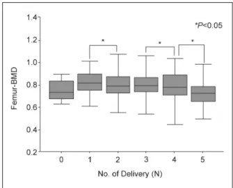
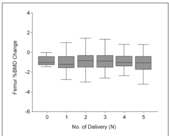
The Effect of Parity on Bone Mineral Density in Korean Postmenopausal Women and the Change of Bone Mineral Density after 2-year Alendronate Treatment
전체 글
수치
관련 문서
The purpose of this study was to evaluate the effect of these substances on bone regeneration by applying these materials to bone defects after cyst
A and E, In control group, a small amount of new bone was observed at the margin of bone defect (40×); B and F, In experimental group 1, a large amount of new bone was formed
3 Mean percentages of bone implant contact ratio in the control group and experimental groups at 6 and 12 weeks after placement of the
The purposes of the present study are to evaluate the clinical outcomes and radiological outcomes including bone fusion and subsidence that occurred after
Methods to overcome insufficient bone due to poor bone quality, the pneumatization of a maxillary sinus and other anatomical limitations of implant placement
Alveolar ridge augmentation with titanium mesh and a combination of autogenous bone and anorganic bovine bone: a 2-year prospective study.. Corinaldesi G, Pieri F, Sapigni
Histologic evaluation of early human bone response to different implant surfaces2. Histologic evaluation of human bone integration on machined and
of mineralized cancellous bone allograft(Puros) and anorganic bovine bone matrix(Bio-oss) for sinus augmentation: Histomorphometry at 26 to 32 weeks after
