http://www.ecevr.org/ 169
CLINICAL
EXPERIMENTAL VACCINE
RESEARCH
Introduction
Rabies causes death in many mammals, including pigs. Over 98% of human rabies comes from dogs [1], and rabies in animals has been transmitted by dogs and various wild carnivores, including foxes, wolves, raccoon dogs, skunks, mongooses, badgers, ferret badgers, and bats, all around the world [2]. Pig rabies is uncommon, represent- ing only 0.1%-1.1% of animal rabies [3,4]. Nevertheless, there have reports of the iden- tification of rabies in pigs in China, United States, and Brazil [5-7]. The main clinical signs in a pig bitten by a rabid dog in China are known to be hyperexcitation, roaming, attacks on other pigs, and attempts to jump pen walls to bite pigs [4]. In the United States, clinical signs in pigs with rabies transmitted by wild animals were fever, aggres- sion, restlessness, ptyalism, anorexia, head rubbing, progressive paralysis, depression, and vocalization [6]. Pig rabies results not only in economic losses but also in a serious
© Korean Vaccine Society.
This is an Open Access article distributed under the terms of the Creative Commons Attribution Non-Com- mercial License (http://creativecommons.org/licenses/
by-nc/4.0) which permits unrestricted non-commercial use, distribution, and reproduction in any medium, pro- vided the original work is properly cited.
K O R E A N V A C C I N E S O C I E T Y
K O R E A N K O R E A N A C C I N E O C I E T Y V
S
Clin Exp Vaccine Res 2016;5:169-174 http://dx.doi.org/10.7774/cevr.2016.5.2.169 pISSN 2287-3651 • eISSN 2287-366X
Dong-Kun Yang, Ha-Hyun Kim, Sung-Suk Choi, Seong Heon Lee, In-Soo Cho
Viral Disease Division, Animal and Plant Quarantine Agency, MAFRA, Gimcheon, Korea Received: June 2, 2016
Revised: June 25, 2016 Accepted: June 30, 2016
Corresponding author: Dong-Kun Yang, PhD, DVM Viral Disease Division, Animal and Plant Quarantine Agency, 177 Hyeoksin 8-ro, Gimcheon 39660, Korea
Tel: +82-54-912-0785, Fax: +82-54-912-0812 E-mail: yangdk@korea.kr
No potential conflict of interest relevant to this article was reported.
This work was supported financially by a grant (B1543083-2016-18-01) from Animal, and Plant Quarantine Agency, Ministry of Agriculture, Food and Rural Affairs (MAFRA), Republic of Korea.
Purpose: Rabies viruses (RABV) circulating worldwide in various carnivores occasionally cause fatal encephalitis in swine. In this study, the safety and immunogenicity of a recombi- nant rabies virus, the ERAGS strain constructed with a reverse genetics system, was evalu- ated in domestic pigs.
Materials and Methods: Growing pigs were administered 1 mL (108.0 FAID50/mL) of the ERAGS strain via intramuscular (IM) or oral routes and were observed for 4 weeks’ post-inoculation.
Three sows were also inoculated with 1 mL of the ERAGS strain via the IM route. The safety and immunogenicity in swine were evaluated using daily observation and a virus-neutralizing assay (VNA). Fluorescent antibody tests (FAT) for the RABV antigen and reverse transcriptase- polymerase chain reaction (RT-PCR) assays for the detection of the nucleocapsid (N) gene of RABV were conducted with brain tissues from the sows after necropsy.
Results: The growing pigs and sows administered the ERAGS strain did not exhibit any clinical sign of rabies during the test period test and did develop VNA titers. The growing pigs inocu- lated with the ERAGS strain via the IM route showed higher VNA titers than did those receiv- ing oral administration. FAT and RT-PCR assays were unable to detect RABV in several tissues, including brain samples from the sows.
Conclusion: Our results suggest that the ERAGS strain was safe in growing pigs and sows and induced moderate VNA titers in pigs.
Keywords: Rabies, Vaccines, Swine
A recombinant rabies virus
(ERAGS) for use in a bait vaccine
for swine
public health concern. The most effective ways to prevent pig rabies would probably involve blocking the access of wild an- imals and preparing an effective vaccine.
Animal rabies cases in South Korea have increased in cat- tle, dogs, and raccoon dogs since 1993. Veterinary authorities made a decision to distribute a rabies bait vaccine to high- risk rabies regions in South Korea as a kind of eradication program [8]. Since 2000, large amounts of bait vaccines have been distributed annually in two provinces, Gyeounggi-do and Gangwon-do, which have been designated as rabies risk regions, with a view to blocking the transmission of animal rabies. The amount of bait vaccine has increased gradually, and it reached 970,000 doses in 2016. In addition to the distri- bution of a bait vaccine, a new and safe rabies vaccine is need- ed for domestic pets and wild animals. Previously, we report- ed on the construction of the recombinant ERAG3G strain containing an Arg-to-Glu amino acid substitution at position 333 of the Evelyn-Rokitnicki-Abelseth (ERA) glycoprotein [9].
The safety and efficacy of the ERAG3G strain was assessed in mice. A single immunization with the ERAG3G strain con- ferred complete protection in mice and also induced a pro- tective immune response in dogs, cats, and raccoon dogs [9,10]. Recently, we constructed a new rabies vaccine candi- date, the ERAGS strain, which contains two mutations, at po- sitions 194 and 333 of the G protein of ERA strain. The World Health Organization (WHO) recommends that rabies vac- cines not cause any adverse symptoms in target and non-tar- get species [1]. The safety of rabies vaccine candidates should be evaluated in rodents, wild animals, and other domestic species [11]. In accordance with this recommendation, the safety and efficacy have already been investigated in mice and raccoon dogs. In this study, we sought to accumulate fundamental data about the safety and immunogenicity of the ERAGS strain in growing pigs and sows, acting as target and non-target animals, and to prepare for the expanded use of the ERAGS strain in pigs susceptible to rabies. Thus, we evaluated whether the new rabies vaccine strain (ERAGS) was safe and whether it induced sufficient immunogenicity in pigs.
Materials and Methods
Cells and viruses
Using a reverse genetics system, a recombinant rabies viruses (RABV), the ERAGS strain, was constructed to serve as a rabies vaccine candidate, as described previously [9]. The ERAGS
strain was propagated in murine neuroblastoma (NG108-15) cells maintained in Dulbecco’s modified Eagle’s medium (DMEM) supplemented with 5% of fetal bovine serum (FBS).
The titer of the ERAGS strain was 108.0 FAID50/mL. BHK-21 cells were grown in DMEM with 10% heat-inactivated FBS and antibiotic-antimycotic solution (GenDEPOT, Katy, TX, USA). BHK21 cells and a fixed RABV, CVS11 strain, were used for the fluorescent assay virus-neutralizing (FAVN) test.
Safety and immunogenicity of the ERAGS strain in swine Four-month-old pigs that were sero-negative against RABV were divided into three groups. Group 1, consisting of six pigs, was inoculated with the ERAGS strain (1 mL, 108.0 FAID50/mL) via the intramuscular (IM) route. Group 2, consisting of six pigs, was administered the same strain and dose via the oral route. Group 3, consisting of three sows, was inoculated with the ERAGS strain at the same dose but using an IM route of administration. Group 4 consisted of three pigs as a control group that received no treatment. All pigs were monitored daily for clinical signs of rabies, such as abnormal behavior, nervous prostration, anxiety, agitation, aggression, and pa- ralysis for 4 weeks’ post-administration. Following inocula- tion, at 2 and 4 weeks, blood was collected from all pigs, in- cluding the controls. The serum titers, expressed in Interna- tional Units per milliliter (IU/mL), were compared with those of a rabies-positive standard serum. The minimum protective titer was determined to be 0.5 IU/mL of FAVN [1]. After fin- ishing the safety and immunogenicity testing in group 3, all sows were euthanized and tissue samples, including the ce- rebrum, cerebellum, midbrain, spleen, liver, kidney, and lym- phoid tissue, were obtained to check for rabies infection.
Serological assay
A virus-neutralizing antibody (VNA) test, the FAVN test, was performed with blood samples from all pigs [12,13]. Briefly, a positive reference serum from WHO, adjusted to 0.5 IU/mL, was used as a positive control. Serum samples (50 µL) and the positive and negative controls were distributed in four consecutive wells and then serially diluted three-fold. The RABV (CVS-11 strain) at ~100 FAID50/50 μL was then added to each well. After a 60-minute incubation at 37°C, 50 μL of BHK-21 cell suspension containing 4×10⁵ cells/mL was add- ed to each well, and the microplates were incubated for 72 hours in a humidified incubator with 5% CO2 at 37°C. The cells were fixed in cold acetone (-20°C) for 20 minutes. After three successive washes with phosphate-buffered saline (PBS,
pH 7.2), the cells were reacted with a specific monoclonal an- tibody (Median Diagnostics, Chuncheon, Korea) against the rabies N protein for 45 minutes at 37°C; they were then stained with fluorescein isothiocyanate−conjugated goat-anti mouse IgG+IgM (KPL, Gaithersburg, MD, USA). After washing with PBS, the microplates were air-dried and examined at ×200 using a fluorescence microscope (Nikon, Tokyo, Japan). The titers of serum samples, expressed in IU/mL, were compared with those of the positive standard.
Identification of RABV with a fluorescent antibody test The fluorescent antibody test (FAT) was performed using brain samples (cerebrum, cerebellum, and midbrain) of sows ac- cording to the procedure described by the World Organiza- tion for Animal Health (OIE) [11]. Smears of brain tissues on slides were fixed in cold acetone (20°C) for 20 minutes. After three successive washes with PBS, the slides were incubated with a monoclonal antibody against RABV for 45 minutes at 37°C and then stained with the same conjugate mentioned above. After rinsing with PBS, the slides were examined un- der cover slips at ×200 using a fluorescence microscope. Pos- itive and negative controls were run together with the test sam- ples. Slides showing specific fluorescence were confirmed as positive.
Identification of RABV with reverse transcriptase−polymerase chain reaction
Total RNA was extracted from the homogenates of seven tis- sue samples (cerebrum, cerebellum, midbrain, spleen, liver, kidney, and lymph node) with an RNA extraction kit (Bioneer, Daejeon, Korea) according to the manufacturer’s protocol.
The extracted RNA was eluted in 50 μL of RNase- and DNase- free water. Reverse transcriptase−polymerase chain reaction (RT-PCR) was carried out to detect RABV genomic sequences using specific primer sets (RABVDNF and RABVDNR) that amplify the N gene of RABV (Table 1). RT-PCR was performed
in a TPersonal 48 thermal cycler (Biometra, Horsham, PA, USA) with a reaction mixture containing 5 µL of denatured RNA, 1 µL of each primer (50 pmol), 5 µL of 5× buffer (12.5 mM MgCl2), 1 µL of dNTP mix, 1 µL of an enzyme mix (re- verse transcriptase and Taq polymerase), and 11 µL of dis- tilled water. The cycling program was as follows: cDNA syn- thesis at 42°C for 30 minutes, followed by 45 cycles of 95°C for 15 seconds, 55°C for 15 seconds, 72°C for 15 seconds, and a final extension at 72°C for 5 minutes. The RT-PCR products were visualized by electrophoresis in 1.8% agarose gels con- taining ethidium bromide. Samples showing a 467 bp band were considered positive.
Statistical analysis
Data are expressed as means±standard deviations. Statistical analyses for VNA titers were conducted with the IBM SPSS version 19.0 for Windows (IBM Corp., Armonk, NY, USA).
Differences between groups were analyzed with one-way ANOVA followed by Tukey’s post hoc test. For more detailed investigations, the unpaired Student’s t-test was individually performed at each time point. A p-value of <0.05 was consid- ered to indicate statistical significance.
Results
Safety and immunogenicity of the ERAGS strain in swine The growing pigs and sows showed no clinical sign of rabies during the experimental period irrespective of whether the ERAGS was administered orally or via the IM route. As shown in Table 2, unvaccinated pigs also remained in good health during the observation period. As shown in Fig. 1, all growing pigs in group 1 inoculated with the ERAGS strain IM showed VNA titers of 2.6-7.9 IU/mL (mean, 5.92 IU/mL) against RABV at 4 weeks’ post-inoculation. However, there were no signifi- cant difference in the mean titers in the ERAGS-vaccinated groups of pigs at 2 and 4 weeks after the first vaccination (p<
Table 1. Oligonucleotide sequences of primers used to amplify the nucleocapsid gene of the rabies virus in RT-PCR
Primer Nucleotide sequences
(5’ → 3’) Rabies
gene
Size of amplicon
(bp) RABVDNF GCA GAT AGG ATA GAG CARa) A N 467 RABVDNR AAA GTG AAT GAG ATT GAA C
RT-PCR, reverse transcriptase−polymerase chain reaction.
a)R: A or T.
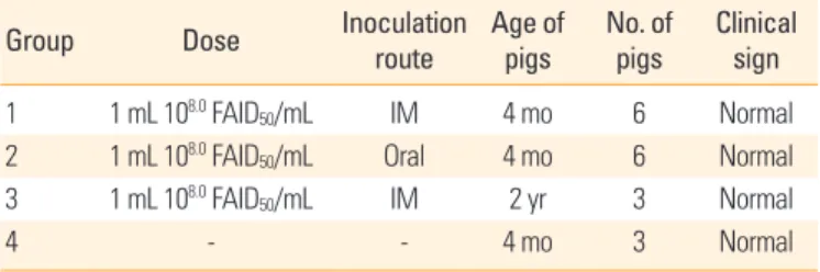
Table 2. Clinical signs in domestic pigs administered the ERAGS strain via intramuscular or oral route
Group Dose Inoculation
route Age of pigs No. of
pigs Clinical sign 1 1 mL 108.0 FAID50/mL IM 4 mo 6 Normal 2 1 mL 108.0 FAID50/mL Oral 4 mo 6 Normal 3 1 mL 108.0 FAID50/mL IM 2 yr 3 Normal
4 - - 4 mo 3 Normal
IM, intramuscular inoculation.
Fig. 1. Protective effect of antibody elicited by administration of the ERAGS in pigs. At 2 and 4 weeks after administration, high levels of neutralizing antibody titers were induced in growing pigs and sows inoculated via intramuscular (IM) and oral routes versus the control group that received no treatment. No pig inoculated with the ERAGS strain showed any clinical sign related to rabies. Each bar represents the mean ±standard error of the mean of six or three independent samples. Different lower-case letters indicate significant differences (*p <0.05, Tukey’s post hoc test). FAVN, fluorescent assay virus- neutralizing.
10
8
6
4
2
0 0 2 4
Weeks after administration
FAVN titer (IU/mL)
a a a a a a
b*
b*
b*
b*
b* b* Growing pig (IM)
Growing pig (Oral) Sow (IM) Control
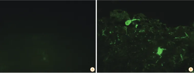
Fig. 2. Identification of the rabies viruses (RABV) antigen using fluorescent antibody tests in brain samples obtained from sows inoculated with the ERAGS strain. There were no positive reactions to the RABV in the sow brain samples (A), and specific fluorescence was shown in the posi- tive control sample (B).
A B
0.05, Tukey’s post hoc test) (Fig. 1). Group 2, administered the ERAGS strain orally, showed moderate VNA titers, 0.29-7.9 IU/mL (mean, 2.41 IU/mL). Half (3/6) of group 2 showed pro- tective VNA titers (0.8-7.9 IU/mL) at 2 weeks’ post-adminis- tration, and four pigs showed protective VNA titers (0.5-7.9 IU/mL) at 4 weeks’ post-administration. Two pigs in group 2 showed slight sero-conversion (0.29 IU/mL) but did not reach
VNA titers of a protective level. The mean VNA titer of group 2 was higher than those for the sera obtained from the con- trol group (p<0.05, unpaired t-test) (Fig. 1). All sows in group 3, inoculated with ERAGS strain via the IM route, developed protective VNA titers, of 0.8-4.6 IU/mL (mean, 2.7 IU/mL) at 4 weeks’ post-inoculation. The three pigs in group 4 remained sero-negative against RABV throughout the test, confirming that no contact transmission occurred between vaccinated animals and the control group.
Detection of RABV in several sow tissues
After performing necropsies on the three sows, tissue sam- ples, including cerebrum, cerebellum, midbrain, spleen, liver, kidney, and lymphoid tissues, were obtained. The brain sam- ples (cerebrum, cerebellum, and midbrain) were subjected to the FAT and RNA was extracted from seven tissue samples.
The brain samples obtained from sows inoculated with the ERAGS strain were negative (Fig. 2). The extracted RNAs were subjected to RT-PCR amplifying the N gene of RABV. All sam- ples were negative by RT-PCR (Fig. 3).
Discussion
Before commercialization of a new recombinant rabies vac- cine, the safety of the vaccine candidate should be evaluated in target and non-target animals so that all warm-blooded animals, including pigs, that are susceptible to rabies are test- ed [1,12]. Domestic pigs may be classified as target animals
because an outbreak of pig rabies caused by a rabid dog was reported in China [4]. In our study, high VNA titers were ob- tained when the ERAGS strain was inoculated into growing pigs and sows via an IM injection. After vaccination with a high dose (108.0 FAID50/mL), six growing pigs and three sows exhibited no clinical sign due to the ERAGS strain. At the time of sacrifice, 35 days following inoculation of the ERAGS strain, clearance of the ERAGS strain from brain samples ob- tained from the three sows that were inoculated was con- firmed by the absence of RABV antigens and gene signals ac- cording to FAT or RT-PCR.
The glycoprotein of RABV seems to be important in stimu- lating a humoral immune response against the rabies vaccine [14]. All growing pigs and sows vaccinated via the IM route developed significant VNA titer levels, ranging from 0.87 to 7.9 IU/mL, indicating that pigs inoculated with the ERAGS strain can be protected against virulent wild RABV, given that WHO considers a 0.5 IU/mL rabies VNA titer to be the minimum protective antibody level in carnivores [15,16]. The mean VNA titer of growing pigs was a little higher than that of sows, sug- gesting that a higher and more rapid VNA antibody response was induced in lighter animals [17].
Since the first outbreak of rabies in Korea was reported in 1907, 16,146 rabies cases have been identified in dogs, cats, cattle, raccoon dogs, horses, and goats [18]. Until 1982, dogs were primarily involved in the transmission of rabies, but, since 1993, raccoon dogs have been responsible for transmis- sion of rabies to other animals in South Korea [19]. Rabies bait vaccine has been distributed as a preventative measure to high-risk rabies regions since 2000.
In the development process of a rabies bait vaccine, the SAD Bern and SAD B19 strains, isolated by successive pas- saging and simple cloning from the ERA strain, provoked
specific mortality in field mice and the meadow vole [20]. A similar study on the safety of the SAG2 strain was conducted in several non-target animals, including wild boar, in areas where bait containing the SAG2 strain was distributed [21].
The results showed that the safety of the SAG2 vaccine was satisfactory. In our study, the oral route was used for growing pigs because this will be the relevant route if wild boar con- sume bait containing the ERAGS strain. The growing pigs ad- ministered a high viral titer of the ERAGS strain via the oral route did not exhibit any clinical signs of rabies during the test period and showed sero-conversion at 4 weeks’ post ad- ministration. However, 33.3% (2/6) of the pigs did not devel- op a protective level of antibodies, indicating that pigs need to be administered the virus twice or more. It has been re- ported that the oral route required more Aujeszky diseases virus vaccine than the intranasal route [22]. Additionally, the presence of antibodies aga inst RABV has been confirmed in wild boar living in Slovenia where anti-rabies vaccination oc- curred in 2005-2006 [23]. As oral administration of the ERAGS strain in growing pigs did induce an immunogenic response, wild boar consuming the bait may have VNA titers against RABV. Tests of the safety and immunogenicity of the ERAGS strain in target animals are required before commercializa- tion. Thus, further studies concerning the use of the ERAGS as a rabies vaccine will be conducted in pets and domestic animals.
In conclusion, we assessed the safety and immunogenicity of a newly constructed rabies vaccine strain, which was ad- ministered via oral and IM routes to growing pigs and sows.
No pig inoculated with ERAGS exhibited any clinical sign of rabies over 28 days, and RABV was not detected in tissue sam- ples by FAT or RT-RCR, suggesting that the ERAGS strain may be safe in pigs. Additionally, the vaccinated pigs developed Fig. 3. Identification of the rabies viruses (RABV) using reverse transcriptase−polymerase chain reaction in tissue samples obtained from sows inoculated with ERAGS strain. There were no positive reactions to the RABV in the seven tissue samples. M, 1-kb ladder; lanes 1 to 7, cerebrum, cerebellum, midbrain, spleen, liver, kidney, and lymphoid node, respectively; P, positive sample; N, negative sample.
500 bp
467 bp
significant VNA titers against RABV, indicating that the ERAGS strain may be immunogenic in swine. Thus, the ERAGS strain is a new, prospective candidate for a rabies vaccine for pigs.
ORCID
Dong-Kun Yang http://orcid.org/0000-0001-5765-3043 Ha-Hyun Kim http://orcid.org/0000-0001-6473-0035 Sung-Suk Choi http://orcid.org/0000-0001-5124-9736 Seong Heon Lee http://orcid.org/0000-0002-6024-3798 In-Soo Cho http://orcid.org/0000-0002-2692-3191
References
1. World Health Organization. WHO Expert Consultation on Rabies. World Health Organ Tech Rep Ser 2005;931:1-88.
2. Yang DK, Park YN, Hong GS, et al. Molecular character- ization of Korean rabies virus isolates. J Vet Sci 2011;12:57- 63.
3. Baer GM, Olson HR. Recovery of pigs from rabies. J Am Vet Med Assoc 1972;160:1127-8.
4. Jiang Y, Yu X, Wang L, et al. An outbreak of pig rabies in Hunan province, China. Epidemiol Infect 2008;136:504-8.
5. Luo Y, Zhang Y, Liu X, et al. Complete genome sequence of a highly virulent rabies virus isolated from a rabid pig in south China. J Virol 2012;86:12454-5.
6. DuVernoy TS, Mitchell KC, Myers RA, Walinski LW, Tins- ley MO. The first laboratory-confirmed rabid pig in Mary- land, 2003. Zoonoses Public Health 2008;55:431-5.
7. de Macedo Pessoa CR, Cristiny Rodrigues Silva ML, de Barros Gomes AA, et al. Paralytic rabies in Swine. Braz J Microbiol 2011;42:298-302.
8. Kim YR. Prophylaxis of human hydrophobia in South Ko- rea. Infect Chemother 2014;46:143-8.
9. Yang DK, Nakagawa K, Ito N, et al. A single immunization with recombinant rabies virus (ERAG3G) confers com- plete protection against rabies in mice. Clin Exp Vaccine Res 2014;3:176-84.
10. Yang DK, Kim HH, Jo HY, Kim HW, Choi SS, Cho IS. Safety and immunogenicity of a recombinant rabies virus strain (ERAG3G) in Korean raccoon dogs. J Bacteriol Virol 2015;
45:250-5.
11. Faber M, Dietzschold B, Li J. Immunogenicity and safety of recombinant rabies viruses used for oral vaccination of stray dogs and wildlife. Zoonoses Public Health 2009;56:
262-9.
12. World Organization for Animal Health. Manual of diag- nostic tests and vaccines for terrestrial animals. 6th ed.
Paris: World Organization for Animal Health; 2012. p.263- 82.
13. Brochier B, Blancou J, Thomas I, et al. Use of recombinant vaccinia-rabies glycoprotein virus for oral vaccination of wildlife against rabies: innocuity to several non-target bait consuming species. J Wildl Dis 1989;25:540-7.
14. Brown LJ, Rosatte RC, Fehlner-Gardiner C, et al. Oral vac- cination and protection of striped skunks (Mephitis me- phitis) against rabies using ONRAB(R). Vaccine 2014;32:
3675-9.
15. Cliquet F, Aubert M, Sagne L. Development of a fluores- cent antibody virus neutralisation test (FAVN test) for the quantitation of rabies-neutralising antibody. J Immunol Methods 1998;212:79-87.
16. Wright E, Temperton NJ, Marston DA, McElhinney LM, Fooks AR, Weiss RA. Investigating antibody neutralization of lyssaviruses using lentiviral pseudotypes: a cross-spe- cies comparison. J Gen Virol 2008;89(Pt 9):2204-13.
17. Nandi S, Kumar M. Development in immunoprophylaxis against rabies for animals and humans. Avicenna J Med Biotechnol 2010;2:3-21.
18. Joo YS, Lee JH, Lee KK, Bang HA, Lee WC. Retrospective study of extensive vaccination programs for canine rabies control and public health in Korea. Jpn J Infect Dis 2011;
64:513-5.
19. Yang DK, Kim HH, Lee KW, Song JY. The present and fu- ture of rabies vaccine in animals. Clin Exp Vaccine Res 2013;
2:19-25.
20. Artois M, Guittre C, Thomas I, Leblois H, Brochier B, Bar- rat J. Potential pathogenicity for rodents of vaccines intend- ed for oral vaccination against rabies: a comparison. Vac- cine 1992;10:524-8.
21. Masson E, Cliquet F, Aubert M, et al. Safety study of the SAG2 rabies virus mutant in several non-target species with a view to its future use for the immunization of foxes in Europe. Vaccine 1996;14:1506-10.
22. Jakubik J. Comparative susceptibility of rabbits, rats, mice and pigs to infection with Aujeszky diseases virus (ADV) in the development of an efficacy test for ADV vaccines.
Zentralbl Veterinarmed B 1977;24:764-6.
23. Vengust G, Hostnik P, Cerovsek M, Cilensek P, Malovrh T.
Presence of antibodies against rabies in wild boars. Acta Vet Hung 2011;59:149-54.