https://doi.org/10.5624/isd.2018.48.2.103
Introduction
Cone-beam computed tomography(CBCT) is an im- aging technology using ionizing radiation that was in- troduced into dentistry in the late 1990s.1,2 CBCT has revolutionized how patients are evaluated in virtually all dental specialties due to its lower cost and greater acces- sibility than conventional computed tomography(CT).3
Although the radiation dose of CBCT is usually lower than that of conventional CT, it is generally higher than
that of conventional 2-dimensional dental radiography.4,5 The radiation dose of CBCT depends on both the equip- ment(with variation across manufacturers) and the expo- sure parameters used by the operator.3,4
In dentistry, the most important adverse effects on health derived from ionizing radiation are stochastic.
These effects occur without a specific threshold; that is, even minimal doses of radiation are associated with a risk of inducing cancer or hereditary effects. The probability of these effects occurring, but not their severity, is propor- tional to the radiation dose.4,6
Due to the risks involved in the use of ionizing radia- tion, all necessary measures must be taken to minimize radiation exposure.5 This is especially important in pedi- atric patients, who are more susceptible to the effects of radiation due to their longer life expectancy and the great-
Uses of cone-beam computed tomography in San José, Costa Rica
Lucía Barba1, Ana Luisa Berrocal2, Alejandro Hidalgo1,*
1Specialization Program in Oral and Maxillofacial Imaging, Graduate School, Universidad de Talca, Talca, Chile
2Department of Diagnostic and Surgical Sciences, Faculty of Dentistry, Universidad de Costa Rica, San José, Costa Rica
ABSTRACT
Purpose: To analyze cone-beam computed tomography(CBCT) use, indications, and exposure parameters in San José, Costa Rica.
Materials and Methods: A cross-sectional study was performed. All CBCT examinations over a period of 6 months at 2 radiological centers in San José, Costa Rica were evaluated. The examinations were performed with Veraview EPOC X550 and Veraviewepocs 3D R100 equipment. The patients’ age and sex, clinical indication for CBCT, region of interest(ROI), repeat examinations, specialty of the referring dentist, field-of-view(FOV), tube voltage (kV), tube current(mA), and radiation dose(μGy) were evaluated. Patients were classified by age as children(≤12 years), adolescents(13-18 years), and adults(≥19 years).
Results: The mean age of the 526 patients was 49.4 years. The main indications were implant dentistry and dental trauma. The most frequent ROIs were posterior, while anterior ROIs were much less common. The highest percentage of repeat examinations was in children. Fifty-six percent of the referring dentists were specialists. The most commonly used FOV was small. The mean tube voltage and current were 79.8kV and 7.4mA for Veraview EPOC X550 and 89.9kV and 6mA for Veraviewepocs 3D R100, respectively. The mean doses for children, adolescents, and adults were 6.9μGy, 8.4μGy, and 7.8μGy, respectively.
Conclusion: Although CBCT was most commonly used in adults for implant dentistry, most repeat examinations were in children, and the highest mean dose was in adolescents. Additional dose optimization efforts should be made by introducing low-dose protocols for children and adolescents.(Imaging Sci Dent 2018; 48: 103-9)
KEY WORDS: Cone-Beam Computed Tomography; Radiation Dosage; Radiation Protection
Copyright ⓒ 2018 by Korean Academy of Oral and Maxillofacial Radiology
This is an Open Access article distributed under the terms of the Creative Commons Attribution Non-Commercial License(http://creativecommons.org/licenses/by-nc/3.0) which permits unrestricted non-commercial use, distribution, and reproduction in any medium, provided the original work is properly cited.
Imaging Science in Dentistry·pISSN 2233-7822 eISSN 2233-7830
*Lucía Barba was supported and funded by the University of Costa Rica through its program Becas de Posgrado al Exterior.
Received January 22, 2018; Revised April 19, 2018; Accepted May 2, 2018
*Correspondence to : Prof. Alejandro Hidalgo
Specialization Program in Oral and Maxillofacial Imaging, Graduate School, Universidad de Talca, Avenida Lircay S/N, Talca 3460000, Chile
Tel) 56-71-2200476, Fax) 56-71-2200476, E-mail) ahidalgo@utalca.cl
er radiosensitivity of developing organs and tissues.7 In 2012, Guide No. 172 was published by the Euro- pean Commission with the objective of providing evi- dence-based information on the use of CBCT in various clinical situations, with the goal of benefiting both cli- nicians and patients. This guide promotes the use of ap- propriate exposure parameters for dose reduction, while maintaining image quality.4
Since CBCT is a new technology, few studies are avail- able on exposure parameters in different age groups and for different types of CBCT equipment.8 Two recent stud- ies have evaluated the use of CBCT in children in differ- ent populations.8,9 Both studies found that most CBCT examinations were performed with a small field-of-view (FOV), which implies that this aspect of optimization had been performed properly. However, Hidalgo-Rivas et al.8 did not find any statistically significant difference between radiation doses in the age groups studied. They suggested that greater efforts should be made to reduce exposure in the youngest individuals as part of optimizing the radiation dose in CBCT examinations.
Only a single study performed in Latin America was found, which evaluated patients under 25 years of age in Chile.10 CBCT arrived in Costa Rica in 2008, but to date no studies have evaluated the use of this technology in Costa Rica. The purpose of the present study was to ana- lyze CBCT use, indications, and exposure parameters at 2 radiological centers in Costa Rica to gather information that may be useful for the implementation of radiological protection strategies.
Materials and Methods
This research was approved by the Bioethics Commit- tee of the University of Talca(folio 2017-06-AH).
The study was cross-sectional and retrospective. Data were collected from all patients, along with their CBCT examinations, at 2 radiological centers in San José, Costa Rica over a period of 6 months(July to December 2016).
The same maxillofacial radiologist managed both radio- logical centers. The equipment used for all the examina- tions was Veraview EPOC X550 Type EX1(J. Morita, Kyoto, Japan)(60-80kV, 1-10mA) or Veraviewepocs X550-EX1 Type 3D R100(J. Morita, Kyoto, Japan)(60-90 kV, 1-10mA).
Only examinations with complete information regard- ing the variables under study were analyzed. These vari- ables included patient information(age, sex, clinical indi- cation, region of interest[ROI], repeat examinations, and
the specialty of the referring dentist) and exposure param- eters(field of view[FOV], tube voltage[kV], tube current [mA], and radiation dose[μGy]).
Patient information was obtained from the research da- tabase, which was constructed by the maxillofacial radiol- ogist who managed both radiological centers using the centers’ records. Exposure parameters were obtained by the main investigator from the metadata files. All the in- formation collected was recorded on a spreadsheet in Ex- cel 2010(Microsoft Corporation, Redmond, WA, USA).
Patients were classified by age as children(≤12 years), adolescents(13-18 years), and adults(≥19 years).
The clinical indications for the examinations were cat- egorized according to the classification of uses of den- tal CBCT adapted from the European Guidelines.8 This classification divides indications according to the recom- mended FOV. For localized indications, a small FOV is recommended, while for generalized indications a medi- um or large FOV is recommended.4 Examinations with a clinical indication that did not correspond to these guide- lines were recorded as having ‘other’ indications. When an examination had more than 1 indication, each indica- tion was recorded separately. In the classification of indi- cations by age group, indications corresponding to fewer than 5% of all examinations were recorded as ‘uncom- mon.’ Temporomandibular joint(TMJ) examinations were considered as a single examination per patient, regardless of the number of exposures used.
The ROI of each examination was classified as follows:
1) anterosuperior(AS): maxillary canine and incisor re- gion(primary and/or permanent), 2) posterosuperior(PS):
distal to the maxillary canines(left and right), 3) anteroin- ferior(AI): mandibular canine and incisor region(primary and/or permanent), and 4) posteroinferior(PI): distal to the mandibular canines(left and right). The TMJ exam- inations were excluded from the ROI analysis, as they did not correspond to dental areas. Examinations with ‘other’
indications were excluded from the ROI analysis, as mul- tiple individual indications were anticipated for this cate- gory.
Repeat examinations were defined as multiple exam- inations performed for the same indication on the same day because the initial image obtained was unsuitable for diagnosis. This decision was made by the radiographer at each center, both of whom had more than 8 years of expe- rience with CBCT. Repeat examinations and their causes were recorded according to the classification of quality standards for CBCT images from Guide No. 172.4 The number of repeat examinations was not included in the
total number of examinations in the study.
The referring dentist’s specialty was recorded accord- ing to the specialties recognized by the College of Dental Surgeons of Costa Rica. Specialties with 10 or fewer re- ferred examinations were classified as ‘other’ specialties.
The FOV was classified by diameter as small(<8cm) or medium(≥8cm and <14cm).11,12
Regarding the radiation dose, the weighted computed tomography dose index(CTDIw) provided by the equip- ment after each exposure was recorded. For TMJ exam- inations, the dose was defined as the sum of the doses of all examinations performed. For repeat examinations, the dose was recorded as the sum of the examination dose and the repeat exposure doses.
The total number of patients was used for analyses by age group and sex. For the other results, the total number of examinations was used, since the same patient could have undergone more than 1 indicated examination. The distributions of examinations by age group and sex, and clinical indications by age group were calculated. ROIs were analyzed across the entire study group, as well as by age group and clinical indication. The cause and percentage of repeat examinations were recorded for the study group as a whole and by age group. The distribution of examina- tions by the referring dentist’s specialty was also calcu- lated. The distribution of FOVs was analyzed across the overall study group and by age group, and the frequency of clinical indications was calculated based on Guide No.
172 according to FOV size.4 The mean tube voltage and current of each CBCT apparatus was calculated, and the radiation dose was compared among the age groups.
The maxillofacial radiologist managing the centers assigned an identifying code to each patient for internal use. This code replaced the patient’s personal information in the research database. This database was given to the main investigator by the maxillofacial radiologist. Once data input was completed, the main investigator eliminat- ed each identifying code and replaced it with a correlative number. In this way, the research database was complete- ly anonymized, and it was impossible to obtain personal data from it.
Results
Of the examinations performed in the period studied (n =943), 317 were excluded due to incomplete infor- mation. A total of 586 examinations and 599 indications were obtained from 526 patients.
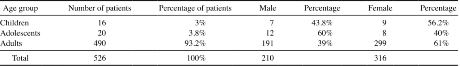
The mean age of the study group was 49.4±17.4 years (range, 6-82 years), and 60.1% percent of patients were women and 39.9% were men. The distribution of the sub- jects by age group and sex is presented in Table 1.
Of the total number of examinations, 2.9%(n =17) were performed in children, 4.1%(n=24) in adolescents, and 93%(n=545) in adults.
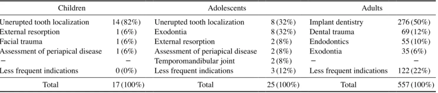
The clinical indications were implant dentistry(46.6%), dental trauma(12%), endodontics(9.3%), exodontia(7.3%), assessment of periapical disease(4.8%), unerupted tooth localization(4.7%), periodontal assessment(3.8%), TMJ (1.8%), bony pathosis(1.8%), external resorption(1.5%), facial trauma(1.2%), and other indications(5%). No indi- cations for surgical/orthodontic management, dental car- ies diagnosis, or cleft palate were found. The clinical in- dications by age group are presented in Table 2.
Regarding the ROI(n =376), the most common was the posterior region(79.2%), with PI accounting for the greatest proportion(44.1%), followed by PS(35.1%).
The anterior ROIs(20.8%) were mostly AS(16.8%), fol- lowed by AI(4%). The most common ROI in adults was PI(46.1%), followed by PS(36.1%), AS(13.4%), and AI (4.4%). In adolescents, there was no difference between the AS, PI, and PS ROIs(33.3% each). In children, 100%
of the examinations were requested for the AS region.
Regarding the most frequent ROI for each clinical in- dication, the PS ROI was most commonly requested for endodontics(49.1%) and dental trauma(41.2%). The PI ROI was most commonly requested for exodontia(75%) and implant dentistry(43.5%), and the AS ROI was most commonly used for unerupted tooth localization(83.3%).
Overall, repeat examinations were required in 4.9% of cases(n=29). In 55.2% of these cases(n=16), the repeat examinations were required because the ROI was exclud- ed from the scan volume, while 44.8%(n=13) of the re-
Table 1. Distribution of subjects by age group and sex
Age group Number of patients Percentage of patients Male Percentage Female Percentage
Children 16 3% 7 43.8% 9 56.2%
Adolescents 20 3.8% 12 60% 8 40%
Adults 490 93.2% 191 39% 299 61%
Total 526 100% 210 316
peat examinations were because of motion artifacts. The distribution of repeat examinations by age group was as follows: children, 11.8%(n=2); adolescents, 4.2%(n=1);
and adults, 4.8%(n=26).
The referring dentist was a general dentist in 44% of cases(n=258) and a specialist in 56%(n=328). Of the specialists, 22.3%(n=73) were prosthodontists, 19.5%
(n=64) endodontists, 16.8%(n=55) periodontists, 13.7%
(n=45) oral and maxillofacial surgeons, 9.8%(n=32) orthodontists and specialists in dentofacial orthopedics, 7.3%(n=24) oral implantologists, and 3.4%(n=11) spe- cialists in advanced general dentistry. Other specialties accounted for 7.3% of the examinations(n=24).
The most commonly used FOV was small(56.8%, n=333), followed by medium(43.2%, n=253). In chil- dren, a medium FOV was used in 58.8% of examinations (n=10), while in adolescents and adults, a small FOV was used in 70.8%(n=17) and 56.7%(n=309) of exam- inations, respectively. The frequency of clinical indica- tions according to FOV size is shown in Figure 1.
For the Veraview EPOC X550 equipment, the mean tube voltage was 79.8±4.1kV and the mean tube current was 7.4±1.9mA. For the Veraviewepocs 3D R100 equip- ment, the mean tube voltage was 89.9±0.7kV and the mean tube current was 6±0.2mA.
The radiation dose and standard deviation by age group were 6.9±2.0μGy in children, 8.4±9.0μGy in adoles- cents, and 7.8±3.5μGy in adults.
Discussion
In the present study, CBCT use was evaluated at 2 ra- diological centers in San José, Costa Rica. This study is the first of its kind to analyze data from Costa Rica.
A sizable proportion of the examinations evaluated were excluded due to incomplete data. This may indicate that the referring dentists did not provide complete writ- ten information in the radiographic requests, or that the database of the radiological centers was incomplete. It is important to emphasize the need for referring dentists to provide enough clinical information. Doing so enables the maxillofacial radiologist to perform the most appro- priate examination for the patient’s needs,4 and to comply as closely as possible with the principle of justification for radiological protection.4 In the absence of detailed information, the maxillofacial radiologist should select the most appropriate examination based on patient indica- tions or be guided by the specialty of the referring dentist.
At the same time, it is important that radiological centers maintain regular and complete records of their patients.
Doing so provides maxillofacial radiologists with easy access to information about the previous examinations undergone by each patient and allows them to carry out quality control of the procedures.4
The majority of the procedures were carried out in adults, while the percentage of examinations in children was very low. This agrees with other studies, which have reported the total percentages of examinations to be 3.5%
Table 2. Clinical indications by age group
Children Adolescents Adults
Unerupted tooth localization 14(82%) Unerupted tooth localization 8(32%) Implant dentistry 276(50%)
External resorption 1(6%) Exodontia 8(32%) Dental trauma 69(12%)
Facial trauma 1(6%) External resorption 2(8%) Endodontics 55(10%)
Assessment of periapical disease 1(6%) Assessment of periapical disease 2(8%) Exodontia 35(6%)
- - Temporomandibular joint 2(8%) - -
Less frequent indications 0(0%) Less frequent indications 3(12%) Less frequent indications 122(22%)
Total 17(100%) Total 25(100%) Total 557(100%)
Fig. 1. Frequency of clinical indications according to field-of-view (FOV) size. UTL: unerupted tooth localization, ER: external re- sorption, PA: periodontal assessment, APp: assessment of periapi- cal disease, DT: dental trauma, E: endodontics, Ex: exodontia, ID:
implant dentistry, BP: bony pathosis, TMJ: temporomandibular joint, FT: facial trauma. LI: localized indications GI: generalized indications
Small FOV Medium FOV
in children under 12 years10 and 5.2% in children under 13.8 In the present investigation, no patient under 6 years of age was seen, corresponding to previous investiga- tions.9,10 This situation is in accordance with the principle of justification, since a CBCT examination may not be necessary in patients under 6 years of age, because it may not be possible to clinically intervene in patients in this age group.
Regarding ROI by clinical indication, a posterior ROI was most commonly requested for the most common in- dications for adults, in order to evaluate the proximity of anatomical structures, root anatomy, and fractures in multiradicular teeth.4,13,14 All examinations for exodontia in adolescents were requested for the third molars. This could be explained by the fact that there is evidence to support the use of CBCT to evaluate the proximity of third molars to the mandibular canal, due to the possibil- ity of damaging the inferior alveolar nerve and causing neurosensory alterations.4,15 This is in line with our re- sults. For children, examinations for the most frequent in- dications were requested with the AS ROI, in agreement with other studies.8,10 Surgical indications, orthodontic in- dications, and cleft palate are pathologies that are mostly evaluated in public hospitals, which could explain the ab- sence of examinations with these indications in the pres- ent study.
The low rate of repeat examinations in the present study is in accordance with Guide No. 172, which recommends that the rate of repeat examinations should be maintained below 5%.4 Repeat examinations due to patient move- ment during examinations can be reduced by reducing the exposure time and by stabilizing and securing the patient’s head.16,17 In addition, it is important for the cli- nician to record the presence of any medical conditions that may affect movement. In this way, the maxillofacial radiologist can consider modifying the exposure parame- ters, according to the equipment, to reduce the possibility of motion artifacts.17 For repeat examinations due to a mismatch between the ROI and the FOV, Guide No. 172 recommends taking a scout view and using available vi- sual aids, such as light indicators, to confirm positioning in equipment that has these characteristics.4
As recommended by Guide No. 172, an audit should be carried out at intervals of no more than 6 months, in which one would expect to find a reduction in the repeat examination rate of 50%.4 In the present study, the highest rate of repeat examinations by age group was in children.
This result agrees with those of previous studies, in which higher percentages of motion artifacts were found in pa-
tients younger than 15 years and older than 60 years.18 This result is notable due to the effects of radiation and the increased radiation dose that accompanies repeat ex- aminations. It is therefore advisable to keep a record of repeat examinations because doing so makes it possible to determine the factors that cause patients to move, which allows them to be corrected before performing an exam- ination.4,18
Specialist dentists requested the most examinations.
This information is particularly relevant for raising aware- ness of the importance of providing adequate clinical information by correctly filling out the radiology request form.
Small FOVs predominated in the present study, with a percentage slightly higher than 50%, unlike the values of around 80% reported in previous studies.8,9 For some localized clinical indications, such as unerupted tooth lo- calization, periodontal assessment, implant dentistry, and bony pathosis, a medium FOV was most commonly used in this study. However, in this regard, the recommenda- tion of Guide No. 172 is to use a small FOV for localized indications.4 In the present study, a medium FOV was used most frequently in children, even though the most common clinical indication was unerupted tooth local- ization, for which a small FOV is recommended.4 The FOV is an important parameter in terms of radiation dose.
The same FOV size can cover a larger area in a smaller patient, so the smallest FOV should be selected accord- ing to the clinical indication of the examination. Doing so allows observation of only the area of interest, there- by yielding a reduction in dose.4,19,20 Additional efforts should be made at the centers included in this study to adjust the FOV to be as small as possible, while allowing adequate observation of the area of interest.
The equipment used in the present study allowed both tube voltage and tube current to be varied. However, both parameters remained fixed for most examinations. There is a linear relationship between the tube current and radi- ation dose.21 It is well known that changes in the radiation dose are directly proportional to the tube current, so that a decrease in the tube current directly leads to a decrease in the patient dose.21 Since a 10% dose reduction is consid- ered clinically relevant,22 a 10% reduction in tube current would be an important objective for dose optimization.
In the present study, the exposure time was not recorded separately, because it was fixed in the equipment that was studied(9.4 seconds). Tube current and voltage are oper- ator-dependent factors and should be kept as low as pos- sible to reduce the radiation dose, while maintaining the
diagnostic quality of the image.6
Although a decrease in tube current produces an in- crease in the noise in CBCT images, it is possible to de- crease tube current while maintaining acceptable image quality for diagnosis.23,24 It has been shown that such a decrease can be achieved, in some cases, by using lower parameters than those recommended by the manufactur-
er.23,24 In the present study, no variation was found in the
tube voltage and current in children, despite evidence that such variation may reduce the dose in pediatric patients while maintaining diagnostic image quality.24 Due to variation in the CBCT equipment on the market and the wide variety of exposure parameters, it is not possible to standardize specific settings for tube voltage and current for all equipment types and all patients. More research is needed to determine specific protocols for each equip- ment type, indication, and patient category. Optimization efforts should be made using the pediatric parameters of the equipment when available.20
The lowest average radiation dose was found in child- ren, while the highest was in adolescents. The fact that adolescents recorded a higher dose than adults could not be explained by the number of TMJ examinations or the number of repeat examinations. It is suggested that these centers should evaluate their protocols and establish dif- ferent protocols for adults, adolescents, and children.
The doses were measured in CTDIw, which combines the measurements of the center and the periphery of a phantom to generate a weighted dose index,6,11 so the dose measurements were not a direct evaluation of the effective dose received by each patient. As the CTDIw describes an average dose, it can be inferred that children receive a higher dose than is recorded by the equipment.
This has been shown in several investigations, in which the use of the same exposure parameters for both children and adults produced a greater effective radiation dose in children.6,20 This higher dose can be explained because a small FOV covers a greater area in children than in adults, and the thyroid gland is therefore located closer to the primary beam. In addition, the other organs are closer to the surface, so they receive a larger dose.20 Therefore, the use of CBCT in children must be justified, meaning that the benefits must outweigh the risk.20,4
The information obtained in the present investigation on the use of CBCT in San José, Costa Rica is expected to be used as a basis for implementing radiological pro- tection strategies. It is important to emphasize that refer- ring dentists and those responsible for radiological centers should follow the principles of justification and optimi-
zation to protect patients. Further research is required to reduce doses, especially in children, while maintaining diagnostic quality.
In conclusion, although CBCT was mostly used in adults for implant dentistry, most repeat examinations were in children, and the highest mean dose was in adolescents.
Additional dose optimization efforts should be made, and low-dose protocols should be introduced for children and adolescents.
Acknowledgements
The first author acknowledges the University of Costa Rica for funding her postgraduate studies.
References
1. Mozzo P, Procacci C, Tacconi A, Martini PT, Andreis IA. A new volumetric CT machine for dental imaging based on the cone-beam technique: preliminary results. Eur Radiol 1998; 8:
1558-64.
2. Arai Y, Tammisalo E, Iwai K, Hashimoto K, Shinoda K. Devel- opment of a compact computed tomographic apparatus for dental use. Dentomaxillofac Radiol 1999; 28: 245-8.
3. Pauwels R. Cone beam CT for dental and maxillofacial imag- ing: dose matters. Radiat Prot Dosimetry 2015; 165: 156-61.
4. European Commission. Radiation protection 172: cone beam CT for dental and maxillofacial radiology: evidence-based guidelines. Luxembourg: European Commission Directorate for Energy; 2012.
5. FDI World Dental Federation. FDI policy statement on radia- tion safety in dentistry: adopted by the FDI General Assembly:
13 September 2014, New Delhi, India. Int Dent J 2014; 64:
289-90.
6. ICRP, Rehani MM, Gupta R, Bartling S, Sharp GC, Pauwels R, et al. Radiological protection in cone beam computed tomo- graphy(CBCT). ICRP Publication 129. Ann ICRP 2015; 44:
9-127.
7. White SC, Scarfe WC, Schulze RK, Lurie AG, Douglass JM, Farman AG, et al. The Image Gently in Dentistry campaign:
promotion of responsible use of maxillofacial radiology in dentistry for children. Oral Surg Oral Med Oral Pathol Oral Radiol 2014; 118: 257-61.
8. Hidalgo-Rivas JA, Theodorakou C, Carmichael F, Murray B, Payne M, Horner K. Use of cone beam CT in children and young people in three United Kingdom dental hospitals. Int J Paediatr Dent 2014; 24: 336-48.
9. Van Acker JW, Martens LC, Aps JK. Cone-beam computed tomography in pediatric dentistry, a retrospective observational study. Clin Oral Investig 2016; 20: 1003-10.
10. Arancibia B, Schilling J, Schilling A, Correa-Beltrán G, Hid- algo A. Usos de tomografía computarizada de haz cónico en menores de 25 años en Talca, Chile. Rev Cubana Estomatol 2017; 54: 1-11.
11. Pauwels R, Beinsberger J, Collaert B, Theodorakou C, Rogers
J, Walker A, et al. Effective dose range for dental cone beam computed tomography scanners. Eur J Radiol 2012; 81: 267- 12. Nemtoi A, Czink C, Haba D, Gahleitner A. Cone beam CT: a 71.
current overview of devices. Dentomaxillofac Radiol 2013;
42: 20120443.
13. Special Committee to Revise the Joint AAE/AAOMR Po- sition Statement on use of CBCT in Endodontics. AAE and AAOMR Joint Position Statement: use of cone beam computed tomography in endodontics 2015 Update. Oral Surg Oral Med Oral Pathol Oral Radiol 2015; 120: 508-12.
14. Tyndall DA, Price JB, Tetradis S, Ganz SD, Hildebolt C, Scarfe WC. Position statement of the American Academy of Oral and Maxillofacial Radiology on selection criteria for the use of radiology in dental implantology with emphasis on cone beam computed tomography. Oral Surg Oral Med Oral Pathol Oral Radiol 2012; 113: 817-26.
15. Adibi S, Paknahad M. Comparison of cone-beam computed tomography and osteometric examination in preoperative assessment of the proximity of the mandibular canal to the apices of the teeth. Br J Oral Maxillofac Surg 2017; 55: 246- 16. Schulze R, Heil U, Gross D, Bruellmann DD, Dranischnikow 50.
E, Schwanecke U, et al. Artefacts in CBCT: a review. Dento- maxillofac Radiol 2011; 40: 265-73.
17. Donaldson K, O’Connor S, Heath N. Dental cone beam CT image quality possibly reduced by patient movement. Dento-
maxillofac Radiol 2013; 42: 91866873.
18. Spin-Neto R, Wenzel A. Patient movement and motion arte- facts in cone beam computed tomography of the dentomaxil- lofacial region: a systematic literature review. Oral Surg Oral Med Oral Pathol Oral Radiol 2016; 121: 425-33.
19. Kiljunen T, Kaasalainen T, Suomalainen A, Kortesniemi M.
Dental cone beam CT: a review. Phys Med 2015; 31: 844-60.
20. Theodorakou C, Walker A, Horner K, Pauwels R, Bogaerts R, Jacobs R, et al. Estimation of paediatric organ and effec- tive doses from dental cone beam CT using anthropomorphic phantoms. Br J Radiol 2012; 85: 153-60.
21. Kalender W. Computed tomography: fundamentals, system technology, image quality, applications. 3rd ed. Erlangen:
Publicis Corporate Pub.; 2011.
22. Pernicka F, McLean ID, International Atomic Energy Agency.
Dosimetry in diagnostic radiology: an international code of practice. Technical reports series No. 457. Vienna: Interna- tional Atomic Energy Agency; 2007.
23. Pauwels R, Seynaeve L, Henriques JC, de Oliveira-Santos C, Souza PC, Westphalen FH, et al. Optimization of dental CBCT exposures through mAs reduction. Dentomaxillofac Radiol 2015; 44: 20150108.
24. Hidalgo Rivas JA, Horner K, Thiruvenkatachari B, Davies J, Theodorakou C. Development of a low-dose protocol for cone beam CT examinations of the anterior maxilla in children. Br J Radiol 2015; 88: 20150559.