The impact of portal vein resection on outcome of hilar cholangiocarcinoma
Ki Beom Kim
1, Dong Wook Choi
1, Jin Seok Heo
1, In Woong Han
1, Sang Hyun Shin
1, Yunghun You
1, and Dae Joon Park
21Department of Surgery, Samsung Medical Center, Sungkyunkwan University School of Medicine,
2Department of Surgery, Ewha Womans University Mokdong Hospital, Ewha Womans University School of Medicine, Seoul, Korea
Backgrounds/Aims: Portal vein resection (PVR) with major hepatic resection can increase the rate of curative resection for hilar cholangiocarcinoma (HC). However, the oncologic role and safety of PVR is still debatable. This study aims to analyze PVR in terms of safety and therapeutic effectiveness. Methods: We retrospectively analyzed 235 patients who had undergone major hepatic resection for HC with curative intent, including patients with PVR (PVR, n=35) con- sisting of PV invasion (PVR-A, n=9), No PV invasion (PVR-B, n=26); and patients without PVR (No PVR, n=200).
Results: There was no significant difference in the 30-day mortality or postoperative morbidity between PVR and No PVR (2.9% vs. 1.0%; p=0.394 and 34.3% vs. 35.0%; p=0.875). The rate of advanced HC (T3: 40% vs. 12%; p<0.001 and nodal metastasis: 60% vs. 28%; p<0.001) was higher in PVR compared to No PVR. There was no significant difference in the 5-year overall survival rates and disease-free survival between PVR-A vs. PVR-B vs. No PVR. In multivariate analysis, estimated blood loss >600 ml (p=0.010), T3 diseases (p=0.001), nodal metastasis (p=0.001) and poor differentiation (p=0.002) were identified as independent risk factors for survival. Conclusions: PVR does not increase postoperative mortality or morbidity. It showed a similar oncologic outcome, despite a more advanced disease state in patients with HC. Given these findings, PVR should be actively performed if necessary, after careful patient selection. (Ann Hepatobiliary Pancreat Surg 2021;25:221-229)
Key Words: Hilar cholanagiocarcinoma; Portal vein resection; Mortality; Morbidity; Oncologic outcome
Received: August 28, 2020; Revised: November 26, 2020; Accepted: November 27, 2020 Corresponding author: Dong Wook Choi
Department of Surgery, Samsung Medical Center, Sungkyunkwan University School of Medicine, 81 Irwon-ro, Gangnam-gu, Seoul 06351, Korea Tel: +82-2-3410-3462, Fax: +82-2-3410-6980, E-mail: dw7722.choi@samsung.com
Copyright Ⓒ 2021 by The Korean Association of Hepato-Biliary-Pancreatic Surgery
This is an Open Access article distributed under the terms of the Creative Commons Attribution Non-Commercial License (http://creativecommons.org/
licenses/by-nc/4.0) which permits unrestricted non-commercial use, distribution, and reproduction in any medium, provided the original work is properly cited.
Annals of Hepato-Biliary-Pancreatic Surgery ∙ pISSN: 2508-5778ㆍeISSN: 2508-5859
INTRODUCTION
Hilar cholangiocarcinoma (HC) is known to account for 60% of all biliary tract malignancy.
1Adjuvant treatment for HC does not appear to have any practical treatment effect,
2and curative surgical resection is accepted as the best option for treatment. Despite the high morbidity rate after liver resection,
3,4the extent of resection for HC has been extended from resection of the bile duct with af- fected liver parenchyma to the radical resection accom- panying major hepatectomy. Over the past decades, sev- eral tertiary institutions have demonstrated that hepatec- tomy combined with bile duct resection can lead to im- proved oncologic outcomes.
5-8Recently, attempts have been made to extended surgery
with portal vein resection (PVR) to overcome the high re-
currence rate
2and to obtain a clear resection margin. The
tumor growth of HC acts as an obstacle to achieving a
clear margin because of anatomical features of the hilar
portion adjacent to the vascular structure, such as the hep-
atic artery or the portal vein. Theoretically, HC’s tumor
progression is likely to anatomically invade vascular struc-
tures such as the portal vein. A multicenter study reported
that approximately 32% of patients with PVR had a
pathological tumor invasion in the portal vein.
9Although
some institutions have proposed PVR as a standard treat-
ment for HC under the concept of no-touch resection, it
is still debatable whether PVR has a superior oncologic
effect. Numerous studies to date have shown conflicting
results for perioperative risk, mortality, and the oncologic
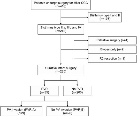
Fig. 1. Patients selection.
role brought about by PVR.
10-15In this regard, the purpose of this study is to verify the therapeutic role of PVR for HC. To this end, we com- pared perioperative morbidity, mortality rate, and long-term survival outcomes between patients who received PVR and those who did not. In addition, we analyzed the risk factors affecting the prognosis of HC after surgery.
MATERIALS AND METHODS
Patients selection
We retrospectively analyzed 418 patients who had un- dergone surgery for HC during January 2005 through December 2016 at a single center. Of these patients, we excluded 176 patients with Bismuth type Ⅰ and Ⅱ and included 242 patients with Bismuth type Ⅲa, Ⅲb and Ⅳ.
Of these 242 patients, we excluded 7 patients (4 with pal- liative surgery, 2 with biopsy only, and 1 with R2 re- section). We enrolled the remaining 235 patients for anal- ysis in this study. Among the finally enrolled 235 patients, 35 received PVR and 200 did not receive PVR. Of the 35 PVR patients, 9 (25.7%) were found to have portal vein invasion on the final pathological examination (PVR-
A), and 26 (74.3%) were found to have no portal vein invasion (PVR-B) (Fig. 1). This study was approved by Institutional Review Board of Samsung Medical Center, Seoul, Republic of Korea (approval number: 2020-05-167).
Preoperative management
If preoperative hyperbilirubinemia (total bilirubin level above 3.0 gm/dl) was present before surgery, we per- formed preoperative bile drainage (PBD). The procedures we performed were percutaneous transhepatic biliary drainage, endoscopic retrograde biliary drainage, and en- doscopic nasobiliary drainage. For hypertrophy of the fu- ture remnant liver after liver resection, preoperative portal vein embolization (PVE) was performed in patients with a future remnant volume of less than 20% as measured in CT liver volumetry. We evaluated the volumetry again 2 to 3 weeks following PVE, and we undertook surgical treatment when the future remnant liver volume was 20%
or more of the total liver volume.
Surgical management
We established our strategy for liver resection and PVR
after considering the Bismuth type and the vascular tumor
invasion as identified in the preoperative imaging. We made our final decision based on intraoperative findings.
We resected the proximal bile duct along with the liver parenchyma as an en-bloc specimen. We executed PVR by using methods such as wedge resection with patch graft (1 patient) or segmental resection and end-to-end anastomosis (34 patients). We also performed hepatic ar- tery resection and anastomosis in 5 patients. To confirm that the patency of the portal vein flow to the remnant liver, we took an intraoperative Doppler sonogram before the end of surgery. We performed transection of the liver parenchyma according to the method we had described in our previous study.
16We executed a Roux-en-Y bilioen- teric anastomosis to maintain bile flow from the remnant liver to the alimentary tract. We routinely performed lymph node dissection to achieve radical surgery. The ex- tent of the Lymph node dissection was as follows; lymph nodes around the celiac trunk, the common hepatic artery, peripancreatic area and hepatoduodenal ligament.
Pathologic examination and adjuvant treatment After histological examination, we performed tumor TNM staging, using the AJCC, 7th edition.
17We micro- scopically determined the margin status of the ductal mar- gin and the radial margin. The ductal margin was eval- uated by the cut margins of the distal bile duct and the proximal bile duct. We defined R0 as being pathologically tumor-free at the margins mentioned above. We defined R1 as having the presence of invasive carcinoma. This in- stitution considers carcinoma in situ at the margin to be R0. We did not provide adjuvant treatment to all cohorts.
But, after considering TNM staging, margin status, and patient performance in multidisciplinary approach, we provided adjuvant treatment to 65 (27.7%) patients.
Investigated variables and definition
The analyzed variables were age, sex, body mass index (BMI), ASA class, follow-up duration, comorbidity, PBD, PVE, preoperative laboratory tests, adjuvant treatment, op- erating time, estimated blood loss (EBL), type of liver re- section, positive resection margin, T stage by AJCC 7th edition, tumor size, node metastasis, differentiation type, perineural invasion, lymphovascular invasion, 30-day mor- tality, and postoperative complications. We divided post- operative complications into general complications and
procedure-related complications. We described the former according to the Clavien-Dindo classification.
18The latter consisted of portal vein thrombosis, liver failure, bile leak, bleeding, intraabdominal fluid collection, intraabdominal abscess and wound problems. Liver failure defined as the development of severe acute liver damage accompanied by hepatic encephalopathy and synthetic dysfunction (INR of ≥1.5) in patients without cirrhosis or liver disease and its cutoff is an illness duration of <26 weeks. And pre- operative bilirubin was defined as the last bilirubin level before operation.
Statistical analysis
We performed statistical analyses by using the PASW Statistics version 23.0 (SPSS, IBM Corp., Armonk, NY, USA). We used the Kaplan-Meier method to calculate the overall survival rate (OSR) and median survival times.
We performed the log rank test to compare survival curves. We included all recurrences and tumor-related deaths in the disease-free survival rates (DFS) analysis. We also performed univariate and multivariate Cox regression analysis to identify prognostic factors for OSR and DFS.
In univariable analysis, we considered p<0.1 to be signi- ficant. We included parameters with p<0.1 in a multi- variable Cox proportional hazards regression analysis to identify the risk factors for prognosis. Statistical signifi- cance was indicated at p<0.05 in multivariate analysis.
RESULTS
Demographics and perioperative details Table 1 summarizes the demographics and perioper- ative details of the PVR group (n=35) and the No PVR group (n=200). The proportion of preoperative biliary drainage was significantly higher in PVR than in No PVR (85.7% vs. 61.5%; p=0.006). There was no significant dif- ference in the proportion of preoperative PVE between the two groups (22.8% vs. 18.5%; p=0.642). The median val- ue of the estimated blood loss of the PVR group was sig- nificantly greater than that of No PVR group (1143.2±
1034.51 vs. 688.0±442.3 ml; p<0.001). Right hepatectomy
was performed more frequently in the PVR than in the
No PVR group (94.3% vs. 72%; p=0.001). In detail, right
trisectionectomy (45.7% vs. 19%), extended right hemi-
hepatectomy (22.9% vs. 28%), right hemihepatectomy (20%
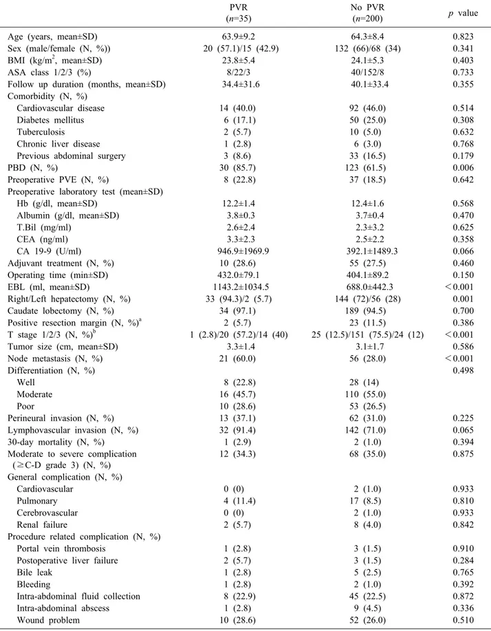
Table 1. Demographics and perioperative details
PVR (n=35)
No PVR
(n=200) p value
Age (years, mean±SD) 63.9±9.2 64.3±8.4 0.823
Sex (male/female (N, %)) 20 (57.1)/15 (42.9) 132 (66)/68 (34) 0.341
BMI (kg/m2, mean±SD) 23.8±5.4 24.1±5.3 0.403
ASA class 1/2/3 (%) 8/22/3 40/152/8 0.733
Follow up duration (months, mean±SD) 34.4±31.6 40.1±33.4 0.355
Comorbidity (N, %)
Cardiovascular disease 14 (40.0) 92 (46.0) 0.514
Diabetes mellitus 6 (17.1) 50 (25.0) 0.308
Tuberculosis 2 (5.7) 10 (5.0) 0.632
Chronic liver disease 1 (2.8) 6 (3.0) 0.768
Previous abdominal surgery 3 (8.6) 33 (16.5) 0.179
PBD (N, %) 30 (85.7) 123 (61.5) 0.006
Preoperative PVE (N, %) 8 (22.8) 37 (18.5) 0.642
Preoperative laboratory test (mean±SD)
Hb (g/dl, mean±SD) 12.2±1.4 12.4±1.6 0.568
Albumin (g/dl, mean±SD) 3.8±0.3 3.7±0.4 0.470
T.Bil (mg/ml) 2.6±2.4 2.3±3.2 0.625
CEA (ng/ml) 3.3±2.3 2.5±2.2 0.358
CA 19-9 (U/ml) 946.9±1969.9 392.1±1489.3 0.066
Adjuvant treatment (N, %) 10 (28.6) 55 (27.5) 0.460
Operating time (min±SD) 432.0±79.1 404.1±89.2 0.150
EBL (ml, mean±SD) 1143.2±1034.5 688.0±442.3 <0.001
Right/Left hepatectomy (N, %) 33 (94.3)/2 (5.7) 144 (72)/56 (28) 0.001
Caudate lobectomy (N, %) 34 (97.1) 189 (94.5) 0.700
Positive resection margin (N, %)a 2 (5.7) 23 (11.5) 0.386
T stage 1/2/3 (N, %)b 1 (2.8)/20 (57.2)/14 (40) 25 (12.5)/151 (75.5)/24 (12) <0.001
Tumor size (cm, mean±SD) 3.3±1.4 3.1±1.7 0.586
Node metastasis (N, %) 21 (60.0) 56 (28.0) <0.001
Differentiation (N, %) 0.498
Well 8 (22.8) 28 (14)
Moderate 16 (45.7) 110 (55.0)
Poor 10 (28.6) 53 (26.5)
Perineural invasion (N, %) 13 (37.1) 62 (31.0) 0.225
Lymphovascular invasion (N, %) 32 (91.4) 142 (71.0) 0.065
30-day mortality (N, %) 1 (2.9) 2 (1.0) 0.394
Moderate to severe complication (≥C-D grade 3) (N, %)
12 (34.3) 68 (35.0) 0.875
General complication (N, %)
Cardiovascular 0 (0) 2 (1.0) 0.933
Pulmonary 4 (11.4) 17 (8.5) 0.810
Cerebrovascular 0 (0) 2 (1.0) 0.933
Renal failure 2 (5.7) 8 (4.0) 0.842
Procedure related complication (N, %)
Portal vein thrombosis 1 (2.8) 3 (1.5) 0.910
Postoperative liver failure 2 (5.7) 3 (1.5) 0.284
Bile leak 1 (2.8) 5 (2.5) 0.765
Bleeding 1 (2.8) 2 (1.0) 0.392
Intra-abdominal fluid collection 8 (22.9) 45 (22.5) 0.872
Intra-abdominal abscess 1 (2.8) 9 (4.5) 0.336
Wound problem 10 (28.6) 52 (26.0) 0.510
aResection margin positive means invasive carcinoma at margin
bAccording to AJCC 7th edition
BMI, body mass index; PBD, preoperative bile drainage; PVE, portal vein embolization; T. bili, total bilirubin; CEA, carcinoem- bryonic antigen; CA19-9, cancer antigen 19-9; EBL, estimated blood loss; C-D grade, Clavien-Dindo classification
Fig. 2. Kaplan-Meier survival analysis (A) 5-year overall survival rate (OSR) of PVR and No PVR (B) 5-year disease-free survival rates (DFS) of PVR and No PVR (C) 5-year OSR for PVR-A, PVR-B and No PVR (D) 5-year DFS for PVR-A, PVR-B and No PVR.
vs. 25.5%), left trisectionectomy (2.9% vs. 3.5%), extended left hemihepatectomy (8.6% vs. 16%) and left hemihepa- tectomy (0% vs. 8%) were performed. T3 stage and node metastases were observed more frequently in the PVR than in the No PVR group (40% vs. 12%; p<0.001 and 60% vs. 28%; p<0.001). There was no significant differ- ence in proportion of positive resection margins between the two groups (5.7% vs. 11.5%; p=0.386).
The 30-day mortality developed in one (2.9%) case in PVR group and two (1.0%) cases in No PVR group (p=0.394). There was no significant difference in pro- portion between the two groups having portal vein throm- bus (2.8% vs. 1.5%; p=0.910), liver failure (5.7% vs.
1.5%; p=0.284), bile leakage (2.8% vs. 2.5%; p=0.765), bleeding (2.8% vs. 1.0%; p=0.392) and intraabdominal
fluid collection (22.9% vs. 22.5%; p=0.872).
Survival analysis
There was no significant difference in the 5-year OSR between the PVR and No PVR groups (37.7% vs. 42.6%;
p=0.2300) (Fig. 2A). The 5-year DFS of the PVR and No PVR groups was 29.6% and 27.7% (p=0.379) (Fig. 2B).
Fig. 2C, D shows a 5-year OSR and DFS for PVR-A, PVR-B and No PVR groups. There was no significant dif- ference in the 5-year OSR and DFS between the three groups. The 5-year OSR for PVR-A, PVR-B and No PVR groups were 33.3%, 40.4% and 42.6%, respectively (p=
0.479). The 5-year DFS of the three groups was 44.4%,
22.9% and 27.7%, respectively (p=0.576).
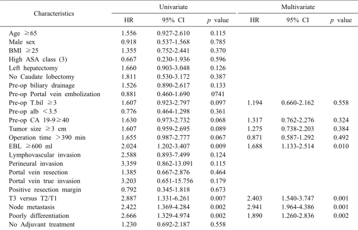
Table 2. Univariate and multivariate analysis of risk factor for overall survival rate (OSR)
Characteristics Univariate Multivariate
HR 95% CI p value HR 95% CI p value
Age ≥65 1.556 0.927-2.610 0.115
Male sex 0.918 0.537-1.568 0.785
BMI ≥25 1.355 0.752-2.441 0.370
High ASA class (3) 0.667 0.230-1.936 0.596
Left hepatectomy 1.660 0.903-3.048 0.126
No Caudate lobectomy 1.811 0.530-3.172 0.387
Pre-op biliary drainage 1.526 0.890-2.617 0.133 Pre-op Portal vein embolization 0.881 0.460-1.690 0741
Pre-op T.bil ≥3 1.607 0.923-2.797 0.097 1.194 0.660-2.162 0.558
Pre-op alb <3.5 0.776 0.464-1.298 0.361
Pre-op CA 19-9≥40 1.630 0.973-2.732 0.068 1.317 0.762-2.276 0.324
Tumor size ≥3 cm 1.607 0.959-2.695 0.089 1.275 0.738-2.203 0.384
Operation time >390 min 1.655 0.987-2.777 0.067 0.871 0.587-1.292 0.492
EBL ≥600 ml 2.024 1.202-3.407 0.009 1.688 1.133-2.514 0.010
Lymphovascular invasion 2.588 0.893-7.499 0.124
Perineural invasion 3.359 0.862-13.091 0.115
Portal vein resection 1.385 0.667-2.876 0.464
Portal vein true invasion 3.203 0.651-15.756 0.179 Positive resection margin 0.792 0.345-1.818 0.673
T3 versus T2/T1 2.887 1.331-6.261 0.007 2.403 1.540-3.747 0.001
Node metastasis 2.422 1.369-4.284 0.002 2.941 1.964-4.386 0.001
Poorly differentiation 2.666 1.329-4.974 0.002 1.890 1.260-2.836 0.002
No Adjuvant treatment 1.230 0.692-2.187 0.558
HR, hazard ratio; CI, confidence interval; BMI, body mass index; T. Bili, total bilirubin; CA19-9, cancer antigen 19-9; EBL, estimated blood loss
Risk factor for overall survival rate (OSR)
Table 2 summarizes the risk factor for overall survival rate (OSR). In univariate analysis, preoperative bilirubin
≥3 mg/dl, preoperative CA 19-9 ≥40, tumor size ≥3 cm, operation time >390 min, EBL ≥600 ml, T3 versus T2/T1, node metastasis and poor differentiation sig- nificantly affected, OSR (hazard ratio [HR]=1.607, 95%
confidence interval [CI] 0.923-2.797; p=0.097, HR=1.603, 95% CI 0.973-2.732; p=0.068, HR=1.607, 95% CI 0.959- 2.695; p=0.089, HR=1.655, 95% CI 0.987-2.777; p=0.067, HR=2.024, 95% CI 1.202-3.407; p=0.009, HR=2.887, 95%
CI 1.331-6.261; p=0.007, HR=2.422, 95% CI 1.369-4.284;
p=0.002 and HR=2.666, 95% CI 1.329–4.974; p=0.002).
EBL >600 ml (HR=1.688, 95% CI 1.133-2.514; p=
0.010), T3 versus T2/T1 (HR=2.403, 95% CI 1.540-3.747;
p=0.001), node metastasis (HR=2.941, 95% CI 1.964-4.386;
p=0.001), and poor differentiation (HR=1.890, 95% CI 1.260-2.836; p=0.002) were identified as independent risk factors in multivariate analysis. PVR and portal vein true invasion were not independent risk factors for OSR.
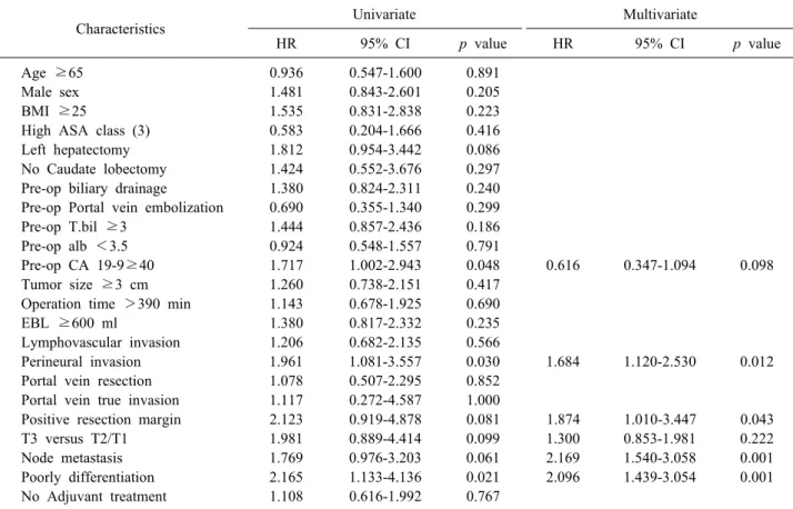
Risk factor for disease free survival (DFS) Table 3 summarizes the risk factor for disease free sur- vival (DFS). In univariate analysis, preoperative CA 19-9
≥40, perineural invasion, positive resection margin, T3 versus T2/T1, node metastasis and poor differentiation sig- nificantly affect DFS (HR=1.717, 95% CI 1.002-2.943; p=
0.048, HR=1.961, 95% CI 1.081-3.557; p=0.030, HR=
2.123, 95% CI 0.919-4.878; p=0.081, HR=1.981, 95% CI 0.889-4.414; p=0.099, HR=1.769, 95% CI 0.976-3.203;
p=0.061, and HR=2.165, 95% CI 1.133-4.136; p=0.021).
Perineural invasion (HR=1.684, 95% CI 1.120-2.530;
p=0.012), positive resection margin (HR=1.874, 95% CI 1.010-3.447; p=0.043), node metastasis (HR=2.169, 95%
CI 1.540-3.058; p=0.001), and poor differentiation (HR=
2.096, 95% CI 1.439-3.054; p=0.001), were identified as
independent risk factors in multivariate analysis. PVR and
portal vein true invasion were not independent risk factor
for DFS.
Table 3. Univariate and multivariate analysis of risk factor for disease free survival (DFS)
Characteristics Univariate Multivariate
HR 95% CI p value HR 95% CI p value
Age ≥65 0.936 0.547-1.600 0.891
Male sex 1.481 0.843-2.601 0.205
BMI ≥25 1.535 0.831-2.838 0.223
High ASA class (3) 0.583 0.204-1.666 0.416
Left hepatectomy 1.812 0.954-3.442 0.086
No Caudate lobectomy 1.424 0.552-3.676 0.297
Pre-op biliary drainage 1.380 0.824-2.311 0.240 Pre-op Portal vein embolization 0.690 0.355-1.340 0.299
Pre-op T.bil ≥3 1.444 0.857-2.436 0.186
Pre-op alb <3.5 0.924 0.548-1.557 0.791
Pre-op CA 19-9≥40 1.717 1.002-2.943 0.048 0.616 0.347-1.094 0.098
Tumor size ≥3 cm 1.260 0.738-2.151 0.417
Operation time >390 min 1.143 0.678-1.925 0.690
EBL ≥600 ml 1.380 0.817-2.332 0.235
Lymphovascular invasion 1.206 0.682-2.135 0.566
Perineural invasion 1.961 1.081-3.557 0.030 1.684 1.120-2.530 0.012
Portal vein resection 1.078 0.507-2.295 0.852
Portal vein true invasion 1.117 0.272-4.587 1.000
Positive resection margin 2.123 0.919-4.878 0.081 1.874 1.010-3.447 0.043
T3 versus T2/T1 1.981 0.889-4.414 0.099 1.300 0.853-1.981 0.222
Node metastasis 1.769 0.976-3.203 0.061 2.169 1.540-3.058 0.001
Poorly differentiation 2.165 1.133-4.136 0.021 2.096 1.439-3.054 0.001
No Adjuvant treatment 1.108 0.616-1.992 0.767
HR, hazard ratio; CI, confidence interval; BMI, body mass index; T. Bili, total bilirubin; CA19-9, cancer antigen 19-9; EBL, estimated blood loss
DISCUSSION
Several previous studies have reported that the pro- portion of Bismuth type III and IV in HC are at least 31%.
9,19Extended hepatectomy should be performed for curative resection of this type of advanced HC. The prob- lem is that such aggressive surgical excision increases morbidity and mortality after surgery. Furthermore, fre- quent tumor invasion into the portal vein because of the anatomical characteristics of HC has been recognized as an important factor limiting R0 resection. Nevertheless, various efforts have been made to achieve R0 resection through extended resection with PVR in several large centers. Two recent meta-analyses on the suitability of PVR have been conducted in terms of oncologic and sur- gical outcomes. Among these, one meta-analysis that W.
Chen et al.
20conducted included 13 studies (9 Asian groups, 2 USA groups, and 2 European groups). The results of this analysis showed that the PVR groups had a worse OSR than did the No PVR groups (HR=1.90, 95% CI
1.59-2.28; p<0.00001). This can be explained by the re- sult of the PVR group having more LN metastases (HR=
1.50, 95% CI 1.06-2.13; p=0.02) and a higher proportion of perineural invasion (HR=2.95, 95% CI 1.80-4.84; p<
0.0001) than did the No PVR group. The fact that the cu- rative resection was done in a smaller proportion in the PVR group (HR=0.65, 95% CI 0.46-0.91; p=0.0) also seems to have brought about the worse survival outcomes. How- ever, there was no significant difference between the two groups in postoperative mortality and morbidity. Another meta-analysis reported by Bai et al.
21was based on 21 ret- rospective studies and 2403 patients. This study showed a similar trend compared to the analysis of Chen et al.
20The lymphatic invasion (HR=1.14, 95% CI 1.02-1.28;
p=0.02) and perineural invasion (HR=1.31, 95% CI 1.05-
1.64; p=0.01) were more frequently observed in the PVR
group. The rate of curative resection was lower in the
PVR group than in No PVR group (HR=0.89, 95% CI
0.75-0.99; p=0.03). Patients undergoing PVR experienced
a higher postoperative mortality and a worse OSR (HR=
1.52, 95% CI 1.06-2.18; p=0.02 and HR=0.67, 95% CI 0.49-0.91; p<0.001). Postoperative morbidity was not significantly different between the two groups (HR=1.06, 95% CI 0.94-1.02; p=0.35). However, one cannot make these results into a simple generalization that the surgical oncologic outcome of the PVR group is worse than that of the No PVR group. This is because the tendency to have more advanced disease in the patient group under- going PVR likely caused these results. We urgently need a prospective randomized control study because the above-mentioned meta-analysis used prior retrospective studies. On the other hand, the findings that there were no differences in morbidity in both the PVR and the No PVR groups in two meta-analyses shows us that PVR is acceptable in terms of safety.
In the current study, advanced T stage and LN meta- stases were observed more frequently in the PVR group than in the No PVR group (40% vs. T3: 12%; p<0.001 and 60% vs. 28%; p<0.001), which is in the same con- text as the previous reports. Nonetheless, there was no significant difference in OSR, DFS, 30-day mortality or postoperative morbidity in either group. Of note is that unlike the previous meta-analyses, the prognosis of pa- tients undergoing PVR is not inferior to that of the No PVR group. Considering that the achievement of R0 re- section is an important factor for a good prognosis of HC,
22a surgical strategy that increases the possibility of a negative margin rate through PVR needs to be consid- ered for the long-term survival of the selective cases of HC. Although PVR was not an independent risk factor for OSR and DFS in this study, the findings that R1 status is an independent risk factor for DFS also supports this view.
Among the patients in this study who underwent PVR, the rate of microscopic portal vein invasion was 25.1%.
There were no significant differences in OSR and DFS be- tween patients with or without microscopic portal vein in- vasion. A meta-analysis performed by Abbas et al.
23reported that true microscopic portal vein invasion did not signifi- cantly affect survival using the previous seven retrospec- tive studies (HR=1.59, 95% CI 0.75-3.35; p=0.22).
10-12,15,24-26The percentage of patients having pathologically confirmed disease with microscopic portal vein invasion in these seven studies ranged from 22% to 88%. Microscopic vas- cular invasion was not found to have a significant effect
on survival in a multicenter study of 305 cases consisting of cohorts in four USA and three European institutions.
Our study also revealed that microscopic portal vein in- vasion is not a risk factor for OSR or DFS.
This study has some limitations. First, the absence of a protocol for adjuvant treatment is likely to have acted as a bias in the analysis of DFS. Although we determined the type of adjuvant treatment considering TNM staging and various oncologic factors, oncologist and radiologist’s preferences may have been involved. Second, the number of PVR patients, which is relatively small compared to the No PVR patients, may have made a solid statistical analysis difficult. Finally, as a selection bias, patients in the PVR group were selected for patients with operability, so it is considered that there is a limit to comparison with the non-PVR group. However, this study has implications for comparable survival to No PVR patients when PVR was performed in operable patients with only PV invasion.
In the future, large-scale prospective randomized con- trolled trials will minimize bias in studies that may occur in the patient selection process and will be necessary to validate the outcome of the PVR.
PVR does not increase postoperative mortality or morbidity. Although the PVR group has a more advanced disease state than the No PVR group, the PVR showed similar oncological results compared to the No PVR.
Given these findings, PVR should be actively performed if necessary, after careful patient selection.
CONFLICT OF INTEREST
Ki Beom Kim, Dong Wook Choi, Jin Seok Heo, In Woong Han, Sang Hyun Shin, Yunghun You and Dae Joon Park have none to declare.
ORCID
Ki Beom Kim: https://orcid.org/0000-0002-0563-8375
Dong Wook Choi: https://orcid.org/0000-0002-0751-7841
Jin Seok Heo: https://orcid.org/0000-0001-6767-2790
In Woong Han: https://orcid.org/0000-0001-7093-2469
Sang Hyun Shin: https://orcid.org/0000-0002-2533-4491
Yunghun You: https://orcid.org/0000-0002-0684-7523
Dae Joon Park: https://orcid.org/0000-0002-1285-9248
AUTHOR CONTRIBUTIONS
Conceptualization: DWC, KBK, JSH, IWH, SHS, YY, DJP. Data curation: KBK, YY, DJP. Methodology: DWC, KBK, JSH, IWH, SHS, YY, DJP. Project administration:
DWC, JSH, IWH, SHS. Visualization: KBK. Writing - original draft: KBK. Writing - review & editing: DWC, KBK, JSH, IWH, SHS, YY, DJP.
REFERENCES
1. Khan SA, Thomas HC, Davidson BR, Taylor-Robinson SD.
Cholangiocarcinoma. Lancet 2005;366:1303-1314.
2. Ito F, Cho CS, Rikkers LF, Weber SM. Hilar cholangiocar- cinoma: current management. Ann Surg 2009;250:210-218.
3. Seyama Y, Kubota K, Sano K, Noie T, Takayama T, Kosuge T, et al. Long-term outcome of extended hemihepatectomy for hilar bile duct cancer with no mortality and high survival rate.
Ann Surg 2003;238:73-83.
4. Sano T, Shimada K, Sakamoto Y, Yamamoto J, Yamasaki S, Kosuge T. One hundred two consecutive hepatobiliary resections for perihilar cholangiocarcinoma with zero mortality. Ann Surg 2006;244:240-247.
5. Lazaridis KN, Gores GJ. Cholangiocarcinoma. Gastroenterology 2005;128:1655-1667.
6. Blechacz B, Gores GJ. Cholangiocarcinoma: advances in patho- genesis, diagnosis, and treatment. Hepatology 2008;48:308-321.
7. Blechacz B, Komuta M, Roskams T, Gores GJ. Clinical diag- nosis and staging of cholangiocarcinoma. Nat Rev Gastroenterol Hepatol 2011;8:512-522.
8. Deoliveira ML, Schulick RD, Nimura Y, Rosen C, Gores G, Neuhaus P, et al. New staging system and a registry for perihilar cholangiocarcinoma. Hepatology 2011;53:1363-1371.
9. de Jong MC, Marques H, Clary BM, Bauer TW, Marsh JW, Ribero D, et al. The impact of portal vein resection on outcomes for hilar cholangiocarcinoma: a multi-institutional analysis of 305 cases. Cancer 2012;118:4737-4747.
10. Neuhaus P, Jonas S, Bechstein WO, Lohmann R, Radke C, Kling N, et al. Extended resections for hilar cholangiocarcinoma. Ann Surg 1999;230:808-818; discussion 819.
11. Ebata T, Nagino M, Kamiya J, Uesaka K, Nagasaka T, Nimura Y. Hepatectomy with portal vein resection for hilar cholangio- carcinoma: audit of 52 consecutive cases. Ann Surg 2003;238:720- 727.
12. Miyazaki M, Kato A, Ito H, Kimura F, Shimizu H, Ohtsuka M, et al. Combined vascular resection in operative resection for hilar
cholangiocarcinoma: does it work or not? Surgery 2007;141:581- 588.
13. Hirano S, Kondo S, Tanaka E, Shichinohe T, Tsuchikawa T, Kato K. No-touch resection of hilar malignancies with right hep- atectomy and routine portal reconstruction. J Hepatobiliary Pancreat Surg 2009;16:502-507.
14. Hirano S, Kondo S, Tanaka E, Shichinohe T, Tsuchikawa T, Kato K, et al. Outcome of surgical treatment of hilar chol- angiocarcinoma: a special reference to postoperative morbidity and mortality. J Hepatobiliary Pancreat Sci 2010;17:455-462.
15. Hemming AW, Mekeel K, Khanna A, Baquerizo A, Kim RD.
Portal vein resection in management of hilar cholangiocarci- noma. J Am Coll Surg 2011;212:604-613; discussion 613-616.
16. Song SC, Choi DW, Kow AW, Choi SH, Heo JS, Kim WS, et al. Surgical outcomes of 230 resected hilar cholangiocarcinoma in a single centre. ANZ J Surg 2013;83:268-274.
17. Edge SB. AJCC cancer staging manual. 7th ed. New York:
Springer, 2010.
18. Dindo D, Demartines N, Clavien PA. Classification of surgical complications: a new proposal with evaluation in a cohort of 6336 patients and results of a survey. Ann Surg 2004;240:205- 213.
19. Kawarada Y, Das BC, Naganuma T, Tabata M, Taoka H.
Surgical treatment of hilar bile duct carcinoma: experience with 25 consecutive hepatectomies. J Gastrointest Surg 2002;6:617-624.
20. Chen W, Ke K, Chen YL. Combined portal vein resection in the treatment of hilar cholangiocarcinoma: a systematic review and meta-analysis. Eur J Surg Oncol 2014;40:489-495.
21. Bai T, Chen J, Xie ZB, Ma L, Liu JJ, Zhu SL, et al. Combined portal vein resection for hilar cholangiocarcinoma. Int J Clin Exp Med 2015;8:21044-21052.
22. Kloek JJ, Ten Kate FJ, Busch OR, Gouma DJ, van Gulik TM.
Surgery for extrahepatic cholangiocarcinoma: predictors of survival. HPB (Oxford) 2008;10:190-195.
23. Abbas S, Sandroussi C. Systematic review and meta-analysis of the role of vascular resection in the treatment of hilar cholan- giocarcinoma. HPB (Oxford) 2013;15:492-503.
24. Muñoz L, Roayaie S, Maman D, Fishbein T, Sheiner P, Emre S, et al. Hilar cholangiocarcinoma involving the portal vein bi- furcation: long-term results after resection. J Hepatobiliary Pancreat Surg 2002;9:237-241.
25. Song GW, Lee SG, Hwang S, Kim KH, Cho YP, Ahn CS, et al. Does portal vein resection with hepatectomy improve survival in locally advanced hilar cholangiocarcinoma? Hepatogastroen- terology 2009;56:935-942.
26. Nagino M, Nimura Y, Nishio H, Ebata T, Igami T, Matsushita M, et al. Hepatectomy with simultaneous resection of the portal vein and hepatic artery for advanced perihilar cholangiocarci- noma: an audit of 50 consecutive cases. Ann Surg 2010;252:115- 123.