임상병리검사과학회지
:제 29권 제 1 호
1997.PCR 방법에 의한 Shigella 속 균주와
enteroinvasive E. coli 검 출방법 의 확렵
원자력병원 임상병리과 1 고려대학교 생명공학원2 이윤상1.2
.홍석일 1. 강운형 1. 박영인 2.
Detection of Shigella s Pf cies and enteroinvasive E. coli by PCR
Lee,
Yun-
Sang
1.2.,HOng
, Seok-
Il1•,Kang
,WOOn-HYUngl.
,Park
,YOUng In
21J
ePt. 01 Clinical Pathology, Korea Cancer Center Hospital 2Graduate School 01 Biotechnology, Korea University, Seoul, KoreaInvasive Shigella species and enteroinvasive E. coli (EIEC) are enteropathogenic for humans causing dysenteries with abdominal pain and frequent stools containing blood and mucus. To identify these microorganisms, biochemical and serological methods have been used in most clicinal laboratories. However, these tests have difficulties in distinguishing each strains and can not be performed in a routine laboratory procedure on large numbers of stools. Therefore, isolates thought to be invasive Shigella species or EIEC are required to be confirmed by animal inoculation or tissue culture studies. Recent discovery of a few virulence plasmid-encoded loci from has provided the altemative approach such as hybridization assay or polymerized chain reaction (PCR) to identify these strains.
In this study, the optimal thermocycler conditions was established and the sensitivity of PCR was estimated by the cell numbers as well as by the amount of DNA for each standard strains: Shigella dysenteriae (ATCC 9752), Shigella flexneri (ATCC 9403), E.
coli 029 (ATCC 43892), and E. coli 0124 (ATCC 43893). In addition, one strain of Shigella flexneri strain, two strains of Shigella sonnei and E. coli 0157 strain isolated from patients stools were investigated.
PCR was performed with DNA extracted from suspension of each organism diluted in the range of 108 to 102 cells or with total cellular DNA in the concentration of 101 to 10-6
μ
g/μ
using primer Sh-1 and Sh-2, foIlowed by the detection of 320bp DNA fragment.The most suitable reaction conditions consisted of 40 cycles of 1 min at 94.C (denaturation), 1min at 55.C (annealing), and subsequently 1 min at 72.C (extension).
The sensitivity estimated by the ce11 number was 104 for Shige11a dysenteriae (A TCC 9752), Shigella flexneri (A TCC 9403), and E. coli 029 (A TCC43892) and 100 for E. coli 0124 (ATCC 43893). The organisms
is이ated
from the patient stools exhibited sensitivity of 106 for al1 Shigella flexneri strain, 107 for one Shigella sonnei strain and 108 for another Shigella sonnei strain. However, no bands were produced from the E. coli 0157 strain. On the other hand, the sensitivity estimated by amount of DNA was10-
2μg for
Shigella dysenteria (ATCC 9752) and E. coli 029 (ATCC 43892), and10-
3μg for
Shigella flexneri (ATCC 9403) and E. coli 0124 (ATCC 43893) For the organism obtained from patient specimens the sensitivity was 10-3 for one a11 Shigella flexneri strain, 10-1 for one Shigella sonnei strain and 101 for another Shigella sonnei strain. No bands were from the E. coli 0157 strain.PCR can be used as good alternative method to identify invasive Shigella species and EIEC, since this method is sensitive, and rapid, and does not require selective enrichment or high ce11 numbers. In the future, more studies with clinical specimens are required to evaluate the conditions of PCR examined in this investigation.
Kev words Invasive shige/la, enteroinvasive E. coli
1 .
서 론
이 질균
(Shigellasp.)과
enteroinvasive E.coli-
(EIEC)은 예로부터 어린아이에서부터 노인 에 이르기까지 광범위하게 감염되는 설사질환의 원인균으로 일반적으로 위생상태가 나쁘고 인구 밀도가 조밀한 곳에서 많이 발생되며 특히
1-5세 사이의 소아에게서는 심한 설사를 나타내는 것으로 알려져 있다(1) 이질질환은 salmone11a증 과는 달리 환자의 혈액에서 분리되는 일이 거의 없고 대변이 주 가검물이 되고 있어 검증을 어 렵게 하고 있다. 환자의 검체물로부터 원인균을 찾아내어 질환을 확정하는 것이 정확한 치료의 필수적인 선행조건이므로 위의 여러문제에도 불 구하고 임상적 측면에서 빠르고 간편한 검증법 이 다양하게 시도되고 있다(2,3)
1. 이질균의 특징
Shigella는 그람음성 간균 (Grarn
negativebacilli)으로서 현미경에 의한 관찰로는 다른 장내세균들과 구별되는 특징은 관찰하기 어려
우며 통성혐기성이고 호염성을 가진다(4,5) Shigella감염증의 발생빈도를 지역별로 살펴 보면, 선진국에서는
Shigellasonnei가 가장 많 고 그 다음이
Shigella flexneri, Shigellabφ'dii,
Shigelladysenteriae순으로 나타난다.
그러나 위생시설이 나쁜 개발도상국 또는 후진 국에서는 거꾸로 S. dysenteriae와 S. boydii가 가장 많이 나타나며 그 다음이
S. flexneri, S.sonnez순서로 나타난다. 우리나라에서는 B군인
S.
flexneri가 거의 대부분을 차지하고 그 다음 이
S.sonn낌 이 며,
S.dysenter따e에 의 한 감 염증은 거의 볼 수 없다(6)
2. 이질균 감염증세
shigella
감염에 의한 증상범위는 증상이 없
는 경우에서 부터 고열, 오한, 경련, 복통, 후 중, 설사 및 혈변을 나타내는 심한 경우까지 다양하다.
가장 정확한 진단은 대변배양을 통하여 병원
성 shigella를 검출하는 것이므로 대변을 이용
하여 균체를 검색하는 것이 일반적인 방법인데
shigella는 대변속의 산에 감수성이 높기 때문 에 될 수 있는 한 가검물을 빨리 배지에 접종 해야 한다. 설사가 시작된 후 3 일이내에 대변 배양을 실시한 경우는 약 90% 정도의 검체물로 부터 배양양성율을 보이나, 일주일 이상 지난 경우는 단지 75% 정도만이 양성으로 나타나고
있다 (7,8)
shigella의 주 병원소가 바로 인간이기 때문 에 shigella는 분변-경구 경로를 통해 사람에게 서 사람으로 전달이 된다. 즉 shigella에 감염 된 환자 또는 보균자의 대변을 통해 배출된 균 은 음식물, 사람의 손 또는 파리 등의 곤충에 의해 다른 사람에게 전파된다(9)
정 등은 우리나라에서의
Shigellasp. 의 월별 발생빈도를 보면 1 년중 5월이 발생빈도가 가장 높았고 2월이 가장 낮은 것으로 나타났으며,
지역적인 발생분포는 충남이 가장 높고 다음으 로 강원, 전북의순으로 보고하였다. 연령에 의 한 분포는 9세 이하가 가장 많았고 다음이
60대 이상이었으며 성별분포는 여성이 남성보다 높았다고 보고하였다(10)
3. Shigella
sp. 와
enteroinvasive E.coli
검출방법
많은
E. coli균주들은 shigella와 밀접하게 연관되어 있으며 shigella와 같이 대장 상피세 포를 뚫고 들어가 위장관계질환을 야기시킬 수 있다. 이러한 균주는 O항원 생산을 담당하는 큰 플라스미드(120-140 megadalton)를 가지고 있으며 (lLl2), 때로는 장관의 점막을 침입하여 shigella에 의한 이질증세와 비슷한 증상을 보 이기도 하며 이 경우 사망율도 높다. 현재까지
Shigella sp, enteroinvasive E. coli
의 효과적 인 검출방법은 대변을 배양하여 생화학적인 특 성을 보는것으로서
methyl red반응 양성,
Voges-Proskauer
반응 음성
, urease가수분해 음성
, lysine decarboxylase음성
, glucose양 성, H2S 음성,
gas음성등이며, 그외에도
mo-tility
음성 또는 이 세 균을
guinea pig의 눈속 에 접종후 충혈된 것을 보는 방법이다(13,14)
본 실 험 에 서 는
Shigella sp.와
enteroinvasive E.coli의 검색시에 소요되는 시간과 인력을 줄일 수 있는 효율적인 방법으로
polymerase chain reaction (PCR)분석법을 이용한
IAL (invasive associate lows) loci범 위 에 서
320bpband을 검출하여
Shigellasp. 와
enteroinva- sive E. coli (EIEC)검색을 시도하였다05,16)
ll.
재료 및 실험방법
1 .
재료
1)
사용균주
국립보건원에서 분양받은
S. dysenteriae (ATCC 9752), S. flexneri (ATCC 9403)의 2종 과 미 국에 서 구 입 한
enteroinvasive E. coli 029 (ATCC 43892), E. coli 0124 (ATCC43893) 와 환자의 대변으로부터 분리한
S flexneri, S. sonnei및
E. coli0157를 사용하
였다.
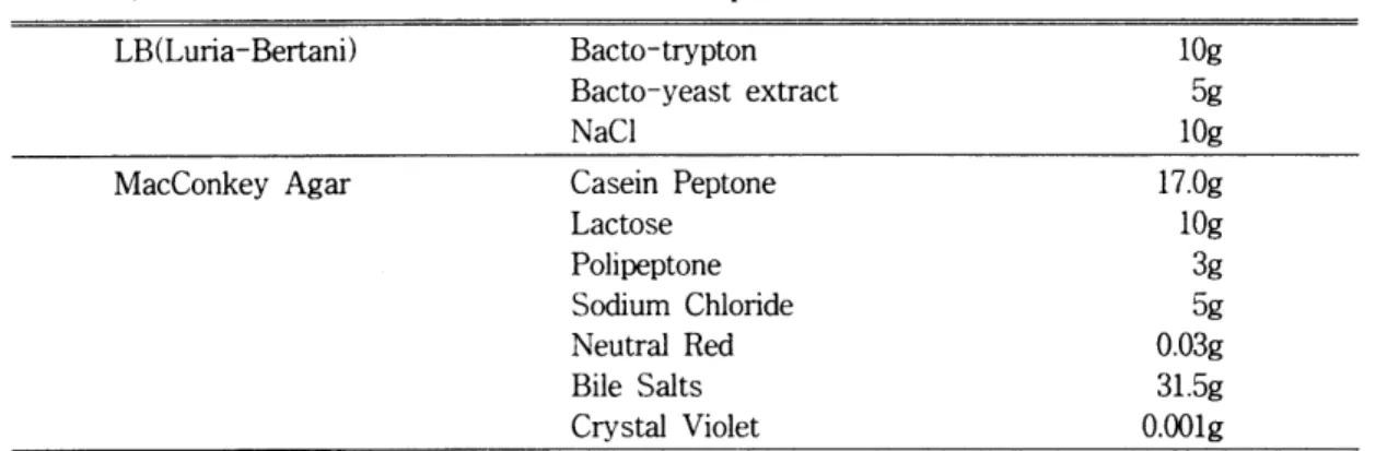
2) 배지류
실험에 사용한 배지의 종류및 조성은
Table1 과 2에 나타내었으며, 사용된 시약인 trγpton 과
yeastextract는 Difo社, NaCl은
Junsei ChemicalCo. 의 제품을 사용하였다.
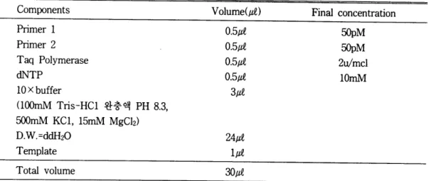
3) 시 악
실험에 사용된
Taq polymerase, 10 X buffer,pnmer는
· 한국생공(주)에서 인공합성한 것을사용하였고 (Table
3)dNTP는
Pharmacia Co.로 부터 구입하여 사용하였다.
시약의 조성은
Table4에 나타내었다.
2.
실험 방법
1)균주배앙
LB
액체배지 값띠에 실험균주 S. dysentriae
(ATCC 9752),S.
j1exneri(A TCC여03),
E. coli 029(ATCC4잃92),
E. coli 0124(ATCC염893) , 환자로 부터 분리 한 균주인
S. j1exneri, S.sonnei
2주와
E. coli0157를 각각 단일 집락을 백금이로 따서 접종시켜 3TC 에서 18시간 정도 배양 하였다.
LB액체배지 따띠에 배양한 세포 액 40μM 를 접종하여 2시간 동안 3TC 에서 배양
한 후
spectrophotometer (Beckman DU 650)을 이용하여 흡광도 600nm에셔
,측정하여 10
8cell을 모은후
102 cello1되도록 6단계 연속희 석 하였다. 10
8-10
2균체수 까지 희석한 균체를 PCR을 하기 위한 시료사용하였다.
DNA The- rmalCycler는
PERKIN ELMERCo. 제품을 사 용하였다.
Table 1. Media used in the culture of bacteria(17). (per liter) LB(Luria-Bertani) Bacto-trypton
Bacto-yeast extract NaCl
MacConkey Agar Casein Peptone
Lactose Polipeptone Sodium Chloride Neutral Red Bile Salts Crystal Violet
Table 2.
M어ia used in the culture of bacteria
(17). (per liter) Triple Sugar Iron Agar(TSI Agar)
Brain Heart Infusion Agar
Pancreatic Digest of Casein Peptic Digest of Animal Tissue Sodium Chloride
Lactose Sucrose Dextrose
Ferrous Ammonium Sulfate Sodium Thiosulfate
Phenol Red Agar
Brain Heart, infusion from (Solids) Peptic Digest of Animal Tissue Pancreatic Digest of Casein Sodium Chloride
Dextrose
Disodium Phosphate Agar
lOg 5g 10g 17.0g lOg 3g 5g O.03g 31.5g O.OOlg
1O.Og 10.0g 5.0g 10.0g 1O.Og l.Og O.2g O.2g O.025g
13.0g 8.0g 5.0g 16.0g
5.0g 2.0g 2.5g 13.5g
Table 3. Primer used for detection of Shigella species and EIEC(8).
Name Primer: Sh-l Primer : Sh-2
Nucleotide sequence, 5’ to 3’
CTG GAT GGT ATG GTG ADD GGA GGC CAA CAA TTA TTT CC Th primers were derived from the i머 loci of the invasion plasmid.
Table 4. The optimum condition of PCR determined for Shigella dysentriae, Shigella flexneri,
E. coli
0'
29, E. coli 0124, Shigella flexneri, Shigella sonneP,2) and, E. coli 0157 DNA.Components Primer 1 Primer 2 Taq Polymerase dNTP
10xbuffer
(100mM Tris-HCl
완충액
PH 8.3 500mM KC1, 15mM MgClz)D.W.=d바120
Template Total volume
Volume{때)
0.5μ 0.5μ
0.5따
0.5μ3μe
24μM 1μM 30μM
Final concentration
밍pM
50p
M
2ψmcl
10mM
PCR was performed for 40 cycles of denaturation at 94 "c for 1 min, annealing at 55.C for lmin, and extension at 72.C for lmin.
2)
DNA추출
DNA
추출은 4종의 세포인
S. dysentriae (ATCC 9752), S. flexneri (ATCC 9403), E.coli 029 (ATCC 43892), E. coli 0124 (ATCC
43893) 와 환자로 부터 분리 한 균주인 s
flexneri, S. sonnei
2주
, E. coli 0157배 양 액 1.5me 을
microcentrifugetube에 넣고
1 xSSCE solution(O.l5M NaCl, 15mm sodium citrate, lmM EDT A)0.5m~을 넣 어 pellet을 현 닥시 켰 다. 다음으로
20% SDS solution25μM을 넣 고 10mg/m~
lysozyme solution50μM을 넣어
3TC에서 30분 동안 반응시킨 후
Proteinase K solution30따을 넣고 55
0C 에서 30분동안 반응
시키고
RNase1μM 을 넣어 20분 동안 반응시켰
다. 다음
Phenol0.5m~ 넣 어 l분간
vortexing한후 microcentrifuge에서 15,α)()rpm의 속도로 5분간 원심분리한 후 상층액을 취하여 새로운
eppendorf
tube에 옮겼다. 여기에 동일한 양의
chloroform을 가하여 추출한 후
3M sodium acetate solution50뼈을 넣 어 주고
isopropyl alcohol0.5ml을 넣 은 후
-200C맹 동실 에 서
30분이상 하여 DNA를 침전시키고
microcen-trifuge
15,아뻐pm의 속도로 10분간 원심분리한
후 상층액을 제거 하였다. 알콜올도 완전히 제
거한 후 멸균 증류수를 넣어 pellet를 녹이고
spectrophotometer(Pharmacia社,
Model LKB ultrospec3) 을 이용하여 260nm에서의 홉광도
를 측정하여 DNA농도를 1μg/1μM 되도록 맞춘
다음 10 1μ g/μ~ -10-
6μg/때까지 희 석 하여 PCR에
사용하였다.
m. 컬 과
국립보건원에서 분양받은 s.
dysentriae (ATCC 9752), S. flexneri (ATCC9403) 와 미 국에 서 구입 한
E. coli 029 (ATCC 43892), E.coli 0124 (A TCC
43893) 을 가지고 연구를 계 속하였다. 온도는 변성
(denaturation) 94.C1 분,
pnmer의 결합(annealing)
55"c에서 1 분,
DNA사슬의 연장(elongation) 72.C 에서 1 분의 조건 으로
40cydes을 실시하였다. 그 결과
Figure1 에서 나타난 것과 같이 검출되는 세포의 최소 농도에 는 조금씩 차이 가 났다. 즉 S.
dysentria (ATCC 9572), S. flexneri (ATCC 9403), E.coli 029 (A TCC
43892) 는
104세 포 농도일 때 까지 검출되었고,
E. coli 0124 (ATCC 43893)는 10
5세포 농도까지 검출되었다. 이와 같이
실험균주의 종류에 따라 검출에 필요한 최소희 석배수가 달랐으나 최소농도
108-104세포수
의 범위내에서 검출이 되었다.
실험균주의 DNA를 PCR한 경우에는
Figure2에서 나타난 것과 같이
S. dysentria (A TCC 9572), E. coli 029 (ATCC43892)는 10-
2μg/μM 농도까지 320bp의 product가 검출되고 S flexneri와
E. coli 0124 (ATCC43893)는
10-3μ g/따 농도까지 product가 검출되었다. 이와같 이 DNA희석을 이용한 방법에서도 실험균주의 종류에 따라 검출되는 것이 다르게 나왔으며 최소농도 10
1μg/따 -10-
3μg/μM 범위내에서 검출 이 되었다.
고려대학교 안암병원에서 구한 환자균주인
S. flexneri, S. sonnei
2주,
E. coli0157 의
plasmid
DNA를 template로 하여 PCR을 실 시
A 8 7 6 5 4 B 8 7 6 5 4 C 8 7 6 5 4 D 8 7 6 5
500bp
F ig. 1. Agarose gel electrophoretic patterns of PCR products of Shigella sp. and enteroinvasive E. coli.
PCR was performed for 40 cycles of denaturation at 94.C for 1 min, annealing at 55.C for 1 min, and extension at 72"C for 1 min.
L~mes
are : A. size marker (lKb ladder) : Shigella dysentriae 108 to 104;B. size marker (lKb ladder) : Shigella Flexn_eri lOö to 10'1;
C. size marker (lKb ladder) : E. coli 029 10ö to 10'1;
D. size marker (lKb ladder) : E. coli 0124 108 to 105;
A 1 -1 -2 B 1 -1 -2 -3 C 1 -1 -2 0 1 -1 -2 -3
500bp-'
F i g. 2. Agarose gel electrophoretic pattems of PCR products of Shigella sp. and enteroinvasive E. coli.
PCR was performed for 40 cycles of denaturation at 94 oC for 1 min, annealing at 55 t for 1 min, and extension at 720C for 1 min.
Lanes are : A. size marker (lKb ladder) : Shigella dysentriae 101 to _10-2;
B. size marker (IKb ladder) :
와úgella
flexneri 101 to 10-3;C. size marker (lKb ladder) : E. coli 029 101 to 10-2;
D. size marker (lKb ladder) : E. coli 0124 101 to 10-3;
A 8 7 6 B 8 7
C 8
D500bp ....
Fig.3. Agarose gel electrophoretic pattems of PCR products of Shigella sp. and enteroinvasive E. coli.
PCR was performed for 40 cycles of denaturation at 940C for 1 min, annealing at 55 oC for 1 min. and extension at 72
t
for 1 min.Lanes are : A. size marker (lKb ladder) :
와ligella
flexneri 108 to 106;B. size marker (IKb ladder) : Shigella sonnei (1) 100 to 10’ ; C. size marker (lKb ladder) : Shigella sonnei (2) 108;
D. size marker (IKb ladder) : E. coli 0157
A 1 -1 -2 -3
B 1 -1 C 1 D...
500bp
-를-Fig.4. Agarose gel electrophoretic pattems of PCR products of Shigella sp. and enteroinvasive E. coli.
PCR was performed for 40 cyc1es of denaturation at 94 DC for 1 min, annealing at 55 t for 1 min, and extension at 72 DC for 1 min.
Lanes are : A. size marker (IKb ladder) : Shigella fiexneri 101 to 10-3; B. size marker (1Kb ladder) : Shigella sonnei (1) 101 to 10-1 ;
C. size marker (IKb ladder) : Shigella sonnei (2) 101;
D. size marker (IKb ladder) : E. coli 0157
하였다. 그 결과
Figure12에 나타난것과 같이 N. 고 활
s. flexneri은
106세 포수까지 product가 나타났
고 s.
sonnei(1)는
107세포수까지 나타났으며 설사질환의 주원인균인 Shigella sp. 와
S. sonnei
(2)은
108세포수에서 나타났으나 E.
enteroinvasive E.coli관계 질환은 과거보다
coli
0157은 product가 나타나지 않았다. 실험 현저하게 감소되었으나 위생시설이 빈약할 때
균주의 DNA희석 실험에서는
S.flexneri은 에 대인접촉, 음식물, 식수로부터 감염되어 이 10-
3μg/따 농도까지 나타났고 s.
sonnei(1)은 차적으로 감염된 가족사이에서의 이환율이 10-
1μg/μM 농도까지,
S. sonnei(2)은 10
1μg/μf 30-50%에 달한다(19) 우리나라에서는 매년 발 농도에서 나타났으며
E. coli0157는 band가 생하는 이질균증 중 85%가
S.flexneri로 대부 나타나지 않았다. 이와같은 결과로 볼 때 검체 분을 차지하고 있으며 병원성 대장균에 의하여
물에 세포는 10
8 celV
mf - 1 04celV
mf범위에서, 발병하는 장질환도 최근들어 많은 연구가 되고
DNA로부터의
Shigella sp, enteroinvasive E.있다. 따라서 장질환의 하나인
Shigellasp. 와
coli
의 존재 확인이 가능하고 DNA농도는
101 enteroinvasive E. coli병원성을 생화학적 및 μg/μf-10하g/μM 범위내에서 검출이 가능하다고 혈청학적으로 검출하여 왔는데 그 방법이 어렵
볼 수 있다. 이와같은 조건을 충족시킨 경우 독 고 시간이 오래 걸린다. 본 논문에서는 아직까
소형
Shigella sp, enteroinvasive E.coli의 확 지 국내에서는 실시하고 있지않은 PCR방법을
인이 PCR방법으로 가능하다고 결론지었다. 이용하여 빠르고 신속하게 이들 병원균의 존재
유무를 제시하고자 하였다.
정 (19) 등은
S. flexneri병원성을 알아보는 방 법연구에 관한 논문에서
guineapig의 한쪽에
6
x10
9 cells/me을 접종하여 각결막염 양성반응
을 보인 8주중 2주만이 24시간내에 초기증상이 확인되었고 나머지 6주는 48시간내에 증상이 나타내었다고 보고 하였다. 또한 Surgaila는 근 래에
S.flexneri의 병원성에는 140 MDa의 plasmid가 관여하며, 이 plasmid가 없는 균주 는
gumeaplg의 결막에 염증을 일으키지 못하 고,
HeLacell도 침투하지 못하는 것으로 해석 하였고 이는 상피세포를 침투하는데 필요한 기 능을 조절하거나 결정하는 것으로 보고하였다 (때) 또한 이들에 의해 제시된 방법에 비교하여 PCR을 이용한 검색은 단 시간내에 경제적으로 병원균을 확인할수 있다는 점에서 활용이 가능 하다고 할 수 있다.
REFERENCES
1. Taylor, D. N. (1
986).
The Role of Shigella spp., Enteroinvasive Escherichia coli, and other Enteropathogens as Ca- uses of Childhood Dysentery in Thailand.j. Infectious Diseases.
153, 1132-1138.
2.
Rowe, B.(1983).
Antigenic relationships(1987).
DNA probes for identification for enteroinvasive Escherichia coli. ]. Clin.Microbiol. 25,
2025-2027.
5. 이건섭. (1989). 진단병원 미생물학. 고려의 학 424-425.
6. 정태화.
(1984). 한국에서 분리된
Salmo- nella, Shigella균 속의
R-Palsmid내 성 에 관한연구
Report of NIH Korea.21
,79-96.
7.
Jawetz, E., Melnick, ]. L., and Adelberg, E.A. (1987).
Review of Medical Microbi- ology.17 , 243-244.
8.
Haider, K., Huq, M. 1., SAmadi, A. R., and Ahmad, K. (1985).
Plamid characteri- zation of Shigella spp.is이ated
from children with shigellosis and asymptoma- tic excretors. ]. Antimicrobial Chemothe- rapy.16
,691-698.
9.
Joklik, W. K.,W피et,
H. P., and Amos D.B. (1
984).
Zinsser Microbiology.18
,614- 616.
10. 정태화. (1986). 한국에서 분리된 장내세균
(Salmonella, Shigella, E.
coli균속)의 병 원 적 역할에 관한 연구.
Report of NIH Ko- æa.21, 73-96 .
• ‘i_ Buysse j. M., Stover C. K., Oaks E. V., Venkatesan M., and kopecko D. j.
(987).
between the enteroinvasive Escherchia Molecular cloning of invasion plasmid coli
0
antigens _ and shigella0
antigens. antigen (ipa) genes fromflexm Shigella j. Clin. Microbiol17, 681-684.
flexneri : analysis of ipa gene products3.
Sethabutr. 0., Venkatesan. M., Murphy. and genetic mopping. ]. Bacterol.169
,G. S., Eampokalap. B., Hoge. C.W., and
껑61-2569.
Echeverria. P. (1993). Detection of
12.
SansJnetti, P. ].,Ko야cko,
D. ]., and Shigellae and Enteroinvasive E. coli by Formal, S. B.(1982).
Involvement of a amplification of the invasion plasmidantigen H DNA sequence in patientys with dysentery. ]. Infectious Diseases.
167
,458-46
1.4.
Gomes, T.A.T., Toledo, M.R.F., Trabulsi, L. R., Wood, P. K., and Morris, ]. G.Plasr:rid in the Invasive Ability of Shigella flexneri. Infection and Immunity.
852-860.
13. 이 명 원. (1982). 한국에 서 분리 된
Shigella균속에 관한 세균학적 역학조사연구.
Re-pαi
of NIH Korea.19
,69-78.
14.
Okamura, N. (1983). HeLa Cell Invasive- ness and 0 Antigenof Shigella flexneri as Separate and Prerequisite Attrigutes of Virulence to Evoke Keratoconjuncti- vitis in Guinea Pigs. Infection and Imrnunity.505-513.
15.
Frankel, G., Riley, L., Giron, ]. A., Valmassoi, ]., Fridmann, A., Stro-ckbine, N., Falkow, S., and Schoolnik, G. K.(1990). Detection of Shigella in feces using DNA amplification. ]. Infect. Dis.
161. 1252-1256.
16.
Venkatesan, M., Buysse, ]. M., Vanden-따ies,
E., and Kopecko, D ]. (1988).
De- velopment and Testing of invasion- Associated DNA Probes for Detectio ofShigella Spp. and Enteroinvasive Es- cherichia coli. ], Clinical Microbiology.