www.ksmkorea.org / www.ksov.org 25
ORIGINAL ARTICLE
J Bacteriol Virol. Vol 50. No 1. March 2020; 50(1): 25-34 https://doi.org/10.4167/jbv.2020.50.1.025
eISSN 2093-0249
JBV
Expression and Purification of Recombinant Mayaro Virus Envelope Glycoproteins E1 and E2 to Develop a Mayaro Virus Detection System
So Yeon Yi
1,2, Kyungah Yoon
3, Jungsun Kwon
1, Kyoon Eon Kim
2, Kyoungsook Park
1,4*, Yong Beom Shin
1,5*1
BioNano Health Guard Research Center, 125 Gwahak-ro, Yuseong-gu, Daejeon 34141, Republic of Korea
2
Department of Biochemistry, College of Natural Sciences, Chungnam National University, 99 Daehak-ro, Yuseong-gu, Daejeon 34134, Republic of Korea
3
Department of Clinical Laboratory Science, Daejeon Health Institute of Technology, Daejeon 34504, Republic of Korea
4
Department of General Education, Daejeon Health Institute of Technology, Daejeon 34504, Republic of Korea
5
Bionanotechnology Research Center, Korea Research Institute of Bioscience and Biotechnology (KRIBB), 125 Gwahak-ro, Yuseong-gu, Daejeon 34141, Republic of Korea
Corresponding Kyoungsook Park Yong Beom Shin
Phone : +82-42-860-4445 +82-42-860-4449 E-mail : [email protected].
Received : January 09, 2020 Revised : March 02, 2020 Accepted : March 04, 2020
No potential conflict of interest relevant to this article was reported.
Copyright © 2020 Journal of Bacteriology and Virology
©This is an Open Access article distributed under the terms of the Creative Commons Attribution Non-Commercial License (http://creativecommons.org/
license/by-nc/3.0/).
Mayaro virus (MAYV) is a mosquito-transmitted alphavirus that produces an acute, usually non-fatal, febrile illness including Mayaro fever. Like other alphaviruses, the MAYV E1 and E2 envelope glycoproteins are major viral surface antigens that play a key role in host recognition and infection. Here, we report expression and purification methods for recombinant MAYV E1 (rE1) and rE2 using a baculovirus system. Enzyme-linked immunosorbent assays (ELISA) revealed that rE1 and rE2 were antigenic and reacted with human anti–MAYV IgG and IgM. Cross-reactivity was also confirmed with human anti-Chikungunya virus (CHIKV) IgG and IgM. Furthermore, we developed an immunochro- matographic strip test (IST) with rE2 to diagnose MAYV infection. Thus, purified rE2 may be valuable tool for rapidly diagnosing MAYV infection.
Key Words: Baculovirus/insect cell system, recombinant envelope glycoprotein 1 (rE1), recombinant envelope glycoprotein 2 (rE2), immunochromatographic strip test (IST), Mayaro virus (MAYV)
INTRODUCTION
Mayaro virus (MAYV) was found on the island of Trinidad and has caused small outbreaks in several parts of tropical South America. MAYV is a single strand positive RNA virus of the alphavirus genus in the family Togaviridae and is similar to chikungunya virus (CHIKV) (1-3). Like other alphaviruses, MAYV is an arbovirus (arthropod-borne virus) which is mostly transmitted by Haemagogus mosquitoes. Recent studies have shown that Aedes aegypti mosquitos can spread MAYV worldwide (4-6).
MAYV infection causes a CHIKV-like, acute, usually non-fatal, febrile illness with a 3-5 day duration that includes fever (known as Mayaro fever), joint pain, headache, rashes, and muscle ache. Some patients develop persistent, terrible joint pain that lasts up to a year after infection (7, 8). MAYV infection can be confirmed by viral isolation, RT-PCR, and serological diagnosis using hemagglutination
JBV
VOL 50. NO 1. March 202026 Copyright © 2020 Journal of Bacteriology and Virology
Journal of
Bacteriology and Virology
inhibition tests and enzyme-linked immunosorbent assays (ELISA); however, these methods are unresponsive for a great number of infectious samples and live virus analysis may require biosafety laboratories. Furthermore, due to the antigenic similarity of MAYV and CHIKV, there is an acute need to develop more specific and precise methods for diagnosing MAYV infection (9).
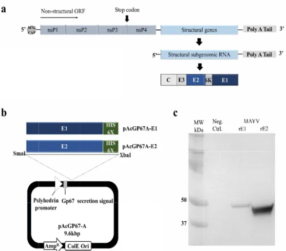
The MAYV genome is a 12kbp long single-stranded RNA containing two open reading frames (ORFs). It encodes four non-structural proteins (nsP1, nsP2, nsP3, and nsP4) and five structural proteins (C, E3, E2, 6K, and E1). The structural envelope glycoproteins E1 and E2 are imbedded in the envelope on the viral surface. E1 mediates the fusion of the virus with the host cell, while E2 is mainly involved in attaching viruses to host cells (10). As for alphaviruses, E1 and E2 are targets of the anti-MAYV antibody response; therefore, E1 and E2 would be useful targets for immunodiagnostic analysis.
In this study, we explain the generation of soluble MAYV E1 and E2 using a baculovirus/insect expression system.
Recombinant envelope proteins E1 (rE1) and rE2 were successfully expressed and purified in soluble forms, with rE2 better expressed and more stable than rE. ELISA revealed that rE1 and rE2 were antigenic and reactive for specifically detecting human anti–MAYV IgG or IgM. Cross-reactivity was also evaluated using human anti–CHIKV IgG or IgM. In particular, rE2 displayed a higher ELISA value than rE1. Furthermore, we developed an immunochromatographic strip test (IST) using rE2 to detect human anti-MAYV IgG or IgM, demonstrating the potential applicability of rE2 in a diagnosis system to detect MAYV infection.
MATERIALS AND METHODS
Construction of recombinant expression vector containing MAYV envelope glycoproteins E1 and E2
Synthetic MAYV DNA encoding E1 (GenBank # KT818520, residues 1~442) or E2 (GenBank # KT818520, residues 1~421) and a C-terminal six histidine tag (His–tag) were obtained from Bioneer. Two enzyme sites, SmaI and XbaI, were created at the 5’ and 3’ ends of the E1 and E2 genes, respectively. E1 and E2 were amplified by PCR using the following primers: E1F, 5’–CCCGGGGGATCCTACGAGCACACG–3’; E1R, 5’–TCTAGATTGCG CCCAAGTCATTGCAGTGCTGGATA–3’; and E2F, 5’–
CCCGGGGGATCCAGCACTGC AAAT–3’; E2R, 5’–TCTAGACGTAGGATGCAGTCCGTAGTAGTATTC–3’ (restriction sites underlined). The PCR products were cloned into corresponding sites in pAcGP67A (Pharmingen). The E1 and E2 genes inserted downstream of the robust polh promoter to create pAcGP67A–E1 and pAcGP67A–E2.
Insect cell culture and transfection
The insect cell line Spodoptera frugiperda (Sf9) was cultured in Insect Sf-900 SFM serum-free media (Invitrogen, Waltham, Massachusetts, USA) at 28°C. Sf9cells were co-transfected with BaculogoldTM DNA (Pharmingen, San Diego, California, USA) and the recombinant vector pAcGP67A–E1 or pAcGP67A–E2, as demonstrated by the manufacturer. Several rounds of culture were performed to amplify the recombinant virus and a high viral titer stock solution was collected. To express the protein on a bigger scale, Trichoplusia ni BTI–TN–5B1–4 (High FiveTM) insect cells (Invitrogen, Waltham, Massachusetts, USA) were grown in Insect Hi-Express serum-free media previous infection with the high titer virus. High FiveTM insect cells were used to express MAYV rEP1 and rEP2 after infection with the recombinant baculovirus.
Purification of recombinant envelope protein (rE1) and rE2
Typically, 1 L of recombinant baculovirus–infected insect cell cultures was harvested approximately 72 h post-infection and the cells were centrifugated at 5000×g for 15 min to recover only the supernatant. The supernatant was dialyzed against binding buffer (50 mM Tris–HCl, pH 8.0, 0.5 M NaCl), loaded onto a Ni2+–nitrilotriacetic acid (Ni-NTA) agarose column
www.ksmkorea.org / www.ksov.org 27 (Qiagen, Hilden, Germany) equilibrated with the binding buffer, and washed with washing buffer (50 mM Tris–HCl, pH 8.0, 0.5 M NaCl, 50 mM imidazole). rE1 and rE2 were eluted with elution buffer (50 mM Tris–HCl, pH 8.0, 0.5 M NaCl, 500 mM imidazole) and the presence of purified protein was confirmed by sodium dodecyl sulfate–polyacrylamide gel electrophoresis (SDS–PAGE). The eluted glycoproteins were dialyzed with Slide–A–Lyzer (3.5 K MWCO; Thermo Scientific, Waltham, Massachusetts, USA) in phosphate buffered saline (PBS, pH 7.4) overnight at 4°C, and stored in PBS (pH 7.4) or 5% (v/v) glycerol at –70°C.
Sodium dodecyl sulfate-polyacrylamide gel electrophoresis (SDS-PAGE) and western blot analyses
The supernatants were separated on NuPAGE Novex 4–12% Bis–Tris SDS-PAGE gels in MES-SDS running buffer (Invitrogen, Waltham, Massachusetts, USA) and visualized by staining with Coomassie brilliant blue. Gel images were obtained using a gel documentation system (AE–9000 E–graph, ATTO Corp, Tokyo, Japan).
For western blot analysis, proteins were separated on SDS-PAGE gels and transferred to polyvinylidenedifluoride (PVDF) membranes using a Trans–Bolt system (Bio–Rad, Hercules, California, USA). The membranes were blocked with 5 % skim milk in 1× PBS containing 0.05% Tween–20 (PBST) for one hour at 25°C, probed with mouse anti–His–tag antibodies (Bethyl Laboratories, Inc., Montgomery, Texas, USA) at a 1:3000 dilution for one hour at 25°C, washed six times with PBST, and incubated with HRP–conjugated goat–anti mouse IgG antibodies at a 1:10,000 dilution for 1 h at 25°C. To visualize antibodies bound to the proteins, ECL luminal kits were used as enzyme substrates and the blots were visualized on a chemiluminescence imaging system (WSE–6200H LuminoGraph II, ATTO Corp, Tokyo, Japan).
Mayaro antigen-specific ELISA
rE1 and rE2 reactivity against human anti–MAYV IgG and IgM was tested (11). A 96-well microtiter plate (Costar 3690, Corning Inc., New York, USA) was coated with 100 ng/well of purified rE1 or rE2 diluted to 1 µg/mL in 0.1 M carbonate–
bicarbonate buffer (pH 9.0) overnight at 4°C. Unbound antigens were discarded and the wells were blocked with 2% BSA in PBS for 60 min at 25°C. After washing, the wells were incubated at 37°C for 2 h with MAYV positive controls (human anti–MAYV IgG or IgM) from an anti–MAYV ELISA kit (Euroimmun, Luebeck, Germany). CHIKV positive controls (human anti–CHIKV IgG or IgM) from an anti-CHIKV ELISA kit (Euroimmun, Luebeck, Germany) were used to test cross-reactivity.
The plates were washed six times with PBST and incubated at 37°C for 1 h with HRP-conjugated anti–human IgG or IgM diluted at 1:10,000. Bound antibodies were detected by adding 3, 3′, 5, 5′–tetramethylbenzidine (TMB, Sigma, St. Louis, Missouri, USA) and optical density was measured at 450 nm using an ELISA microplate reader (Thermo Scientific, Waltham, Massachusetts, USA). Bars represent the average absorbance of three measurements and the error bars indicate standard deviations (SD) of triplicate measurements.
Immunochromatographic Strip Test (IST) method
The IST system in this study used a working solution (PBST) and test strips. Nitrocellulose (NC) membranes, laminated cards, absorbent pads, and sample pads were obtained from Millipore. Red latex beads were supplied by Expedeon. Goat anti–mouse IgG antibodies and other chemicals were purchased from Sigma–Aldrich. The strip sensor was composed of a sample pad, NC membrane, absorption pad, and a backing card. The latex beads were covalently conjugated to rE2 lysine residues, soaked onto the conjugate pad, and dried at 37°C for 2 h. Anti–human IgG (0.1 mg/mL) or IgM (0.1 mg/mL) antibodies were immobilized on the test line and anti–His–tag antibodies (0.1 mg/mL) were deposited on the control line on a NC membrane using a dispenser (DCI100; Zeta Corporation, Gunpo-si, Gyeonggi-do, Korea). It was assembled on a backing card with a 2 mm overlap between each component of the strip sensor. To verify IST performance, we used MAYV positive controls (human anti–MAYV IgG and IgM) from an anti–MAYV ELISA kit (Euroimmun, Luebeck, Germany) and CHIKV positive controls (human anti–CHIKV IgG or IgM) from an anti-CHIKV ELISA kit (Euroimmun, Luebeck, Germany).
JBV
28
Samples (50 Buffer–only color signal custom scrip statistically u
RESULT Construc
To express s an N-termin The MAYV E pAcGP67A selected clo the viral gen
Fig. 1. M of MAY termina pAcGP6 peroxid
Journal of Bacteriology an
0 μL of each) w (PBST with hu intensity of th pt in the Fujif using Origin so
TS
ction of re
secreted versio al gp67 secret E1 and E2 gen vector at Sma ned vectors (p nome via homo
MAYV rE1 and YV genome org al His–tags we
67A–E1- and p ase. Uninfecte
nd Virology
were pipetted uman serum a he test lines w film multi-gau oftware.
combinan
ons of MAYV tion signal pep
es, 442 bp at p aI and XbaI re pAcGP67A–E1 ologous recom
rE2 expressio ganization. (b)
re used to pu pAcGP67A–E2 ed Sf9 cell cult
onto the sam albumin) was a
as quantified uge program
nt baculovi
rE1 and rE2, w ptide for recom positions 1~44 striction sites, or pAcGP67A mbination to p
n using a reco ) Schematic of rify the MAYV -transfected S tivation media
ple pad and m also tested as by calculating (Fuji photo fi
irus to exp
we generated mbinant protei
42 of E1 and 4 respectively ( A–E2 plasmid) produce recom
mbinant bacu f the pAcGP67
V E1 and E2 Sf9 cells using
was used as a
Copyrigh migrated along a negative co the average p lm Co, Ltd., T
press MAY
two recombin n secretion an 421 bp at posit (Fig. 1a and b and wild type mbinant baculo
lovirus/insect 7A–E1 and pAc
proteins. (c) W anti–His–tag a a negative con
t © 2020 Journa g the strip mem ontrol (Neg. C
pixel intensity Tokyo, Japan).
YV rE1 and
nant expressio d a C–termina tions 1~421 of ). Sf9 insect c e viral DNA. Ea ovirus (12, 13)
system. (a) Sch cGP67A–E2 co Western blot a antibodies con ntrol (Neg. Ctr
VOL 50. NO 1
al of Bacteriolog mbrane by cap Ctrl.). For data inside the tes . Data sets w
d rE2 in ins
on constructs i l His 6X tag fo f E2, were inse cells were tran ach gene was
.
hematic repres onstructs gene
analysis of me njugated to ho
l.).
1. March 2020
y and Virology pillary action.
analysis, the t line using a ere analyzed
sect cells
ncorporating or purification.
erted into the nsfected with inserted into
sentation erated. C–
edia from orseradish
www.ksmkor Sf9 cells we baculovirus flask). West calculated m
Overexp recombin
Soluble rE1 baculovirus infection.
rE1 and rE2 terminal His SDS–PAGE ( rE2 were 0.2 rE1 and rE2 immunoblot
Antigeni
To assess th (Euroimmun anti-MAYV cross-reactiv
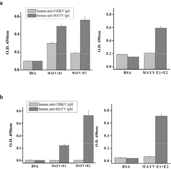
Fig. 2. An MAYV rE MAYV r antibodi the Neg.
rea.org / www.k re directly infe stock. rE1 and tern blot anal molecular mass
ression an nant bacu
and rE2 were (14) and then
secreted into –tag in rE1 an (Fig. 2a), and t
2 mg/L and 2 2 resulted in a
t analysis using
c rE1 and
he antigenicit n, Luebeck, G
ELISA kit. Hu vity with the rE
nalysis of MAY E1 and rE2. Th E1 and rE2. A es conjugated . Ctrl.
ksov.org ected with cel d rE2 expressio lyses revealed ses of rE1 and
nd purifica loviruses
e overexpresse secreted into
the medium w d rE2. The pro their concentr mg/L at a con a protein reco g the anti-His6
rE2 chara
ty of purified Germany) wit man anti–CHI E1 and rE2 MA YV rE1 and rE2 he gel was sta After being tra d to horseradis
l culture medi on by the infe protein band rE2.
ation of M
ed by infectin the extracellu
were purified u oteins were elu rations were e
centration of overy of ~60%
6X antibodies
acterization
rE1 and rE2 h positive (h KV IgG or IgM AYV ELISAs.
2 overexpressio ined with Coo ansferred to a h peroxidase.
ium containing ected Sf9 cells ds at 50 kDa
MAYV rE1
g a new High ular medium. T
using a Ni–NTA uted with bind established usi
1×10⁷ High–Fi
% and purity (Fig. 2b).
n
, we measure uman anti–M M ELISAs (Eur on and purific omassie brillian a PVDF memb Uninfected H
g the recombi was studied o (Fig. 1c) and
and rE2 i
h–Five insect c The highest rE1
A affinity colum ding buffer con ng Bradford a iveTM cells/mL.
of 95 %. Th
ed their antig AYV IgG or roimmun, Lue ation by High nt blue R-250.
brane, the pro igh–FiveTM inse
nant baculovi on a small sca d 48 kDa, cor
n High–Fiv
cell batches w 1 and rE2 yield
mn, which can ntaining 500 m
ssays. The tot The Ni2+ affin e purified pro
genic reactivity IgM) and ne beck, German FiveTM cells. (a (b) Western b oteins were de ect cell cultivat
rus to produc ale (1×10⁶ cells
rresponding w
ve
TMinsec
with high titer ds were obtain
especially bind mM imidazole tal yields of MA nity purificatio oteins were re
y using a mo egative contro ny) were used a) SDS-PAGE o blot analysis o etected by an tion media wa
29 e a high titer s in a 25 cm2 well with the
ct cells by
recombinant ned 72 h post
d with the C–
, analyzed by AYV rE1 and n of secreted ecognized by
odified ELISA ols from the d to evaluate of purified of purified ti–His–tag as used as
JBV
30
As shown in Ctrl. (BSA).
response bu reactivity wi results indic reactive than concluded t
Immuno
To estimate MAYV detec pad, and sam or IgM and a
Fig. 3 deter with stand
Journal of Bacteriology an
n Fig. 3, rE1 an IgG and IgM E ut a very low ith anti-CHIKV cate that both n rE1 with ant hat rE2 would
chromato
the ability of ction. As descr mple pad. Red anti-His-tag an
3. rE1 and rE2 rmined by ELIS 100 ng of pur dard deviations
nd Virology
d rE2 exhibite ELISAs indicate
signal value, V IgG (Fig. 3a)
proteins cou ti–MAYV IgG a d be more suita
graphic St
the IST to det ribed in Fig. 4a
latex beads w ntibodies were
2 reactivity ag SA using anti-M
rified recombi s (SD) of triplic
d significantly ed that rE1 an whilst rE2 sh , but rE2 show ld react with nd IgM and sh able for MAYV
trip Test (IS
ect human an a, the IST cons were labeled w
e immobilized
ainst human a MAYV IgG and nant proteins.
cate measurem
higher reactiv nd rE2 reacted howed higher wed no cross–
anti–MAYV Ig howed no cros V diagnosis th
ST) using
nti–MAYV IgG sisted of a bac with purified rE2
on the test an
anti–MAYV Ig d IgM or anti–
. Bars indicate ments.
Copyrigh vity with huma d with anti-MA
r reactivity tha –reactivity in th gG and IgM in
ss-reactivity fo an rE1.
human M
and IgM, we cking plate, ab
2 and loaded o nd control line
G (a) and IgM CHIKV IgG an e the mean ab
t © 2020 Journa n anti-MAYV AYV IgG and I an rE1. Addit he anti–CHIKV n infected sera
r anti–CHIKV Ig
AYV IgG a
developed a M bsorption pad,
onto the conju s, respectively
M (b). rE1 and d IgM. Each m sorbance at 4
VOL 50. NO 1
al of Bacteriolog IgG and IgM t gM; rE1 show tionally, rE1 d V IgG or IgM E a; however, rE gG and IgM. T
and IgM
MAYV rE2 ant NC membran ugate pad. Ant y.
d rE2 cross–re microplate we 450 nm. Error
1. March 2020
y and Virology than the Neg.
wed a specific displayed low ELISAs. These E2 was more Therefore, we
igen strip for ne, conjugate ti–human IgG
eactivity were ll was coated bars indicate
www.ksmkor First, sample to the conju target comp Complex ac control line anti–MAYV CHIKV ELISA on the test l IgG and IgM
Fig. 4. Sc anti–MA Normaliz control l
rea.org / www.k es including hu ugate pad, hum plexes. When t
cumulation pr and were cap IgG and IgM) A kits (Euroimm
ine of the IST M were tested
chematic illust AYV IgG and Ig zed pixel inten ine intensity. T
ksov.org uman anti–MA man anti–MAY the target com roduced a visib ptured by anti- ) and CHIKV ( mun, Luebeck, within 15 min simultaneousl
tration of the gM (c). BSA w
nsity was qua The graph pre
AYV IgG or IgM YV IgG or IgM mplexes flowed
ble color on th -His-tag antibo (human anti–C , Germany), re n (Fig. 4c). No y. Positive ban
MAYV IST ass was used as a ntified using I sents the ratio
M were dropp in the sample d across the te he test line. Fr odies (Fig. 4b CHIKV IgG or espectively. Hu nonspecific ba nds were only
say (a, b) and a Neg. Ctrl. Qua Image softwar o of signal to n
ed onto the sa e captured red est line, they w ree red latex b ). To verify IST IgM) positive man anti-MAY ands appeared observed in th
an image of M antification of re. Intensity va noise (S/N) on
ample pad. Af latex beads la were captured
eads labeled w T performance
controls from YV IgG and IgM d on the test li he MAYV posit
MAYV IST using f band intensit alues were ca
the test line.
fter the sample abeled with rE
by anti-human with rE2 flowe e, we used MA m the anti-MA
M caused a ba ine and huma tive sample.
g rE2 to detec ty at the strip lculated relati
31 e had moved 2 to produce n IgG or IgM.
ed across the AYV (human YV and anti- nd to appear n anti–CHIKV
ct human test line.
ve to the
JBV
VOL 50. NO 1. March 202032 Copyright © 2020 Journal of Bacteriology and Virology
Journal of
Bacteriology and Virology
DISCUSSION
Mayaro virus is an emerging epidemic prevalent in tropical regions of South America; its range has recently spread into Central America and Caribbean island nations (15). The virus causes acute febrile illness and can cause long term joint pain in some patients after infection. Currently, there are no permitted vaccines or treatments for MAYV infection and transmission (4). MAYV is diagnosed by virus genome-wide detection analysis using viral isolation or a viral standard;
however, this method is limited to viremic phase plasma which usually lasts no longer than 5 days (16).
Recently, the envelope proteins of alphaviruses have been studied to develop a method of MAYV diagnosis (17). MAYV encodes envelope glycoproteins; the E1 protein is primarily required in cell fusion, E2 binds to receptors of cell for entry.
E1 and E2, surface protein, are significant targets for neutralizing antibodies in the host immune response against most alphaviruses (2). Until now, serological assays for commercial MAYV diagnosis have not been available.
In this study, we expressed and purified MAYV rE1 and rE2 in a baculovirus expression system as efficient antigens for serological diagnosis. The E1 and E2 genes of MAYV were cloned into a pAcGP67A vector that used the gp67 secretory signal sequence of the baculovirus protein to secrete the recombinant proteins. The six coding codons of histidine residues downstream of the E1 and E2 genes were translated into polyhistidine peptides at the C–terminals of E1 and E2 for protein purification. Insect cell expression systems are often used for mass production of transformatively modified biologically active proteins. 48 hours after infection, viral infected cells produced a small amount of proteins recognized by anti-His 6X antibodies; the major one had a molecular weight of 50 kDa. When High FiveTM cells were infected, these proteins were nearly entirely secreted into the extracellular medium, with the highest levels obtained 3 days post-infection and decreasing thereafter due to proteases from lysed cells (18). Thus, we obtained rE1 and rE2 secreted into the extracellular medium for 72 h using High Five™ insect cells. Recombinant His–tagged rE1 and rE2 proteins were successfully eluted from the column. rE1 and rE2 purity were found to exceed 95 % in the reducing condition. The yield was 0.2 mg and 2 mg of rE1 and rE2 per liter of media, respectively, with rE1 yield much lower than rE2 yield. Smith JL, et.
al reported that MAYV rE1 and rE2 are expressed as inclusion bodies in bacterial expression system (2). However, to our knowledge, no studies have reported the expression and purification of MAYV rE1 and rE2 in insect cells.
Because infected patient samples were difficult to obtain, purified rE1 and rE2 were tested with MAYV positive controls from human anti–MAYV IgG and IgM ELISA kits (Euroimmun) to confirm their reactivities. The rE1 and rE2 ELISAs were able to detect human anti–MAYV IgG and IgM; in particular, rE2 was more reactive than rE1. Cross-reactivity between related alphaviruses is a major concern in serological diagnostic analysis (19). To solve this common problem, we measured cross–reactivity against human anti–CHIKV antibodies. As shown in Fig. 3, rE2 showed no cross-reactivity with anti–CHIKV IgG/IgM, whilst rE1 exhibited low reactivity with human anti–CHIKV IgG. Therefore, rE2 MAYV ELISA is a suitable tool for diagnosing MAYV infection which can be performed on acute serum samples and can be used for serological investigations.
The IST analysis is a recognized POCT (point–of–care testing) technique. The method enables inexpensive and rapid analysis by users (20). As shown in Fig. 4, the principle of the strip sensor was based on the immune system (antigen–
antibody reaction) between anti-human IgM and IgG (in the test line) and red latex beads labeled with rE2 complex human anti–MAYV IgG and IgM (in samples). When the sample was loaded on the strip, human anti–MAYV IgG or IgM in the samples captured red latex beads labeled with rE2 to form a target complex. During the sample moved to the end of the strip, the target complex is combined with capture antibodies on the test line to form a red signal on the test line. Free red latex beads labeled with rE2 were captured by anti–His–tag antibodies on the control line. We concluded that the strip sensor had no interaction with CHIKV. All results from the IST were checked by ELISA, and the results of the two analysis methods were relatively well matched.
In conclusion, we expressed purified MAYV rE1 and rE2 in insect cells and showed that MAYV ELISA and an IST assay
www.ksmkorea.org / www.ksov.org 33 based on rE1 or rE2 could be useful tools for detecting human anti–MAYV IgG without cross–reactivity with CHIKV, which may be useful for serological screening during possible MAYV outbreaks. Thus, we believe that MAYV rE1 and rE2 have great potential as diagnostic reagents for detecting MAYV infection.
ACKNOWLEDGMENTS
This study was supported by BioNano Health Guard Research Center, funded by the Ministry of Science and ICT (MSIT) of Korea as a Global Frontier Project (Grant number H-GUARD_2013 M3A6B2078950).
REFERENCES
1) Muñoz M, Navarro JC. [Mayaro: a re-emerging Arbovirus in Venezuela and Latin America]. Biomedica 2012;32:286- 302.
2) Smith JL, Pugh CL, Cisney ED, Keasey SL, Guevara C, Ampuero JS, et al. Human Antibody Responses to Emerging Mayaro Virus and Cocirculating Alphavirus Infections Examined by Using Structural Proteins from Nine New and Old World Lineages. mSphere 2018;3.
3) Suhrbier A, Jaffar-Bandjee MC, Gasque P. Arthritogenic alphaviruses--an overview. Nat Rev Rheumatol 2012;8:420-9.
4) Choi H, Kudchodkar SB, Reuschel EL, Asija K, Borole P, Ho M, et al. Protective immunity by an engineered DNA vaccine for Mayaro virus. PLoS Negl Trop Dis 2019;13:e0007042.
5) Powers AM, Aguilar PV, Chandler LJ, Brault AC, Meakins TA, Watts D, et al. Genetic relationships among Mayaro and Una viruses suggest distinct patterns of transmission. Am J Trop Med Hyg 2006;75:461-9.
6) Rodríguez-Morales AJ, Paniz-Mondolfi AE, Villamil-Gómez WE, Navarro JC. Mayaro, Oropouche and Venezuelan Equine Encephalitis viruses: Following in the footsteps of Zika? Travel Med Infect Dis 2017;15:72-3.
7) Auguste AJ, Liria J, Forrester NL, Giambalvo D, Moncada M, Long KC, et al. Evolutionary and Ecological Characterization of Mayaro Virus Strains Isolated during an Outbreak, Venezuela, 2010. Emerg Infect Dis 2015;21:
1742-50.
8) Tesh RB, Watts DM, Russell KL, Damodaran C, Calampa C, Cabezas C, et al. Mayaro virus disease: an emerging mosquito-borne zoonosis in tropical South America. Clin Infect Dis 1999;28:67-73.
9) Esposito DLA, Fonseca BALD. Will Mayaro virus be responsible for the next outbreak of an arthropod-borne virus in Brazil? Braz J Infect Dis 2017;21:540-4.
10) Cheng RH, Kuhn RJ, Olson NH, Rossmann MG, Choi HK, Smith TJ, et al. Nucleocapsid and glycoprotein organization in an enveloped virus. Cell 1995;80:621-30.
11) Tello D, Rodríguez-Rodríguez M, Yélamos B, Gómez-Gutiérrez J, Peterson DL, Gavilanes F. High-yield production of a chimeric glycoprotein based on permuted E1 and E2 HCV envelope ectodomains. J Virol Methods 2015;213:38-44.
12) Masoomi Dezfooli S, Tan WS, Tey BT, Ooi CW, Hussain SA. Expression and purification of the matrix protein of Nipah
JBV
VOL 50. NO 1. March 202034 Copyright © 2020 Journal of Bacteriology and Virology
Journal of
Bacteriology and Virology
virus in baculovirus insect cell system. Biotechnol Prog 2016;32:171-7.
13) Tello D, Rodríguez-Rodríguez M, Yélamos B, Gómez-Gutiérrez J, Ortega S, Pacheco B, et al. Expression and structural properties of a chimeric protein based on the ectodomains of E1 and E2 hepatitis C virus envelope glycoproteins.
Protein Expr Purif 2010;71:123-31.
14) Lombana L, Ortega-Atienza S, Gómez-Gutiérrez J, Yélamos B, Peterson DL, Gavilanes F. The deletion of residues 268-292 of E1 impairs the ability of HCV envelope proteins to induce pore formation. Virus Res 2016;217:63-70.
15) Mackay IM, Arden KE. Mayaro virus: a forest virus primed for a trip to the city? Microbes Infect 2016;18:724-34.
16) Pinheiro FP, Freitas RB, Travassos da Rosa JF, Gabbay YB, Mello WA, LeDuc JW. An outbreak of Mayaro virus disease in Belterra, Brazil. I. Clinical and virological findings. Am J Trop Med Hyg 1981;30:674-81.
17) Fumagalli MJ, de Souza WM, Romeiro MF, de Souza Costa MC, Slhessarenko RD, Figueiredo LTM. Development of an Enzyme-Linked Immunosorbent Assay To Detect Antibodies Targeting Recombinant Envelope Protein 2 of Mayaro Virus. J Clin Microbiol 2019;57.
18) Rodríguez-Rodríguez M, Tello D, Yélamos B, Gómez-Gutiérrez J, Pacheco B, Ortega S, et al. Structural properties of the ectodomain of hepatitis C virus E2 envelope protein. Virus Res 2009;139:91-9.
19) Powers AM, Brault AC, Shirako Y, Strauss EG, Kang W, Strauss JH, et al. Evolutionary relationships and systematics of the alphaviruses. J Virol 2001;75:10118-31.
20) Song C, Li J, Liu J, Liu Q. Simple sensitive rapid detection of Escherichia coli O157:H7 in food samples by label-free immunofluorescence strip sensor. Talanta 2016;156-157:42-7.